NF2 / Merlin · IHC design guide

Design Immunohistochemistry for NF2

Plan NF2 (Merlin) paraffin IHC using the catalog antibody at 0.5–1 μg/mL (datasheet A00279-2). Compare cytoplasmic staining in most tissues with nuclear staining in several tissues (HPA tissue IHC).

Evidence assembled Oct 2026 · For research use; verify linked source records and product datasheet before use
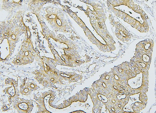
Immunohistochemistry protocol sheet for NF2 (IHC for NF2): expected localisation Cytoplasmic in most tissues; nuclear in several (HPA tissue IHC), antibody A00279-2, validated IHC image, and IHC protocol steps
Printable NF2 IHC protocol sheet — expected localisation Cytoplasmic in most tissues; nuclear in several (HPA tissue IHC), antibody A00279-2, controls and protocol steps. Open the full NF2 IHC guide →

NF2 Immunohistochemistry Experimental Design Guide

Expected localisation, validated protocols, controls and antibodies — the at-a-glance facts below, then the full design guide.

Must know before staining
Expected localisation Cytoplasmic in most tissues; nuclear in several (HPA tissue IHC)
Staining pattern Most tissues show cytoplasmic staining; some show nuclear staining (HPA tissue IHC)
Antigen retrieval Citrate pH 6 HIER, heat-mediated (datasheet A00279-2)
Positive control ⓘ Adrenal gland+4 more · see all
Negative control ⓘ None in HPA (detected in all 45 tissues); use no-primary + isotype controls
Important caveats
Reasons your staining may differ from the expected pattern.
Fixation Keep fixation conditions consistent across sections. (standard IHC practice; not target-specific)
Caveat Phosphorylation can shift nuclear localisation (UniProt)
Regulation Expression regulation is unspecified (UniProt)
Isoform / epitope 10 isoforms; no transmembrane segment; check epitope coverage (UniProt)
Section 1

Recommended NF2 IHC & IF Protocols

The catalog antibody uses citrate pH 6 retrieval (datasheet A00279-2). Three published Merlin IHC protocols provide additional conditions (PMC12605669; PMC10428583; PMC9529776).

Recommended immunohistochemistry (IHC-P) protocol parameters
SampleParaffin-embedded human mammary cancer tissues; fixative not specified (datasheet A00279-2)
FixationImage fixative and duration unreported (datasheet A00279-2); verify before use.
Sectioning4–5 µm sections on charged slides (standard)
DeparaffinisationXylene, graded ethanol series to water (standard)
Antigen retrievalHeat retrieval: Citrate pH 6, 20 min (datasheet A00279-2)
Peroxidase block3% H2O2, 10 min, room temperature (standard)
Blocking10% goat serum (datasheet A00279-2)
Primary antibodyRabbit anti-NF2, 0.5-1μg/ml (datasheet A00279-2)
Primary incubationOvernight at 4 °C (datasheet A00279-2)
DetectionStreptavidin-biotin complex (SABC), DAB chromogen (datasheet A00279-2)
CounterstainHematoxylin, blue, dehydrate and mount (standard)
Expected resultNF2-positive staining in glandular cells of adrenal gland (HPA tissue IHC: High). HPA tissue profile: Cytoplasmic expression in most tissues. Additional nuclear expression in several tissues. No signal in the no-primary control.
💡Decision noteStart with citrate pH 6 heat retrieval for the catalog antibody (datasheet A00279-2); the published high-pH condition was optimized for a different antibody clone (PMC12605669).
Section 2

What Is the Expected NF2 Staining Pattern?

NF2 (merlin) should show mainly cytoplasmic staining across many tissues, with additional nuclear staining in some (HPA tissue IHC: Supported; medium consistency with RNA). High staining is reported in selected glandular, epithelial, neuronal and glial cells (HPA tissue IHC). Membrane-associated staining is also plausible because merlin localizes to cell projections despite having no transmembrane segment (UniProt P35240: localization; topology).

What am I looking at on my slide?
Cytoplasmic staining in breast or colon glandular cells, or bronchial respiratory epithelial cells.This matches cell types scored High by HPA; compare the relevant cells with adjacent tissue structures rather than treating every cell as equally positive (HPA tissue IHC: High).
Nuclear staining accompanies cytoplasmic staining in some cells.This can be compatible with NF2: HPA reports additional nuclear staining in several tissues, and UniProt records nuclear localization. Nuclear staining alone needs comparison with controls and tissue context (HPA tissue IHC; UniProt P35240: localization).
Only an unrelated compartment stains, with no plausible cytoplasmic signal.Question specificity or detection artefact before calling NF2 positive: HPA describes predominantly cytoplasmic tissue staining, while UniProt also permits nuclear and projection-associated localization (HPA tissue IHC; UniProt P35240: localization).
Cells outside the expected tissue pattern stain strongly, including the no-primary control.Consider nonspecific binding or endogenous chromogenic detection activity; a stained no-primary control implicates the detection workflow (standard IHC practice). Check cell identity before calling cross-reactivity because HPA reports broad tissue expression (HPA tissue IHC).
No staining appears in a tissue with HPA High cells.Investigate the run before interpreting absence as loss of NF2. HPA High is an observed cell-level pattern, while its tissue IHC reliability is Supported with medium RNA–staining consistency (HPA tissue IHC).
💡Expected NF2 appearanceCall a convincing positive when the expected cells show clear cytoplasmic signal, with possible nuclear or peripheral signal, at a strength distinguishable from neighboring low-staining cells; uniform signal across unrelated cells or the no-primary control argues against specificity (HPA tissue IHC; UniProt P35240: localization; standard IHC practice).
How each factor affects the staining
Tissue and cell choiceHPA scores adrenal and breast glandular cells, bronchial respiratory epithelium, and several neural cell groups High; smooth muscle cells, chondrocytes and adipocytes are Low, not established negatives (HPA tissue IHC).
Antibody evidenceHPA003097 and CAB005385 are each marked IHC Supported. That status supports use as comparators but does not establish identical staining with another antibody (HPA antibodies: IHC Supported).
Topology and compartmentNF2 has no transmembrane segment; membrane or cell-projection enrichment can still occur alongside cytoplasmic and nuclear localization. Do not require a continuous membrane rim in paraffin sections (UniProt P35240: topology; localization).
Isoforms and phosphorylationUniProt lists ten isoforms, with isoforms 1 and 3 predominant; unphosphorylated NF2 accumulates mainly in nuclei, whereas phosphorylated NF2 is largely non-nuclear. These facts do not identify which form a given stain detects (UniProt P35240: isoforms; localization).
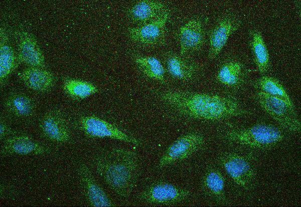
IF/ICC: where should NF2 appear?HPA reports plasma membrane as the main location, with nucleoplasm and cytosol additionally observed; its ICC-IF images include A-431, U-251MG and U2OS. Interpret this as an IF localization reference, not an IHC protocol (HPA subcellular ICC-IF).
Retrieval and detectionAntigen retrieval, blocking and chromogenic detection are general paraffin-IHC workflow variables (standard IHC practice). No supplied source establishes NF2-specific fixation sensitivity or a required retrieval condition.
Why is my staining missing, weak or wrong?
SituationLikely causeNext action
Expected High cells are unstained while the counterstain is visible.A completed counterstain does not verify antigen detection; antibody incubation, retrieval or chromogen development may have failed (standard IHC practice).Check a known-positive section in the same run and review the antibody, retrieval and detection steps; do not infer NF2 absence from one failed run (HPA tissue IHC: High cells; standard IHC practice).
Nearly every structure has diffuse brown signal.Nonspecific binding, excessive detection signal or endogenous activity can obscure cell-level localization (standard IHC practice).Inspect a no-primary control, blocking and development time; then compare the remaining signal with HPA's mainly cytoplasmic pattern (standard IHC practice; HPA tissue IHC).
Only nuclei stain in every cell.Nuclear NF2 is possible, but uniform pan-cellular nuclear staining exceeds what HPA describes as additional staining in several tissues (UniProt P35240: localization; HPA tissue IHC).Compare positive and low-staining cell populations and the no-primary control before assigning specificity; review detection background if the pattern persists (HPA tissue IHC; standard IHC practice).
Strong signal appears in smooth muscle cells or adipocytes.HPA lists these cell types as Low; a strong result may reflect assay conditions, cell misidentification or nonspecific signal (HPA tissue IHC: Low; standard IHC practice).Confirm morphology and compare a same-run High cell population; use a no-primary control to assess detection background (HPA tissue IHC: High and Low; standard IHC practice).
A faint peripheral rim is the only staining.Projection or membrane association is plausible, but a thin rim alone cannot establish NF2 identity in chromogenic IHC (UniProt P35240: localization; standard IHC practice).Check whether the rim follows identifiable cells and whether expected cytoplasmic staining appears in a High tissue; compare control sections (HPA tissue IHC; standard IHC practice).
Two antibodies give different nuclear-to-cytoplasmic patterns.NF2 has multiple isoforms and phosphorylation-linked localization; the supplied sources do not establish either antibody's isoform or phospho specificity (UniProt P35240: isoforms; localization; HPA antibodies).Review each antibody's stated validation and score compartments separately in matched tissue regions; avoid assigning an isoform or phosphorylation state from localization alone (HPA antibodies: IHC Supported; UniProt P35240: localization).

Sample controls for NF2 IHC & IF

🧪Run breast first and expect staining in its glandular cells (HPA: High in breast glandular cells; A00279-2 caption: staining in a paraffin section of human mammary cancer). HPA lists no negative tissue because NF2 is detected in all 45 scored tissues; cells used as internal background references should show no specific DAB signal above the controls, but no cell population is established here as NF2-negative (HPA: no negative tissue rows; NF2 detected in all 45 scored tissues).
Positive control tissue: Adrenal gland (Glandular cells, HPA High)
Negative control tissue: None in HPA: NF2 is detected in all 45 scored tissues. Use a no-primary (secondary-only) and an isotype control instead.
ICC-IF cell lines (HPA subcellular resource): HPA ICC-IF images show NF2 in A-431, U-251MG, U2OS, with annotated localisation: Plasma membrane (enhanced) (HPA subcellular).
Technical controls: Use a no-primary, secondary-only section and a concentration-matched rabbit IgG isotype control; an NF2-knockout sample or antigen-peptide competition can provide a biological specificity control (A00279-2 caption: rabbit primary antibody; standard IHC practice). Check endogenous peroxidase and biotin background in the breast section when using the documented streptavidin-biotin/DAB detection (A00279-2 caption: SABC/DAB; standard IHC practice).
⚠️Feasibility: A target-specific fixation window or fixation effect is unreported, and the selected A00279-2 paraffin-section caption does not state its fixative (A00279-2 caption: fixative unreported). That workflow uses heat retrieval in citrate buffer at pH 6 for 20 minutes, but the supplied evidence does not establish whether retrieval is essential (A00279-2 caption). No supplied comparison establishes whether frozen sections or IF/ICC are easier; assess biotin-related background in breast sections using SABC/DAB, while no breast-specific artefact is documented here (A00279-2 caption: SABC/DAB).

HPA tissue IHC evidence for NF2

Comprehensive Human Protein Atlas IHC scoring per tissue (reliability: Supported — Medium consistency between antibody staining and RNA expression data.). Rows are taken directly from the HPA tissue chart — click any row's HPA link to view the source.

Positive expression · recommended positive controls

TissueCell typeLevelEvidenceSource
Adrenal gland Glandular cells High Protein (IHC) HPA →
Breast Glandular cells High Protein (IHC) HPA →
Bronchus Respiratory epithelial cells High Protein (IHC) HPA →
Caudate Neuronal cells High Protein (IHC) HPA →
Cerebellum Cells in molecular layer High Protein (IHC) HPA →

Undetected expression · recommended negative controls

TissueCell typeLevelEvidenceSource
None in HPA: NF2 is detected in all 45 scored tissues. Use a no-primary (secondary-only) and an isotype control instead.
Section 3

Advanced NF2 IHC Tips

Troubleshoot NF2 (Merlin) staining in paraffin sections by checking retrieval, compartment, controls and scoring against the documented IHC workflow (datasheet A00279-2; HPA tissue IHC).

What retrieval should I start with if NF2 staining is weak?
Start with heat-mediated citrate antigen retrieval at pH 6 for 20 minutes on paraffin sections (datasheet A00279-2). The documented workflow then incubated the IHC-validated antibody at 1 μg/mL overnight at 4°C (datasheet A00279-2). If staining remains weak, check heating uniformity, buffer pH and section adhesion before changing conditions (standard IHC practice). A different retrieval buffer can be tested as a fallback on matched sections, but it has no supplied NF2 validation here (datasheet A00279-2). Compare signal and tissue damage against sections processed with the documented citrate method (standard IHC practice).
Could fixation explain inconsistent NF2 staining between paraffin blocks?
NF2-specific sensitivity to fixation is unknown from the supplied evidence (datasheet A00279-2). The documented image uses paraffin-embedded tissue, but its caption does not state the fixative, so it cannot establish a fixation condition (datasheet A00279-2). Record each block’s fixative and processing history, then compare sections with the same citrate retrieval and detection workflow (standard IHC practice; datasheet A00279-2). Excessive or uneven fixation can alter antigen accessibility in IHC generally; that possibility requires testing on the affected blocks (standard IHC practice). Include a consistently processed reference section in each run to distinguish block variation from staining-run variation (standard IHC practice).
Should I reject NF2 staining outside the cell membrane?
Assess membrane, cytoplasmic and nuclear staining separately instead of rejecting all non-membrane signal (UniProt P35240 localisation; HPA tissue IHC). Tissue IHC reports cytoplasmic expression in most tissues and additional nuclear expression in several tissues (HPA tissue IHC). NF2 also localizes to projection membranes, perinuclear cytoplasm and the nucleus, with phosphorylation associated with different nuclear distributions (UniProt P35240 localisation). Check whether apparent membrane staining follows intact cell borders rather than tissue folds or cut edges (standard IHC practice). Compare each compartment with a no-primary control and the expected cell population before assigning NF2 positivity (standard IHC practice; HPA tissue IHC).
Could isoform choice or epitope masking change the NF2 pattern?
NF2 has 10 listed isoforms, with isoforms 1 and 3 predominant in the supplied tissue record (UniProt P35240 isoforms and tissue specificity). The supplied caption does not identify the catalog antibody’s epitope, so an isoform-specific staining claim is unsupported (datasheet A00279-2). Check the antibody’s documented immunogen and isoform coverage before interpreting a weak compartment or cell population as NF2 loss (standard IHC practice). The FERM domain spans residues 22–311, and phosphorylation is listed at residues 13 and 518; neither fact establishes this antibody’s epitope sensitivity (UniProt P35240 domains and modified residues). Compare matched sections under the documented retrieval conditions when investigating possible masking (datasheet A00279-2; standard IHC practice).
How can I investigate an NF2 IHC pattern with multiplex IF?
Use IF as a separate validation experiment, since the supplied catalog workflow documents chromogenic staining of paraffin sections (datasheet A00279-2). Pair NF2 with a marker for the expected cell population; breast glandular cells are one reported high-expression population (HPA tissue IHC). Select fluorophores after inspecting unstained tissue autofluorescence, and include single-label controls to assess spectral spillover (standard IF practice). NF2 has no transmembrane segment and is reported at membranes and inside cells, so choose permeabilisation according to the antibody’s verified epitope accessibility (UniProt P35240 topology and localisation; standard IF practice). Compare membrane, cytosolic and nuclear IF signals with the compartment-specific IHC observation, without assuming identical staining patterns (HPA subcellular; HPA tissue IHC).
What should I check when NF2 DAB staining looks diffuse?
First compare the stained section with a no-primary control and inspect whether color tracks tissue edges, folds or damaged areas (standard IHC practice). The documented workflow used 10% goat serum block, a biotinylated secondary, streptavidin-biotin detection and DAB (datasheet A00279-2). Check peroxidase blocking and, for this biotin-based workflow, assess endogenous biotin as possible background sources (standard IHC practice; datasheet A00279-2). Keep retrieval, antibody concentration and DAB development consistent across comparison sections before adjusting one variable at a time (standard IHC practice). Require interpretable cellular staining above control background, especially because cytoplasmic NF2 expression is reported across most tissues (HPA tissue IHC; standard IHC practice).
How should I score NF2 staining across tissue sections? ⚠ ANSWER MARKED FOR VERIFICATION
Define the cell population and score cytoplasmic, membranous and nuclear staining separately before reviewing outcomes (HPA tissue IHC; UniProt P35240 localisation; standard IHC practice). For each compartment, record percent positive cells and intensity, then calculate an H-score from 0–300 if graded intensity is reproducible (standard IHC practice). Alternatively, report positive-cell density per mm² of viable tissue when cell abundance is the main question (standard IHC practice). Normalize comparisons to the same eligible cell population, viable tissue area, staining run and control threshold (standard IHC practice). Document the threshold and exclude necrosis, folds and edge staining consistently across sections (standard IHC practice).
When does an unexpected NF2-positive area suggest artefact?
Treat a crisp cellular signal in an expected compartment and cell population as more credible than diffuse color without cell boundaries (UniProt P35240 localisation; HPA tissue IHC; standard IHC practice). Cytoplasmic staining is reported in most tissues, while some show nuclear staining, so nuclear signal alone does not prove artefact (HPA tissue IHC). Check an unexpected positive area for tissue edges, folds, necrosis and staining in the no-primary control (standard IHC practice). Endogenous peroxidase can also produce misleading DAB color if the blocking step is inadequate (standard IHC practice). Resolve discordant areas with matched sections, consistent retrieval and an independently supported cell identity before calling NF2 gain or loss (datasheet A00279-2; standard IHC practice).
Boster reagents

Best NF2 / Merlin IHC Antibodies

A00279-2 has IHC images from paraffin sections of human mammary and intestinal cancers, mouse lung, and rat intestine, plus an IF image from U20S cells (catalog image captions).

Real IHC data IHC analysis of NF2 using anti-NF2 antibody (A00279-2). NF2 was detected in paraffin-embedded section of human mammary cancer tissues. Heat mediated antigen retrieval was performed in citrate buffer (pH6, epitope retrieval solution) for 20 mins. The tissue section was blocked with 10% goat serum. The tissue section was then incubated with 1μg/ml rabbit anti-NF2 Antibody (A00279-2) overnight at 4°C. Biotinylated goat anti-rabbit IgG was used as secondary antibody and incubated for 30 minutes at 37°C. The tissue section was developed using Strepavidin-Biotin-Complex (SABC)(Catalog # SA1022) with DAB as the chromogen.
Anti-NF2/Merlin Antibody ®
Cat # A00279-2

A00279-2 is the pictured card, with paraffin-section IHC in human, mouse, and rat samples and IF in U20S cells (catalog image captions). A00279S10 is listed for IHC and IF in human, mouse, and rat, but has no supplied IHC or IF image and is not among the pictured cards (catalog applications, reactivity, and image listings).

Which to pick: Choose A00279-2 for tissue IHC: its images show paraffin sections across human, mouse, and rat samples, with citrate retrieval at pH 6 and 1 μg/ml primary antibody; the fixative is unreported (A00279-2 IHC image captions). For IF/ICC, A00279-2 has both applications listed and an IF cell image at 2 μg/ml (A00279-2 catalog applications and IF image caption). A00279S10 targets phospho-NF2 S10 and lists human, mouse, and rat reactivity, but has no supplied image; neither antibody has a reported clone (catalog titles, reactivity, image listings, and clone fields).

Each figure is that product's own IHC / IF validation image from its datasheet.

References

  1. UniProt Consortium. UniProt entry P35240 (MERL_HUMAN, Merlin).
  2. Human Protein Atlas. NF2 tissue IHC expression (reliability: Supported).
  3. Human Protein Atlas. NF2 subcellular location (ICC-IF): Mainly localized to the plasma membrane. In addition localized to the nucleoplasm and cytosol..
  4. Human Protein Atlas. NF2 antibody validation summary (2 antibodies).
  5. Merlin immunohistochemistry is a reliable surrogate marker for NF2 gene alterations in meningioma. Histopathology 2025 — PMC12605669.
  6. Neurofibromatosis Type 2-Yes-Associated Protein and Transcriptional Coactivator With PDZ-Binding Motif Dual Immunohistochemistry Is a Reliable Marker for the Detection of Neurofibromatosis Type 2 Alterations in Diffuse Pleural Mesothelioma. Modern pathology : an official journal of the United States and Canadian Academy of Pathology, Inc 2023 — PMC10428583.
  7. Clinical and molecular validation of BAP1, MTAP, P53, and Merlin immunohistochemistry in diagnosis of pleural mesothelioma. Modern pathology : an official journal of the United States and Canadian Academy of Pathology, Inc 2022 — PMC9529776.
  8. NHERF1/EBP50 and NF2 as diagnostic markers for choroid plexus tumors. Acta neuropathologica communications 2016 — PMC4882843.
  9. PubMed PMID:8453669 — UniProt-cited evidence.
  10. PubMed PMID:8379998 — UniProt-cited evidence.
  11. PubMed PMID:9817927 — UniProt-cited evidence.