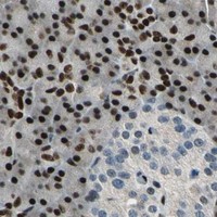
NFIA / Nuclear factor 1 A-type · IHC design guide

Design Immunohistochemistry for NFIA

Plan NFIA paraffin-section IHC around the nuclear staining reported across tissues (HPA tissue IHC). The catalog antibody M03531 has an IHC-P range of 0.5–1 µg/mL (datasheet M03531); score nuclear staining by cell type.

Evidence assembled Oct 2026 · For research use; verify linked source records and product datasheet before use
Immunohistochemistry protocol sheet for NFIA (IHC for NFIA): expected localisation Nuclear staining in tissues (HPA tissue IHC), antibody M03531, validated IHC image, and IHC protocol steps
Printable NFIA IHC protocol sheet — expected localisation Nuclear staining in tissues (HPA tissue IHC), antibody M03531, controls and protocol steps. Open the full NFIA IHC guide →

NFIA Immunohistochemistry Experimental Design Guide

Expected localisation, validated protocols, controls and antibodies — the at-a-glance facts below, then the full design guide.

Must know before staining
Expected localisation Nuclear staining in tissues (HPA tissue IHC)
Staining pattern Nuclear staining across tissues (HPA tissue IHC)
Antigen retrieval EDTA pH 8.0 HIER, heat-mediated (datasheet M03531)
Positive control ⓘ Adipose tissue+4 more · see all
Negative control ⓘ None in HPA (detected in all 45 tissues); use no-primary + isotype controls
Important caveats
Reasons your staining may differ from the expected pattern.
Fixation Keep fixation consistent across sections. (standard IHC practice; not target-specific)
Caveat Staining and RNA show medium consistency (HPA tissue IHC)
Regulation No specific regulator annotated (UniProt)
Isoform / epitope 4 isoforms; epitope coverage is unknown (UniProt)
Section 1

Recommended NFIA IHC & IF Protocols

The catalog antibody protocol is accompanied by published paraffin-section IHC methods for the commissural plate (PMC2910370) and renal cell carcinoma tissue microarrays (PMC10240150).

Recommended immunohistochemistry (IHC-P) protocol parameters
SampleParaffin-embedded human intestinal cancer tissue; fixative not specified (datasheet M03531)
FixationImage fixative and duration unreported (datasheet M03531); verify before use.
Sectioning4–5 µm sections on charged slides (standard)
DeparaffinisationXylene, graded ethanol series to water (standard)
Antigen retrievalHeat retrieval: EDTA pH 8.0 (datasheet M03531); 20 min, 95–100 °C (standard)
Peroxidase block3% H2O2, 10 min, room temperature (standard)
Blocking10% goat serum (datasheet M03531)
Primary antibodyMouse monoclonal (clone 16H11) anti-NFIA, 0.5-1μg/ml (datasheet M03531)
Primary incubationOvernight at 4 °C (datasheet M03531)
DetectionStreptavidin-biotin complex (SABC), DAB chromogen (datasheet M03531)
CounterstainHematoxylin, blue, dehydrate and mount (standard)
Expected resultNFIA-positive staining in adipocytes of adipose tissue (HPA tissue IHC: High). HPA tissue profile: Nuclear expression in all tissues. No signal in the no-primary control.
💡Decision noteStart with heat-mediated EDTA at pH 8.0 (datasheet: M03531); citrate at pH 6.0 is a published alternative (PMC2910370: antigen retrieval).
Section 2

What Is the Expected NFIA Staining Pattern?

NFIA is a nuclear transcription factor without a transmembrane segment (UniProt Q12857: nucleus, topology). In paraffin-section IHC, expect nuclear staining across tissues (HPA: nuclear expression in all tissues), with high staining reported in adipocytes, adrenal glandular cells, bone marrow hematopoietic cells, bronchial respiratory epithelial cells, and selected brain cells (HPA: tissue IHC). HPA rates the tissue profile Enhanced, with medium consistency between antibody staining and RNA expression (HPA: tissue IHC reliability).

What am I looking at on my slide?
Distinct chromogenic signal fills or outlines nuclei of adipocytes, adrenal glandular cells, or bronchial respiratory epithelial cells.This fits the expected compartment and reported high-staining cell populations (UniProt Q12857: nucleus; HPA: High in these cells). Compare signal with the counterstain to confirm that the chromogen belongs to nuclei rather than surrounding cytoplasm (general IHC practice).
Strong staining sits mainly on cell membranes or throughout cytoplasm, while nuclei remain pale.A predominantly nonnuclear IHC pattern conflicts with NFIA's nuclear annotation and HPA's tissue profile (UniProt Q12857: nucleus; HPA: nuclear expression in all tissues). Consider nonspecific staining or detection artefact before scoring it as NFIA; HPA's additional cytosol location comes from ICC-IF (HPA: subcellular).
Staining is concentrated in an unexpected cell population, with little signal in a reported high-staining population on the same section.Check cell identity and compartment first (general IHC practice). Cross-reactivity or endogenous detection activity may explain discordant staining (general IHC practice), but an unlisted cell type alone does not establish a false positive: HPA reports nuclear expression across tissues and lists selected high-staining populations (HPA: tissue IHC).
Brown haze covers nuclei and surrounding tissue, making stained cells hard to distinguish.Diffuse signal cannot support confident nuclear scoring (general IHC practice). Review blocking, antibody concentration, washes, and detection controls as general IHC variables; the supplied HPA and UniProt records do not identify an NFIA-specific cause of diffuse background.
No nuclear signal appears in adipose tissue adipocytes or adrenal glandular cells.These are reported high-staining populations (HPA: High in adipocytes; High in adrenal glandular cells), so absent signal calls for a workflow and control review (general IHC practice). It does not, by itself, establish biological absence or a target-specific fixation effect.
💡Expected NFIA appearanceCall an IHC result positive when discrete nuclear chromogen is visible in a reported high-staining cell population, such as adipocytes or adrenal glandular cells (UniProt Q12857: nucleus; HPA: High in these cells); predominantly membrane or diffuse nonnuclear staining is suspect (HPA: nuclear tissue profile; general IHC practice).
How each factor affects the staining
Nuclear location and topologyNFIA is annotated in the nucleus and has no transmembrane segment (UniProt Q12857: subcellular location, topology). Assess nuclear signal against the counterstain; membrane-dominant staining conflicts with that annotation (general IHC practice).
Breadth of tissue stainingHPA describes nuclear expression in all tissues and low tissue RNA specificity (HPA: tissue IHC, RNA specificity). Its listed High cell populations are useful positive references, but the list is not an exhaustive map of cells that can stain.
Antibody validationHPA006111 and HPA008884 each have Enhanced IHC validation (HPA: antibodies). HPA's Enhanced designation reflects agreement between independent antibodies or orthogonal data; it supports the reported pattern without making every stained cell specific by default (HPA: validation status).
Isoforms and modified residuesUniProt lists four NFIA isoforms and modified residues including phosphoserines at positions 258, 265, 280, 287, and 300 (UniProt Q12857: isoforms, modified residues). No epitope position is supplied, so this record cannot predict isoform coverage or a modification-specific staining change.
IF/ICC Q: What compartment is expected?A: Mainly nucleoplasm, with cytosol listed as an additional location (HPA: ICC-IF subcellular profile). That observation is context for interpreting IF/ICC images; the paraffin-section IHC profile remains nuclear (HPA: tissue IHC).
Why is my staining missing, weak or wrong?
SituationLikely causeNext action
No stain in a reported high-staining tissue.The antibody or detection workflow may have failed (general IHC practice); HPA reports High staining in adipocytes and adrenal glandular cells (HPA: tissue IHC).Run a reported high-staining tissue as a positive control and check reagent order, retrieval conditions, antibody dilution, and detection performance (general IHC practice). The supplied sources give no NFIA-specific retrieval or fixation sensitivity.
Strong cytoplasmic or membrane signal dominates.Nonnuclear signal may reflect nonspecific antibody binding or detection background (general IHC practice); NFIA is nuclear and lacks a transmembrane segment (UniProt Q12857).Compare with a no-primary control, inspect nuclear counterstaining, and reassess antibody concentration and washes (general IHC practice). Do not use the additional cytosol location from ICC-IF to validate membrane-dominant IHC (HPA: subcellular).
Chromogen appears in cells after the primary antibody is omitted.Endogenous detection activity or secondary/detection reagent binding can produce primary-independent signal (general IHC practice).Check the no-primary control and apply the blocking step appropriate to the chromogenic detection system (general IHC practice). Reassess cell-specific NFIA staining only after primary-independent signal is controlled.
Broad tissue haze obscures nuclei.Excess antibody, insufficient blocking, or inadequate washing can raise background (general IHC practice); neither supplied source assigns this haze to an NFIA-specific property.Adjust antibody concentration, blocking, and washes one variable at a time, then compare nuclear contrast with the counterstain and controls (general IHC practice).
Only an unexpected cell population stains.Cell misidentification, cross-reactivity, or endogenous detection activity are possibilities (general IHC practice). HPA's High list is selective despite its all-tissue nuclear profile (HPA: tissue IHC).Confirm cell identity, compare a no-primary control, and examine a reported high-staining population with the same workflow (general IHC practice; HPA: High cell populations). Treat an unlisted cell type as unresolved until the nuclear pattern and controls agree.
Signal varies markedly between sections.Section handling, retrieval, staining, or detection variation can alter apparent intensity (general IHC practice). The supplied NFIA sources do not establish a target-specific fixation effect.Process a reported high-staining control alongside each run and compare matched staining conditions before interpreting intensity differences (general IHC practice; HPA: High in adipocytes and adrenal glandular cells).

Sample controls for NFIA IHC & IF

🧪Run adipose tissue first and assess nuclear staining in adipocytes (HPA: High in adipocytes; UniProt Q12857: nucleus). HPA detects NFIA in all 45 scored tissues, so no negative tissue is supported; use no-primary and isotype controls, and treat any unstained neighboring nuclei on the positive slide as background references rather than verified NFIA-negative cells (HPA: no negative rows; standard IHC practice).
Positive control tissue: Adipose tissue (Adipocytes, HPA High)
Negative control tissue: None in HPA: NFIA is detected in all 45 scored tissues. Use a no-primary (secondary-only) and an isotype control instead.
ICC-IF cell lines (HPA subcellular resource): HPA ICC-IF images show NFIA in A-431, U-251MG, U2OS, NIH 3T3, with annotated localisation: Nucleoplasm (enhanced) (HPA subcellular).
Technical controls: Include a no-primary, secondary-only slide and a control with nonimmune mouse IgG matched to the primary antibody’s isotype; confirm specificity with NFIA knockout material, or a peptide block if the immunizing peptide is available (M03531 caption: mouse primary; standard IHC practice). Quench endogenous peroxidase before DAB detection and assess background around lipid vacuoles in adipose sections (M03531 caption: DAB detection; standard IHC practice).
⚠️Feasibility: No target-specific fixation window or fixation effect is reported, and the selected M03531 paraffin-section caption does not state a fixative (M03531 caption). That caption demonstrates heat-mediated retrieval in EDTA at pH 8.0, but does not establish whether retrieval is required; it also provides no basis to judge whether frozen sections or IF would be easier (M03531 caption). In adipose tissue, lipid extraction during paraffin processing can leave vacuoles and make peripheral adipocyte nuclei harder to assess (standard IHC practice).

HPA tissue IHC evidence for NFIA

Comprehensive Human Protein Atlas IHC scoring per tissue (reliability: Enhanced — Medium consistency between antibody staining and RNA expression data.). Rows are taken directly from the HPA tissue chart — click any row's HPA link to view the source.

Positive expression · recommended positive controls

TissueCell typeLevelEvidenceSource
Adipose tissue Adipocytes High Protein (IHC) HPA →
Adrenal gland Glandular cells High Protein (IHC) HPA →
Bone marrow Hematopoietic cells High Protein (IHC) HPA →
Breast Adipocytes High Protein (IHC) HPA →
Bronchus Respiratory epithelial cells High Protein (IHC) HPA →

Undetected expression · recommended negative controls

TissueCell typeLevelEvidenceSource
None in HPA: NFIA is detected in all 45 scored tissues. Use a no-primary (secondary-only) and an isotype control instead.
Section 3

Advanced NFIA IHC Tips

Troubleshoot NFIA staining in paraffin sections by checking retrieval, nuclear localisation, background and scoring against the catalog antibody’s tissue image and reference patterns.

What retrieval should I start with for NFIA in paraffin sections?
Start with heat-mediated antigen retrieval in EDTA at pH 8.0 for paraffin sections (datasheet M03531). The selected tissue image used that retrieval before incubation with 1 μg/ml primary antibody overnight at 4°C (caption M03531). If nuclear staining is weak, compare retrieval duration or heating intensity on matched sections while keeping the EDTA buffer and antibody concentration fixed (standard IHC practice). Check tissue integrity and staining in a positive reference section after each change; stronger diffuse staining without clearer nuclei suggests excessive retrieval or background rather than improved NFIA detection (UniProt Q12857: nucleus; standard IHC practice).
How should I troubleshoot fixation when NFIA nuclear staining is weak?
The selected paraffin-section caption does not state its fixative, and target-specific fixation sensitivity for NFIA is unknown (caption M03531). Record the fixative, time before fixation, fixation duration and tissue thickness for each specimen, then compare similarly processed sections before changing antibody conditions (standard IHC practice). Poor nuclear morphology or uneven staining may reflect processing differences, so inspect the counterstain and include a well-preserved reference section in the same run (standard IHC practice). Do not attribute a weak result to an NFIA-specific response to fixation solely because the protein is nuclear or phosphorylated (UniProt Q12857: nucleus and modified residues).
Where should convincing NFIA staining appear in a tissue section?
Prioritise staining within intact nuclei: NFIA is annotated as nuclear, and tissue IHC shows nuclear expression across tissues (UniProt Q12857: nucleus; HPA tissue IHC: nuclear expression in all tissues). Its main subcellular location is nucleoplasm, although cytosol is listed as an additional location (HPA subcellular: nucleoplasm enhanced; cytosol). Compare signal with a nuclear counterstain and score cells only when the chromogen can be assigned confidently to a nucleus (standard IHC practice). Diffuse cytoplasmic colour without a reproducible nuclear pattern warrants checks for background, section damage and detection artefacts before it is interpreted as NFIA localisation (standard IHC practice; UniProt Q12857: nucleus).
Can alternative splicing or phosphorylation explain inconsistent NFIA staining?
NFIA has 4 annotated isoforms and multiple modified residues, including phosphoserines at positions 258, 265, 280, 287 and 300 (UniProt Q12857: isoforms and modified residues). The supplied evidence does not map the catalog antibody’s epitope, so isoform coverage and modification sensitivity cannot be assigned to it (supplied M03531 caption; UniProt Q12857). Compare matched sections across processing conditions and seek an independently characterised antibody with a known epitope if isoform-specific interpretation matters (standard IHC practice). Treat differences in nuclear intensity as staining observations until epitope recognition and biological expression have been established independently (standard IHC practice).
How can I adapt NFIA localisation checks to multiplex IF?
For multiplex IF, pair NFIA with an independently validated marker of the expected cell type, such as a glial-cell marker when examining cerebral cortex, and use a nuclear counterstain to assign signal to cells (HPA tissue IHC: high in cerebral-cortex glial cells; standard IF practice). Choose spectrally separated fluorophores and assess an unstained section for tissue autofluorescence before selecting the NFIA channel; a longer-wavelength channel may improve contrast (standard IF practice). Permit access to the nuclear epitope with controlled permeabilisation after fixation, since NFIA is nuclear and has no transmembrane segment (UniProt Q12857: nucleus and topology; standard IF practice). Validate IF staining and controls independently of the paraffin-section chromogenic result (caption M03531; standard IF practice).
What should I check if DAB staining is diffuse or widespread?
The selected tissue procedure used a 10% goat-serum block, biotinylated goat anti-mouse secondary antibody for 30 minutes at 37°C, and an avidin-biotin detection complex with DAB (caption M03531). If colour appears outside nuclei, run primary-omission and detection-only controls to locate nonspecific secondary, endogenous enzyme or detection-system signal (standard IHC practice; UniProt Q12857: nucleus). Include a peroxidase block in the chromogenic workflow and consider endogenous biotin when using avidin-biotin detection (standard IHC practice; caption M03531: detection method). Optimise block, antibody concentration, washes and DAB development one variable at a time, judging improvement by clearer nuclear contrast rather than stronger overall colour (standard IHC practice).
How should I score NFIA staining across different tissue samples? ⚠ ANSWER MARKED FOR VERIFICATION
Score NFIA in intact nuclei within a predefined cell population and tissue region, because the expected tissue pattern is nuclear (HPA tissue IHC: nuclear expression in all tissues). Report the percentage of positive nuclei and, when intensity is reproducible, an H-score from 0–300 based on proportions at 0–3 intensity levels (standard IHC practice). For spatial comparisons, positive nuclei per mm² can supplement the percentage, provided counts are normalised to viable tissue area or total eligible nuclei (standard IHC practice). Apply one threshold, counterstain review and exclusion rule across batches, and document controls and acquisition settings before comparing scores (standard IHC practice).
How do I distinguish true NFIA signal from staining artefacts?
A credible result shows reproducible staining in intact nuclei, consistent with NFIA’s nuclear annotation and the tissue IHC pattern (UniProt Q12857: nucleus; HPA tissue IHC: nuclear expression in all tissues). Check whether signal belongs to the expected cell population using morphology and an appropriate cell marker when identity is uncertain (standard IHC practice). Exclude section edges, folds and necrotic regions from interpretation, and investigate diffuse cytoplasmic colour or broad DAB deposition with primary-omission and detection controls (standard IHC practice; UniProt Q12857: nucleus). Endogenous peroxidase or avidin-biotin detection artefacts can mimic positivity, so require nuclear contrast and clean controls before calling a specimen positive (standard IHC practice; caption M03531: detection method).
Boster reagents

Best NFIA / Nuclear factor 1 A-type IHC Antibodies

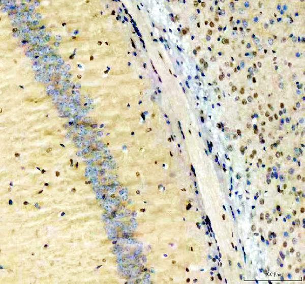
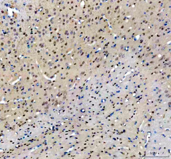
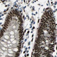
Validated anti-NFIA antibodies have IHC data from human intestinal cancer and tonsil sections (M03531 image captions), mouse and rat brain sections (PB10080 image captions), and IF/ICC data from A431 cells (M03531 IF caption).

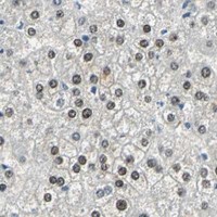
Real IHC data IHC analysis of NFIA using anti-NFIA antibody (M03531). NFIA was detected in paraffin-embedded section of human intestinal cancer tissue. Heat mediated antigen retrieval was performed in EDTA buffer (pH8.0, epitope retrieval solution). The tissue section was blocked with 10% goat serum. The tissue section was then incubated with 1μg/ml mouse anti-NFIA Antibody (M03531) overnight at 4°C. Biotinylated goat anti-mouse IgG was used as secondary antibody and incubated for 30 minutes at 37°C. The tissue section was developed using Strepavidin-Biotin-Complex (SABC) (Catalog # SA1021) with DAB as the chromogen.
Anti-NFIA Antibody ® (monoclonal, 16H11)
Cat # M03531
Real IHC data IHC analysis of NFIA using anti-NFIA antibody (PB10080). NFIA was detected in a paraffin-embedded section of mouse brain tissue. Heat mediated antigen retrieval was performed in EDTA buffer (pH 8.0, epitope retrieval solution). The tissue section was blocked with 10% goat serum. The tissue section was then incubated with 2 μg/ml rabbit anti-NFIA Antibody (PB10080) overnight at 4°C. Peroxidase Conjugated Goat Anti-rabbit IgG was used as secondary antibody and incubated for 30 minutes at 37°C. The tissue section was developed using HRP Conjugated Rabbit IgG Super Vision Assay Kit (Catalog # SV0002) with DAB as the chromogen.
Anti-NFIA Antibody ®
Cat # PB10080

M03531 has IHC images from paraffin-embedded human intestinal cancer and tonsil sections, plus an IF/ICC image from A431 cells (M03531 image captions). PB10080 has IHC images from paraffin-embedded mouse and rat brain sections; its application list includes IHC and its reactivity list includes human, mouse, and rat (PB10080 image captions; catalog applications and reactivity).

Which to pick: For human paraffin-section IHC, choose mouse monoclonal M03531, shown in human intestinal cancer and tonsil sections (M03531 catalog host and clone; M03531 IHC captions). For IF/ICC, choose M03531, which lists both applications and has an A431 cell IF image (M03531 catalog applications; M03531 IF caption). For mouse or rat paraffin-section IHC, choose rabbit PB10080, shown in brain sections from both species; its reactivity list also includes human, but its IHC images show mouse and rat tissue (PB10080 catalog host and reactivity; PB10080 IHC captions). The fixative is unreported for both antibodies' IHC images (M03531 and PB10080 IHC captions).

Each figure is that product's own IHC / IF validation image from its datasheet.

References

  1. UniProt Consortium. UniProt entry Q12857 (NFIA_HUMAN, Nuclear factor 1 A-type).
  2. Human Protein Atlas. NFIA tissue IHC expression (reliability: Enhanced).
  3. Human Protein Atlas. NFIA subcellular location (ICC-IF): Mainly localized to the nucleoplasm..
  4. Human Protein Atlas. NFIA antibody validation summary (2 antibodies).
  5. Molecular regulation of the developing commissural plate. The Journal of comparative neurology 2010 — PMC2910370.
  6. Identification and validation of NFIA as a novel prognostic marker in renal cell carcinoma. The journal of pathology. Clinical research 2023 — PMC10240150.
  7. Multiple intrinsic factors act in concert with Lhx2 to direct retinal gliogenesis. Scientific reports 2016 — PMC5015061.
  8. NFIA haploinsufficiency is associated with a CNS malformation syndrome and urinary tract defects. PLoS genetics 2007 — PMC1877820.
  9. PubMed PMID:10718198 — UniProt-cited evidence.
  10. PubMed PMID:14702039 — UniProt-cited evidence.
  11. PubMed PMID:16710414 — UniProt-cited evidence.