This website uses cookies to ensure you get the best experience on our website.


- Table of Contents
This guide covers paraffin-section chromogenic IHC for NFKBIA, with cytoplasmic staining expected in most tissues (HPA tissue IHC). It addresses stimulation-dependent degradation (UniProt) and the catalog antibody’s 1:50 IHC dilution (datasheet M01139).

Expected localisation, validated protocols, controls and antibodies — the at-a-glance facts below, then the full design guide.
| Expected localisation | Cytoplasmic staining in most tissues (HPA tissue IHC) | |
| Staining pattern | Cytoplasmic signal in glandular and hematopoietic cells (HPA tissue IHC) | |
| Antigen retrieval | Citrate pH 6 HIER, heat-mediated (datasheet M01139) | |
| Positive control | Adrenal gland+4 more · see all | |
| Negative control | Cervix+4 more · see all |
| Fixation | Matched tissue-IHC evidence does not establish the fixation claim. Validate the specimen-specific method before use. (selected-SKU IHC image M01139) | |
| Caveat | Immune activation may reduce signal through degradation (UniProt) | |
| Regulation | Inflammatory stimulation drives degradation (UniProt) | |
| Isoform / epitope | One 1–317 chain; no isoforms or processing annotated (UniProt) |
The catalog antibody’s IHC-P protocol (datasheet M01139) is accompanied by published NFKBIA chromogenic IHC methods for glioma and mouse myocardial sections (PMC4199416; PMC7176878).
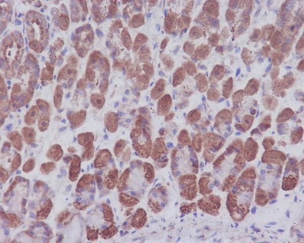
| Sample | Paraffin-embedded mouse stomach tissue; fixative not specified (datasheet M01139) |
| Fixation | Image fixative and duration unreported (datasheet M01139); verify before use. |
| Sectioning | 4–5 µm sections on charged slides (standard) |
| Deparaffinisation | Xylene, graded ethanol series to water (standard) |
| Antigen retrieval | Heat retrieval: Citrate pH 6, 20 min (datasheet M01139) |
| Peroxidase block | 3% H2O2, 10 min, room temperature (standard) |
| Blocking | 10% goat serum (datasheet M01139) |
| Primary antibody | Rabbit monoclonal (clone HO-14) anti-NFKBIA, 1:50 recommended; image 1ug/ml (datasheet M01139) |
| Primary incubation | Overnight at 4 °C (datasheet M01139) |
| Detection | Streptavidin-biotin complex (SABC), DAB chromogen (datasheet M01139) |
| Counterstain | Hematoxylin, blue, dehydrate and mount (standard) |
| Expected result | NFKBIA-positive staining in glandular cells of adrenal gland (HPA tissue IHC: Medium). HPA tissue profile: Cytoplasmic expression in most tissues. No signal in the no-primary control. |
NFKBIA should show predominantly cytoplasmic staining in many cell types, including glandular and hematopoietic cells that HPA rates Medium (HPA: cytoplasmic expression in most tissues; Medium in appendix glandular cells and bone marrow hematopoietic cells). Nuclear staining may occur because the protein shuttles between cytoplasm and nucleus (UniProt P25963: subcellular location). NFKBIA has no transmembrane segment, so a membrane rim is not expected (UniProt P25963 topology). HPA rates its tissue IHC evidence Approved, with medium consistency between staining and RNA data (HPA: tissue IHC reliability).
| Cytoplasmic chromogen in the expected cells, with distinguishable unstained areas. | This fits the predominant tissue pattern (HPA: cytoplasmic expression in most tissues). Medium staining is reported in appendix glandular cells and bone marrow hematopoietic cells (HPA: tissue IHC). Judge each cell population separately; HPA levels describe observations, not an intensity threshold that every section must reproduce. |
| A sharp membrane rim, or strong nuclear-only staining across most cells. | A membrane rim conflicts with the lack of a transmembrane segment (UniProt P25963 topology). Widespread nuclear-only staining departs from HPA's mainly cytosolic IF pattern and cytoplasmic tissue profile (HPA: subcellular IF; tissue IHC). Review the staining pattern and controls before calling it NFKBIA; some nuclear localization remains biologically possible (UniProt P25963: nuclear–cytoplasmic shuttling). |
| Strong signal in a cell population HPA lists as Not detected. | For example, HPA reports kidney glomerular cells and liver cholangiocytes as Not detected (HPA: tissue IHC). Check cell identity and a negative control; antibody cross-reactivity or endogenous chromogenic activity can mimic target staining (general IHC practice). A negative listing for one cell population does not describe the entire organ (HPA: tissue IHC). |
| Diffuse color over stroma, lumina, and cells, without clear cellular borders. | This is difficult to interpret as the reported cytoplasmic pattern (HPA: tissue IHC). Background may reflect nonspecific binding, residual endogenous detection activity, or excessive development (general IHC practice). Compare a section processed without primary antibody and review the detection and development steps before scoring cells (general IHC practice). |
| No signal in a section containing expected positive cells. | First confirm that the section includes the relevant cells: HPA reports Medium staining in breast glandular cells and bone marrow hematopoietic cells (HPA: tissue IHC). Then check the catalog antibody's IHC-P instructions, detection reagents, and a positive control (general IHC practice). An absent signal alone cannot establish absence of NFKBIA. |
| Compartment and cell selection | HPA reports cytoplasmic expression in most tissues, but levels vary by named cell population (HPA: tissue IHC). Use appendix glandular cells or bone marrow hematopoietic cells as examples of reported Medium staining; kidney glomerular cells are listed as Not detected (HPA: tissue IHC). |
| Biological movement | NFKBIA shuttles between cytoplasm and nucleus, and stimulation promotes phosphorylation and degradation while allowing RELA nuclear entry (UniProt P25963: subcellular location and function). Interpret unusual nuclear staining with the experiment's stimulation context; staining alone cannot establish pathway activation. |
| Protein architecture | The recorded chain spans residues 1–317, with no signal peptide, propeptide, or transmembrane segment (UniProt P25963 topology and processing). No supplied evidence supports a shed or membrane-bound staining pattern. The supplied record has no mapped antibody epitope, so epitope-specific retrieval predictions are unavailable. |
| Evidence strength | HPA rates tissue IHC Approved and describes medium consistency with RNA data; the listed antibodies are IHC Approved, while their ICC ratings differ (HPA: tissue IHC reliability; HPA: antibodies). Treat the tissue profile as a guide to expected staining, not proof that every specimen or antibody will match it. |
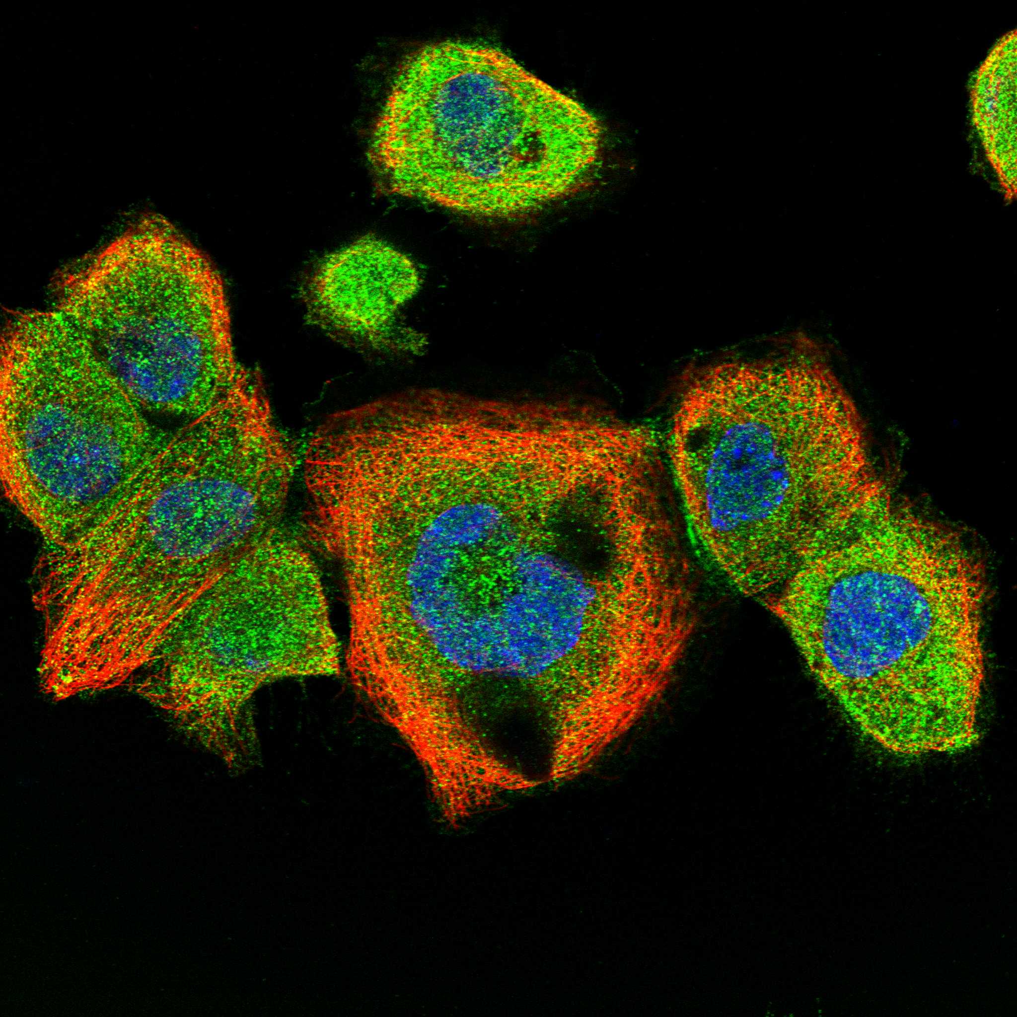
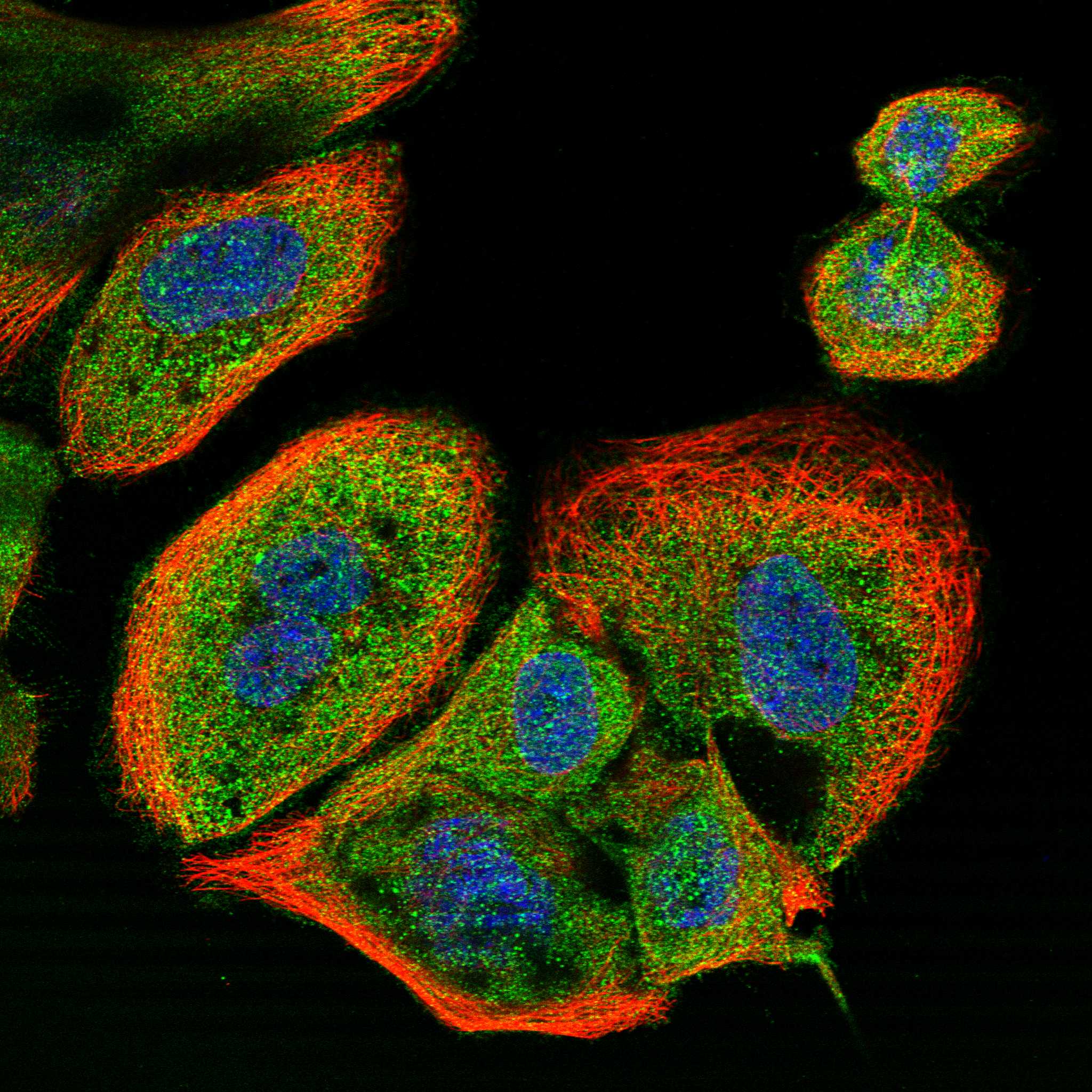
| IF/ICC expectation? | Mainly cytosolic signal is the HPA IF expectation; A-431, U-251MG, U2OS, and HeLa BAC 6210 have ICC-IF images (HPA: subcellular IF). Nuclear localization remains possible through shuttling (UniProt P25963: subcellular location). IF/ICC optimization belongs in its separate guide; these observations do not define an IHC-P protocol. |
| Retrieval and detection | No target-specific fixation sensitivity or retrieval condition is supplied (HPA: tissue IHC; UniProt P25963). Select retrieval and chromogenic detection conditions from the IHC-validated antibody's instructions, then assess controls together with the specimen (general IHC practice). |
| Situation | Likely cause | Next action |
|---|---|---|
| Positive control stains, but the study section appears negative. | The sampled area may lack the cell population being assessed, or its staining may be below visual detection; HPA levels are cell-specific observations (HPA: tissue IHC). | Verify tissue and cell identity on the counterstain, then compare like cell populations and exposure to chromogen with the positive control (general IHC practice). |
| Expected positive tissue and study section are both blank. | A shared staining-step or detection failure is possible; HPA reports Medium staining in bone marrow hematopoietic cells and appendix glandular cells (HPA: tissue IHC). | Check the catalog antibody's IHC-P instructions, reagent activity, incubation steps, and chromogen development; rerun with an appropriate positive section (general IHC practice). |
| Color appears in both the stained section and the no-primary control. | The result may arise from endogenous detection activity or nonspecific detection reagents rather than primary-antibody binding (general IHC practice). | Review the relevant endogenous-activity block and detection controls before changing the primary-antibody conditions (general IHC practice). |
| Nuclear staining dominates and cytoplasm is nearly blank. | This differs from HPA's predominant cytoplasmic tissue pattern, although NFKBIA can shuttle into nuclei (HPA: tissue IHC; UniProt P25963: subcellular location). | Review stimulation context, cell identity, and controls; avoid assigning pathway activation solely from the IHC compartment pattern (UniProt P25963: function; general IHC practice). |
| A continuous membrane rim or staining of an unexpected cell population dominates. | A membrane rim conflicts with NFKBIA topology; unexpected cells may reflect cross-reactivity or chromogenic background (UniProt P25963 topology; general IHC practice). | Compare cellular morphology and no-primary controls, and assess the pattern against HPA's cell-specific observations before scoring it as positive (HPA: tissue IHC; general IHC practice). |
| The section has widespread haze that obscures cell boundaries. | Nonspecific binding or excess chromogen development can obscure a cytoplasmic pattern (general IHC practice; HPA: tissue IHC profile). | Review blocking, washing, antibody concentration, and development time using the catalog antibody's IHC-P instructions; retain a matched control for comparison (general IHC practice). |


Comprehensive Human Protein Atlas IHC scoring per tissue (reliability: Approved — Medium consistency between antibody staining and RNA expression data.). Rows are taken directly from the HPA tissue chart — click any row's HPA link to view the source.
| Tissue | Cell type | Level | Evidence | Source |
|---|---|---|---|---|
| Adrenal gland | Glandular cells | Medium | Protein (IHC) | HPA → |
| Appendix | Glandular cells | Medium | Protein (IHC) | HPA → |
| Bone marrow | Hematopoietic cells | Medium | Protein (IHC) | HPA → |
| Breast | Glandular cells | Medium | Protein (IHC) | HPA → |
| Bronchus | Respiratory epithelial cells | Medium | Protein (IHC) | HPA → |
| Tissue | Cell type | Level | Evidence | Source |
|---|---|---|---|---|
| Cervix | Glandular cells | Not detected | Protein (IHC) | HPA → |
| Esophagus | Squamous epithelial cells | Not detected | Protein (IHC) | HPA → |
| Kidney | Cells in glomeruli | Not detected | Protein (IHC) | HPA → |
| Liver | Cholangiocytes | Not detected | Protein (IHC) | HPA → |
| Oral mucosa | Squamous epithelial cells | Not detected | Protein (IHC) | HPA → |
Troubleshoot NFKBIA staining in paraffin sections by checking retrieval, cell compartment, controls and scoring against the documented IHC evidence.
M01139 has IHC data from paraffin-embedded mouse stomach (IHC image caption) and IF data from HeLa cells (IF image caption); listed reactivity covers human, mouse and rat (catalog reactivity).
M01139 is shown in IHC on paraffin-embedded mouse stomach (IHC image caption). M01139 is also shown in IF on HeLa cells (IF image caption) and is listed for IHC, IF and ICC (catalog applications).
Which to pick: For tissue IHC, choose M01139: its IHC image shows staining in a paraffin-embedded mouse stomach section (IHC image caption); the fixative is unreported (IHC image caption). For IF/ICC, M01139 is listed for both applications (catalog applications), with an IF image from HeLa cells (IF image caption). For human, mouse or rat samples, M01139 is the listed rabbit monoclonal option (catalog host and clone; catalog reactivity); the IHC image specifically documents mouse tissue (IHC image caption).