This website uses cookies to ensure you get the best experience on our website.


- Table of Contents
Plan NIBAN2 staining in paraffin sections with the catalog antibody at 2–5 μg/ml (datasheet A09391-2). Assess cytoplasmic and membranous staining using colon or stomach glandular cells as positive references, while accounting for uncertain tissue staining reliability (HPA tissue IHC).

Expected localisation, validated protocols, controls and antibodies — the at-a-glance facts below, then the full design guide.
| Expected localisation | Cytoplasmic and membranous tissue staining (HPA tissue IHC) | |
| Staining pattern | Most tissues: cytoplasmic and membranous staining (HPA tissue IHC) | |
| Antigen retrieval | EDTA pH 8.0 HIER, heat-mediated (datasheet A09391-2) | |
| Positive control | Appendix+4 more · see all | |
| Negative control | Bone marrow+4 more · see all |
| Fixation | Keep paraffin-section fixation consistent across samples. (standard IHC practice; not target-specific) | |
| Caveat | Tissue staining reliability is uncertain (HPA tissue IHC) | |
| Regulation | Expression regulation is not annotated (UniProt) | |
| Isoform / epitope | 2 isoforms; check epitope coverage (UniProt) |
The catalog antibody protocol is paired with one published NIBAN2 tissue staining protocol (PMC12432436).
| Sample | Paraffin-embedded human placenta tissue; fixative not specified (datasheet A09391-2) |
| Fixation | Image fixative and duration unreported (datasheet A09391-2); verify before use. |
| Sectioning | 4–5 µm sections on charged slides (standard) |
| Deparaffinisation | Xylene, graded ethanol series to water (standard) |
| Antigen retrieval | Heat retrieval: EDTA pH 8.0 (datasheet A09391-2); 20 min, 95–100 °C (standard) |
| Peroxidase block | 3% H2O2, 10 min, room temperature (standard) |
| Blocking | 10% goat serum (datasheet A09391-2) |
| Primary antibody | Rabbit anti-NIBAN2, 2-5 μg/ml (datasheet A09391-2) |
| Primary incubation | Overnight at 4 °C (datasheet A09391-2) |
| Detection | Streptavidin-biotin complex (SABC), DAB chromogen (datasheet A09391-2) |
| Counterstain | Hematoxylin, blue, dehydrate and mount (standard) |
| Expected result | NIBAN2-positive staining in glandular cells of appendix (HPA tissue IHC: High). HPA tissue profile: Cytoplasmic and membranous expression in most tissues. No signal in the no-primary control. |
NIBAN2 staining is expected in the cytoplasm and at cell membranes, including adherens junctions (UniProt Q96TA1; HPA tissue IHC). Strong examples include glandular cells of the colon, stomach, appendix, duodenum, endometrium and gallbladder, and urothelial cells of the urinary bladder (HPA: High). NIBAN2 has no transmembrane segment (UniProt Q96TA1 topology). Treat the tissue pattern as provisional: HPA rates its IHC reliability Uncertain, with medium agreement between staining and RNA expression (HPA tissue IHC).
| Cytoplasmic staining, with some membrane accentuation, in glandular or urothelial cells (HPA tissue IHC). | This fits the reported pattern; colon glandular cells and urinary bladder urothelial cells are examples rated High (HPA tissue IHC). Compare the distribution within the tissue, since HPA reports staining in most tissues but rates the IHC evidence Uncertain (HPA tissue IHC). |
| Predominantly nuclear staining in a paraffin tissue section. | This differs from the principal cytoplasmic and membranous tissue pattern (HPA tissue IHC). Review morphology, counterstain and controls for an artefact (general IHC practice). HPA reports additional nucleoplasmic signal in ICC-IF, so nuclear signal alone is not definitive proof of antibody cross-reactivity (HPA subcellular ICC-IF). |
| Strong signal mainly in cells recorded as Not detected, such as skeletal muscle myocytes (HPA tissue IHC). | This conflicts with that cell-type reference and merits a control check; cross-reactivity or endogenous detection activity are possibilities, not established diagnoses (HPA tissue IHC; general IHC practice). A Not detected entry applies to the named cells, not necessarily every cell in that tissue (HPA tissue IHC). |
| Uniform haze over cells and surrounding tissue, without a discernible cellular pattern. | This is less interpretable than compartment-resolved staining (HPA tissue IHC; general IHC practice). Examine a no-primary control and staining run for nonspecific background or endogenous detection activity before assigning NIBAN2 positivity (general IHC practice). |
| No signal in colon glandular cells included as a positive reference (HPA: High in colon glandular cells). | Check section integrity, retrieval, detection and the run controls before calling the specimen negative (general IHC practice). The HPA High rating makes these cells a useful reference, but HPA's overall IHC reliability is Uncertain (HPA tissue IHC). |
| Cell state and compartment (UniProt Q96TA1). | UniProt reports exclusively cytoplasmic localization in exponentially growing cells and membrane localization at confluency or during telophase; a tissue section need not show identical membrane emphasis in every cell (UniProt Q96TA1). |
| Tissue and cell-type reference (HPA tissue IHC). | HPA rates selected glandular and urothelial cells High, placental cytotrophoblasts Medium, and selected other cell types Not detected. Interpret the named cell type rather than treating an entire organ as uniformly positive or negative (HPA tissue IHC). |
| IHC evidence strength (HPA tissue IHC; HPA antibodies). | HPA calls the tissue IHC profile Uncertain, pending external verification; all four listed HPA antibodies have IHC status Uncertain. Use the pattern as an expectation, not a stand-alone specificity test (HPA tissue IHC; HPA antibodies). |
| Topology and processing (UniProt Q96TA1). | NIBAN2 has no transmembrane segment, signal peptide or annotated propeptide; these annotations provide no basis for predicting a shed extracellular staining pattern or a cleavage-dependent IHC result (UniProt Q96TA1 topology and processing). |
| Isoforms and phosphorylation (UniProt Q96TA1). | UniProt lists two isoforms and multiple modified residues. Antibody epitope and isoform coverage are not supplied here, so neither feature establishes which staining differences this IHC antibody can detect (UniProt Q96TA1). |
| IF/ICC: what localization is reported? (HPA subcellular ICC-IF). | HPA reports enhanced plasma membrane and cytosol localization, plus supported nucleoplasmic localization, in ICC-IF; that assay's compartment evidence should not be treated as an IHC validation result (HPA subcellular ICC-IF; HPA antibodies). |
| Situation | Likely cause | Next action |
|---|---|---|
| Known-positive glandular cells show no chromogenic signal (HPA: High in colon glandular cells). | Target-specific fixation effects are not established by the supplied assay evidence. Verify with a matched IHC source before attributing a result to fixation. | Confirm tissue morphology and run controls, then review the validated IHC workflow, including retrieval and detection, before interpreting a negative result (general IHC practice). |
| Strong nuclear-only staining dominates the section. | It departs from the principal tissue pattern, although ICC-IF reports additional nucleoplasmic localization (HPA tissue IHC; HPA subcellular ICC-IF). | Check the no-primary control, nuclear counterstain and cellular boundaries; require matching cytoplasmic or membranous tissue context before a confident IHC call (general IHC practice; HPA tissue IHC). |
| The whole section has brown haze that obscures cell borders. | Nonspecific background or endogenous detection activity may obscure compartment assignment (general IHC practice). | Review no-primary and detection controls, blocking and reagent handling, then assess whether cell-resolved cytoplasmic or membrane staining becomes visible (general IHC practice; HPA tissue IHC). |
| A nominally negative tissue contains a small stained population. | HPA Not detected entries refer to specified cells, such as bone marrow hematopoietic cells, and do not classify every cell in the organ (HPA tissue IHC). | Identify the stained cell population morphologically and compare that cell type with HPA's entry before labeling the tissue result discordant (HPA tissue IHC; general IHC practice). |
| Staining is strongest in an HPA Not detected cell type (HPA tissue IHC). | Cross-reactivity or endogenous detection activity is possible; HPA's IHC reliability is Uncertain, so the reference alone cannot resolve the cause (HPA tissue IHC; general IHC practice). | Compare positive-reference tissue and no-primary controls from the same run, and seek independent specificity evidence before accepting that signal as NIBAN2 (general IHC practice). |
| Some positive cells show little membrane accentuation. | UniProt describes cytoplasmic localization in growing cells and membrane localization with confluency or telophase; HPA describes both cytoplasmic and membranous tissue expression (UniProt Q96TA1; HPA tissue IHC). | Score cytoplasmic and membrane compartments separately, using the named positive cell types as context; do not require a membrane rim in every cell (HPA tissue IHC; general IHC practice). |
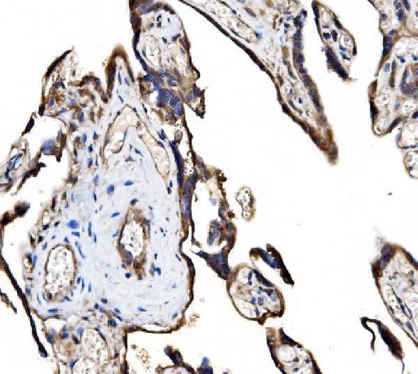
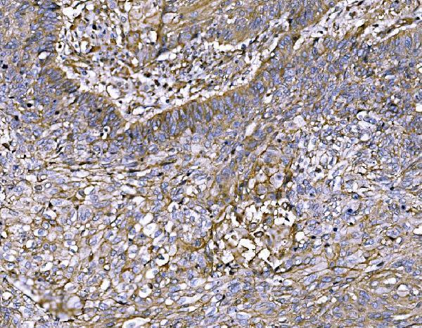


Comprehensive Human Protein Atlas IHC scoring per tissue (reliability: Uncertain — Medium consistency between antibody staining and RNA expression data. Pending external verification.). Rows are taken directly from the HPA tissue chart — click any row's HPA link to view the source.
| Tissue | Cell type | Level | Evidence | Source |
|---|---|---|---|---|
| Appendix | Glandular cells | High | Protein (IHC) | HPA → |
| Colon | Glandular cells | High | Protein (IHC) | HPA → |
| Duodenum | Glandular cells | High | Protein (IHC) | HPA → |
| Endometrium | Glandular cells | High | Protein (IHC) | HPA → |
| Gallbladder | Glandular cells | High | Protein (IHC) | HPA → |
| Tissue | Cell type | Level | Evidence | Source |
|---|---|---|---|---|
| Bone marrow | Hematopoietic cells | Not detected | Protein (IHC) | HPA → |
| Caudate | Glial cells | Not detected | Protein (IHC) | HPA → |
| Cerebellum | Cells in granular layer | Not detected | Protein (IHC) | HPA → |
| Hippocampus | Glial cells | Not detected | Protein (IHC) | HPA → |
| Lymph node | Germinal center cells | Not detected | Protein (IHC) | HPA → |
Use the documented paraffin-section workflow as the starting point, then assess NIBAN2 staining by cell type and compartment (caption A09391-2; HPA tissue IHC).
The catalog antibody has real IHC data from human paraffin sections (A09391-2 image captions); its listed reactivity also includes mouse and rat (A09391-2 catalog). No IF data are supplied (A09391-2 catalog).
A09391-2 was tested by IHC on paraffin sections of human placenta and human lung cancer tissue (A09391-2 image captions). Its listed applications include IHC, while no IF image or IF dilution is supplied (A09391-2 catalog).
Which to pick: Choose A09391-2 for human paraffin-section IHC: its captions show EDTA retrieval at pH 8.0 and primary antibody at 2 μg/ml (A09391-2 image captions); the fixative is unreported (A09391-2 image captions). No SKU in the payload is documented for IF/ICC (A09391-2 catalog). A09391-2 lists mouse and rat reactivity, but the supplied IHC images show human tissue only (A09391-2 catalog; A09391-2 image captions).