NKTR / NK-tumor recognition protein · IHC design guide

Design Immunohistochemistry for NKTR

Plan NKTR paraffin-section IHC around the cytoplasmic staining reported in most tissues (HPA tissue IHC). Start the IHC-validated antibody at 1:100–1:300 (datasheet), and verify the pattern with controls because staining has low concordance with RNA data (HPA tissue IHC).

Evidence assembled Oct 2026 · For research use; verify linked source records and product datasheet before use
Immunohistochemistry protocol sheet for NKTR (IHC for NKTR): expected localisation Cytoplasmic staining in most tissues (HPA tissue IHC), antibody A09881, validated IHC image, and IHC protocol steps
Printable NKTR IHC protocol sheet — expected localisation Cytoplasmic staining in most tissues (HPA tissue IHC), antibody A09881, controls and protocol steps. Open the full NKTR IHC guide →

NKTR Immunohistochemistry Experimental Design Guide

Expected localisation, validated protocols, controls and antibodies — the at-a-glance facts below, then the full design guide.

Must know before staining
Expected localisation Cytoplasmic staining in most tissues (HPA tissue IHC)
Staining pattern Cytoplasmic staining across most tissues (HPA tissue IHC)
Antigen retrieval Citrate pH 6.0 HIER, 95–98 °C, 20 min (rule: cytoplasmic / membrane antigen)
Positive control ⓘ Adrenal gland+4 more · see all
Negative control ⓘ Breast+2 more · see all
Important caveats
Reasons your staining may differ from the expected pattern.
Fixation Matched tissue-IHC evidence does not establish the fixation claim. Validate the specimen-specific method before use. (selected-SKU IHC image A09881)
Caveat Staining has low concordance with RNA data (HPA tissue IHC)
Regulation No expression regulator annotated (UniProt)
Isoform / epitope No isoforms or processing annotated; epitope side unknown (UniProt)
Section 1

Recommended NKTR IHC & IF Protocols

Compare the catalog antibody’s IHC-P protocol with published NKTR IHC conditions from 2 articles (PMC9467399; PMC3307063).

Recommended immunohistochemistry (IHC-P) protocol parameters
SampleParaffin-embedded human brain tissue; fixative not specified (datasheet A09881)
FixationImage fixative and duration unreported (datasheet A09881); verify before use.
Sectioning4–5 µm sections on charged slides (standard)
DeparaffinisationXylene, graded ethanol series to water (standard)
Antigen retrievalHeat-induced epitope retrieval in citrate buffer, pH 6.0, 20 min at 95–98 °C (standard rule: cytoplasmic / membrane antigen)
Peroxidase block3% H2O2, 10 min, room temperature (standard)
Blocking10% normal serum of the secondary host, 30 min, room temperature (standard)
Primary antibodyRabbit anti-NKTR, 1:100 - 1:300 (datasheet A09881)
Primary incubationOvernight at 4 °C (standard)
DetectionHRP-polymer secondary, DAB chromogen 5–10 min (standard)
CounterstainHematoxylin, blue, dehydrate and mount (standard)
Expected resultNKTR-positive staining in glandular cells of adrenal gland (HPA tissue IHC: High). HPA tissue profile: Cytoplasmic expression in most tissues. No signal in the no-primary control.
💡Decision noteStart with citrate pH 6.0 HIER at 95–98 °C for 20 min (page antigen-retrieval specification); assess staining with tissue controls.
Section 2

What Is the Expected NKTR Staining Pattern?

In paraffin-section IHC, expect mainly cytoplasmic NKTR staining across many tissues, including colon glandular cells and kidney tubule cells (HPA tissue IHC). UniProt lists NKTR at the cell membrane but annotates no transmembrane segment (UniProt P30414). HPA rates its tissue IHC profile Approved while reporting low agreement with RNA expression and pending external verification (HPA tissue IHC).

What am I looking at on my slide?
Colon glandular cells or kidney tubule cells show clear cytoplasmic staining, with recognizable cell boundaries.This matches cell types rated High and the predominantly cytoplasmic tissue pattern (HPA tissue IHC). Assess staining within the expected cells against a matched negative control (standard IHC practice).
A paraffin section shows only nuclear staining, with no convincing cytoplasmic signal.That differs from the reported tissue IHC pattern (HPA tissue IHC). Nuclear signal alone does not establish an artefact: nucleoplasm is supported in ICC-IF (HPA subcellular). Check controls and antibody validation before assigning the IHC signal to NKTR (standard IHC practice).
The strongest signal is in breast adipocytes, epididymal glandular cells, or oral squamous epithelial cells.Those specific cell populations were rated Not detected (HPA tissue IHC). Strong staining there warrants a check for cross-reactivity or endogenous detection activity, especially if expected positive cells are weak (standard IHC practice).
Color covers stroma, empty spaces, and several cell types without distinct cell-associated staining.That distribution is difficult to reconcile with the reported cytoplasmic cellular pattern (HPA tissue IHC). Compare a matched negative control; nonspecific antibody binding or detection background can produce diffuse color (standard IHC practice).
Colon glandular cells and kidney tubule cells have no detectable staining.Both were rated High, so a shared absence calls for a run-level check (HPA tissue IHC). Review positive-control performance, retrieval, antibody application, and detection using general IHC controls; the payload gives no NKTR-specific retrieval condition (standard IHC practice).
💡Expected NKTR appearanceCall a positive IHC result when expected cells show distinct, mainly cytoplasmic staining—often High in colon glands or kidney tubules (HPA tissue IHC); broad haze or strong staining in HPA Not detected cell populations needs control-based review (HPA tissue IHC; standard IHC practice).
How each factor affects the staining
Compartment evidenceTissue IHC is predominantly cytoplasmic (HPA tissue IHC); ICC-IF also supports nucleoplasm and enhances plasma membrane and cytosol (HPA subcellular). UniProt lists cell membrane without a transmembrane segment (UniProt P30414). Interpret each assay against its own reported pattern.
IHC antibody validationHPA022120 is Approved for IHC, whereas HPA051576 has no IHC status in the supplied record (HPA antibodies). An Approved status is qualified by low staining-to-RNA consistency and pending external verification for the tissue profile (HPA tissue IHC).
Cell-specific comparisonColon glands, kidney tubules, and testis Leydig cells are rated High; breast adipocytes and oral squamous cells are Not detected (HPA tissue IHC). Compare the named cell populations, since a tissue-wide positive or negative label would obscure the reported pattern.
Target-specific retrieval evidenceTarget-specific fixation effects are not established by the supplied assay evidence. Verify with a matched IHC source before attributing a result to fixation.
Why is my staining missing, weak or wrong?
SituationLikely causeNext action
Expected positive cells and the run's positive control are both blank.A shared processing or detection failure is possible (standard IHC practice); the HPA High calls alone do not identify which step failed (HPA tissue IHC).Verify control tissue, reagent application, retrieval record, and chromogenic detection before interpreting NKTR absence (standard IHC practice).
Colon glands are blank, but the run's positive control stains.A local tissue or sampling issue remains possible (standard IHC practice). HPA reports High staining in colon glandular cells but low overall staining-to-RNA consistency (HPA tissue IHC).Confirm that glandular cells are present and preserved, then compare another reported High population and matched controls (HPA tissue IHC; standard IHC practice).
Color persists in a negative control or spreads over the whole section.Background from nonspecific binding or endogenous detection activity is possible (standard IHC practice). HPA's cellular pattern does not validate diffuse color as NKTR (HPA tissue IHC).Check the negative-control result, blocking and detection steps, and whether color remains confined to cells (standard IHC practice).
Unexpected strong staining appears in a cell population rated Not detected.Cross-reactivity or endogenous detection activity is possible (standard IHC practice); the discrepancy alone cannot prove either cause (HPA tissue IHC).Compare breast adipocytes, epididymal glands, or oral squamous cells with an expected positive population on controlled sections (HPA tissue IHC; standard IHC practice).
IHC staining is mainly nuclear.This conflicts with predominantly cytoplasmic tissue IHC (HPA tissue IHC), although nucleoplasm is supported in ICC-IF (HPA subcellular).Review antibody identity, tissue controls, and cellular localization before scoring it as expected IHC staining (HPA antibodies; standard IHC practice).
IF/ICC Q: Should nucleoplasmic signal be treated as unexpected?A: No; HPA supports nucleoplasm and reports enhanced plasma-membrane and cytosol localization in ICC-IF (HPA subcellular).Interpret IF/ICC against its own localization evidence; use the separate IF/ICC guide for assay design (HPA subcellular).

Sample controls for NKTR IHC & IF

🧪Start with colon: glandular cells should stain (HPA: colon glandular cells High). Use oral mucosa squamous epithelial cells as the negative tissue (HPA: Not detected); on the colon slide, treat neighboring nonglandular cells as an internal background reference only where they are unstained, since no internal-negative cell type is established by the supplied HPA rows.
Positive control tissue: Adrenal gland (Glandular cells, HPA High)
Negative control tissue: Breast (HPA Not detected)
ICC-IF cell lines (HPA subcellular resource): HPA ICC-IF images show NKTR in A-431, U-251MG, U2OS, HeLa, with annotated localisation: Nucleoplasm (supported), Plasma membrane (enhanced), Cytosol (enhanced) (HPA subcellular).
Technical controls: Run a no-primary (secondary-only) slide and a concentration-matched isotype control of the primary antibody’s host species and clonality; add a biological specificity control using NKTR knockout material or competition with the matching peptide (selected A09881 caption: peptide-blocked brain section). For colon chromogenic IHC, quench endogenous peroxidase and check background from any biotin-based detection system, if used (standard IHC practice).
⚠️Feasibility: A target-specific fixation window and retrieval dependency are unreported in the supplied evidence; the selected A09881 paraffin-section caption also leaves the fixative unreported. The supplied evidence does not establish whether frozen sections or IF are easier for NKTR, although ICC-IF images exist (HPA subcellular: A-431, U-251MG, U2OS and HeLa). In colon, assess mucin-associated background when scoring glandular staining (standard IHC practice).

HPA tissue IHC evidence for NKTR

Comprehensive Human Protein Atlas IHC scoring per tissue (reliability: Approved — Low consistency between antibody staining and RNA expression data. Pending external verification.). Rows are taken directly from the HPA tissue chart — click any row's HPA link to view the source.

Positive expression · recommended positive controls

TissueCell typeLevelEvidenceSource
Adrenal gland Glandular cells High Protein (IHC) HPA →
Colon Glandular cells High Protein (IHC) HPA →
Duodenum Glandular cells High Protein (IHC) HPA →
Kidney Cells in tubules High Protein (IHC) HPA →
Small intestine Glandular cells High Protein (IHC) HPA →

Undetected expression · recommended negative controls

TissueCell typeLevelEvidenceSource
Breast Adipocytes Not detected Protein (IHC) HPA →
Epididymis Glandular cells Not detected Protein (IHC) HPA →
Oral mucosa Squamous epithelial cells Not detected Protein (IHC) HPA →
Section 3

Advanced NKTR IHC Tips

Troubleshoot NKTR staining in paraffin sections by checking retrieval, compartment, controls and scoring before interpreting chromogenic signal.

How should I retrieve NKTR in paraffin sections when staining is weak?
Use citrate buffer at pH 6.0 for heat-induced retrieval at 95–98 °C for 20 minutes (page retrieval setting). Let sections cool in retrieval buffer, then wash before blocking; abrupt handling can detach sections (standard IHC practice). If signal remains weak, compare a longer retrieval exposure on adjacent sections while watching for tissue damage and increased background (standard IHC practice). Keep detection and imaging conditions matched across that comparison, and include the peptide-blocked control shown for catalog antibody A09881 (selected A09881 tissue-IHC caption). Score cytoplasmic and membrane-associated signal separately because both are plausible locations for NKTR (HPA tissue IHC; HPA subcellular; UniProt P30414).
How can I assess whether fixation is causing weak NKTR staining?
Target-specific fixation sensitivity for NKTR is unknown from the supplied evidence; the A09881 paraffin-section caption does not state a fixative (selected A09881 tissue-IHC caption). Record the fixative, fixation duration, tissue thickness and processing history for each section before comparing staining (standard IHC practice). On matched sections, hold citrate retrieval at pH 6.0, 95–98 °C for 20 minutes constant while evaluating specimens with different documented fixation histories (page retrieval setting; standard IHC practice). Compare signal with morphology and background, since poor preservation can complicate chromogenic interpretation (standard IHC practice). Do not infer fixation resistance from NKTR phosphorylation sites, topology or tissue staining patterns (UniProt P30414; HPA tissue IHC).
Should NKTR staining be cytoplasmic, membranous or nuclear?
Record cytoplasmic, membranous and nuclear staining separately: tissue IHC describes predominantly cytoplasmic expression, while cell imaging supports nucleoplasm, plasma membrane and cytosol (HPA tissue IHC; HPA subcellular). UniProt assigns NKTR to the cell membrane but reports no transmembrane segment, so a membrane signal alone does not establish membrane-spanning topology (UniProt P30414). At 40× objective magnification, inspect whether chromogen follows intact cell boundaries or pools over folds, edges and damaged areas (standard IHC practice). Compare each compartment against a no-primary control and the peptide-blocked A09881 image, recognising that peptide competition addresses antibody binding within that demonstration (standard IHC practice; selected A09881 tissue-IHC caption). Report the compartment and cell type together rather than combining all brown pixels into one score (standard IHC practice).
How do I troubleshoot an unexpected NKTR epitope staining pattern?
The supplied record lists 0 annotated isoforms and a cyclophilin-type PPIase domain at residues 10–175; neither identifies the A09881 antibody epitope (UniProt P30414; selected A09881 tissue-IHC caption). It also lists phosphorylation sites, including residues 379 and 401, but provides no evidence that these modifications change tissue-IHC binding (UniProt P30414). Check the antibody's documented immunogen or epitope information before attributing a weak compartment to epitope masking (standard IHC practice). Run adjacent sections with the stated peptide competition and a no-primary control to distinguish peptide-sensitive staining from detection background (selected A09881 tissue-IHC caption; standard IHC practice). If an alternative antibody is available, compare independently documented epitopes under matched processing conditions (standard IHC practice).
How should I adapt an NKTR observation to multiplex IF?
Treat IF as a separate assay: A09881 has a paraffin-section chromogenic IHC caption, while the supplied HPA subcellular record reports ICC/IF images from A-431, U-251MG, U2OS and HeLa cells (selected A09881 tissue-IHC caption; HPA subcellular). Pair NKTR with a marker validated for the cell population being examined, and include single-stain controls to check channel overlap (standard IF practice). Choose fluorophores in channels with low measured tissue autofluorescence, using an unstained section to assess that background (standard IF practice). For a cytosolic or nucleoplasmic epitope, evaluate mild detergent permeabilisation; for a surface-accessible epitope, compare an unpermeabilised condition, since NKTR's exact antibody epitope and membrane orientation are unspecified (HPA subcellular; UniProt P30414; standard IF practice).
What controls help identify nonspecific NKTR chromogenic staining?
Begin with a no-primary section and a reagent-only detection control to expose chromogen, secondary-reagent or endogenous-enzyme signal (standard IHC practice). For peroxidase detection, test a 3% hydrogen peroxide block for 10 minutes, then rinse thoroughly before antibody incubation (standard IHC practice). Compare the A09881 staining pattern with its peptide-blocked image, while recognising that peptide competition alone cannot establish every stained cell as NKTR-positive (selected A09881 tissue-IHC caption; standard IHC practice). Reduce excessive primary-antibody concentration or incubation time only in a controlled titration, holding retrieval and development time constant (standard IHC practice). Examine edges, folds and necrotic regions separately from intact cells before assigning a compartment-specific NKTR result (standard IHC practice).
How should I score NKTR staining across paraffin sections? ⚠ ANSWER MARKED FOR VERIFICATION
Define the scored cell population and compartment before viewing treatment groups, then count positive cells among all evaluable cells of that type (standard IHC practice). For intensity scoring, use an H-score from 0–300: sum each intensity grade 0–3 multiplied by its percentage of cells (standard IHC practice). For sparse cells, report positive-cell density per mm² of viable tissue and the percentage positive within the specified population (standard IHC practice). Normalise comparisons to the same cell denominator or viable tissue area, and keep threshold, illumination and chromogen development consistent (standard IHC practice). Report cytoplasmic, membranous and nuclear scores separately because NKTR localisation differs across the supplied tissue and cell imaging records (HPA tissue IHC; HPA subcellular).
When does an NKTR-positive IHC result warrant caution?
Judge a positive result by intact-cell morphology, plausible compartment and agreement with controls; HPA describes cytoplasmic tissue staining and also reports plasma membrane, cytosol and nucleoplasm in cell imaging (HPA tissue IHC; HPA subcellular; standard IHC practice). Strong signal restricted to cut edges, folds or necrosis warrants review as a processing artefact (standard IHC practice). Signal reproduced in a no-primary section warrants investigation of endogenous enzyme activity or detection reagents before attributing it to NKTR (standard IHC practice). Compare the stained cell type with reference patterns, such as high glandular-cell staining in colon and high tubule-cell staining in kidney (HPA tissue IHC). Treat discordance cautiously because the HPA tissue-IHC assessment reports low agreement with RNA expression and pending external verification (HPA tissue IHC).
Boster reagents

Best NKTR / NK-tumor recognition protein IHC Antibodies

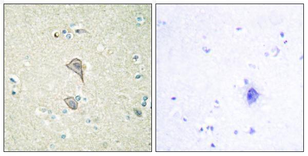
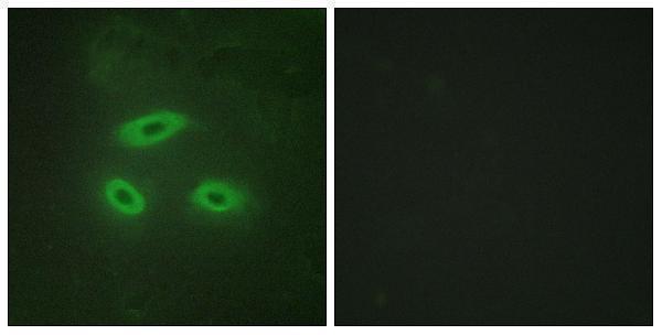
A09881 has IHC data from paraffin-embedded human brain tissue and IF data from HeLa cells (A09881 image captions). Its listed reactivity covers human, mouse, and rat (A09881 catalog reactivity).

Real IHC data Immunohistochemistry analysis of paraffin-embedded human brain tissue, using NKTR Antibody. The picture on the right is blocked with the synthesized peptide.
Anti-NK-tumor recognition protein NKTR Antibody
Cat # A09881

A09881 has an IHC image of paraffin-embedded human brain tissue with peptide blocking (A09881 IHC image caption). A09881 also has an IF image of HeLa cells with peptide blocking, and lists IHC, IF, and ICC applications plus human, mouse, and rat reactivity (A09881 IF image caption; catalog applications and reactivity).

Which to pick: For tissue IHC, choose A09881 for paraffin sections based on its human brain image; the fixative is unreported (A09881 IHC image caption). For IF/ICC, choose A09881 because both applications are listed and its IF image shows HeLa cells (A09881 catalog applications; IF image caption). For cross-species work, A09881 lists human, mouse, and rat reactivity and is rabbit polyclonal, while its supplied IHC image documents human tissue only (A09881 catalog reactivity and clonality; IHC image caption).

Each figure is that product's own IHC / IF validation image from its datasheet.

References

  1. UniProt Consortium. UniProt entry P30414 (NKTR_HUMAN, NK-tumor recognition protein).
  2. Human Protein Atlas. NKTR tissue IHC expression (reliability: Approved).
  3. Human Protein Atlas. NKTR subcellular location (ICC-IF): Mainly localized to the nucleoplasm, plasma membrane and cytosol..
  4. Human Protein Atlas. NKTR antibody validation summary (2 antibodies).
  5. Gene expression profile of human colorectal cancer identified NKTR as a biomarker for liver metastasis. Aging 2022 — PMC9467399.
  6. ACT001 inhibits the proliferation of non-small cell lung cancer cells by upregulating NKTR expression. Thoracic cancer 2022 — PMC9200889.
  7. Identification of stromally expressed molecules in the prostate by tag-profiling of cancer-associated fibroblasts, normal fibroblasts and fetal prostate. Oncogene 2012 — PMC3307063.
  8. CXCL10-induced chemotaxis of ex vivo-expanded natural killer cells combined with NKTR-255 enhances anti-tumor efficacy in osteosarcoma. Molecular therapy. Oncology 2025 — PMC12509742.
  9. PubMed PMID:8421688 — UniProt-cited evidence.
  10. PubMed PMID:17081983 — UniProt-cited evidence.
  11. PubMed PMID:18669648 — UniProt-cited evidence.