NKX2-2 / Homeobox protein Nkx-2.2 · IHC design guide

Design Immunohistochemistry for NKX2-2

Plan paraffin-section NKX2-2 IHC around the expected nuclear location (UniProt) and reported nuclear and cytoplasmic tissue staining (HPA tissue IHC). Use the tissue examples to choose controls and interpret results in light of presumed off-target binding (HPA tissue IHC).

Evidence assembled Oct 2026 · For research use; verify linked source records and product datasheet before use
Immunohistochemistry protocol sheet for NKX2-2 (IHC for NKX2-2): expected localisation Nuclear expected (UniProt); nuclear/cytoplasmic seen (HPA tissue IHC), antibody A04740-2, validated IHC image, and IHC protocol steps
Printable NKX2-2 IHC protocol sheet — expected localisation Nuclear expected (UniProt); nuclear/cytoplasmic seen (HPA tissue IHC), antibody A04740-2, controls and protocol steps. Open the full NKX2-2 IHC guide →

NKX2-2 Immunohistochemistry Experimental Design Guide

Expected localisation, validated protocols, controls and antibodies — the at-a-glance facts below, then the full design guide.

Must know before staining
Expected localisation Nuclear expected (UniProt); nuclear/cytoplasmic seen (HPA tissue IHC)
Staining pattern Nuclear/cytoplasmic staining in CNS and pancreas; CNS glia positive (HPA tissue IHC)
Antigen retrieval EDTA pH 8.0 HIER, heat-mediated (datasheet A04740-2)
Positive control ⓘ Caudate+2 more · see all
Negative control ⓘ Adipose tissue+4 more · see all
Important caveats
Reasons your staining may differ from the expected pattern.
Fixation Keep fixation consistent across sections (standard IHC practice; not target-specific)
Caveat Presumed off-target binding can distort tissue staining (HPA tissue IHC)
Regulation Expression regulation unannotated (UniProt)
Isoform / epitope No annotated isoforms; intact 1–273 chain (UniProt)
Section 1

Recommended NKX2-2 IHC & IF Protocols

The catalog antibody’s IHC protocol (datasheet: A04740-2) is accompanied by published protocols for Ewing sarcoma and neuroendocrine neoplasms (PMC13325908, PMC13518683, PMC8115302).

Recommended immunohistochemistry (IHC-P) protocol parameters
SampleParaffin-embedded human pancreas cancer tissue; fixative not specified (datasheet A04740-2)
FixationImage fixative and duration unreported (datasheet A04740-2); verify before use.
Sectioning4–5 µm sections on charged slides (standard)
DeparaffinisationXylene, graded ethanol series to water (standard)
Antigen retrievalHeat retrieval: EDTA pH 8.0 (datasheet A04740-2); 20 min, 95–100 °C (standard)
Peroxidase block3% H2O2, 10 min, room temperature (standard)
Blocking10% goat serum (datasheet A04740-2)
Primary antibodyRabbit anti-NKX2-2, 2-5 μg/ml (datasheet A04740-2)
Primary incubationOvernight at 4 °C (datasheet A04740-2)
DetectionHRP-conjugated secondary, DAB chromogen (datasheet A04740-2)
CounterstainHematoxylin, blue, dehydrate and mount (standard)
Expected resultNKX2-2-positive staining in glial cells of caudate (HPA tissue IHC: Medium). HPA tissue profile: Nuclear and cytoplasmic expression in several tissues, including CNS and pancreas. No signal in the no-primary control.
💡Decision noteStart with heat-mediated EDTA retrieval at pH 8.0 (datasheet: A04740-2); the published excerpts do not specify retrieval conditions (PMC13325908, PMC13518683, PMC8115302).
Section 2

What Is the Expected NKX2-2 Staining Pattern?

NKX2-2 is a nuclear transcription factor with no transmembrane segment (UniProt O95096). In paraffin-section IHC, expect staining in glial cells of the caudate, cerebral cortex, and hippocampus, where HPA reports medium staining (HPA tissue IHC). HPA also describes nuclear and cytoplasmic staining across several tissues, but rates its tissue IHC antibody Approved with low consistency against RNA data and presumed off-target binding (HPA tissue IHC).

What am I looking at on my slide?
Distinct nuclear staining in glial cells of caudate, cerebral cortex, or hippocampus.This fits the expected compartment and HPA's medium glial staining in those tissues (UniProt O95096; HPA tissue IHC). Score the identified cell population and nuclear signal; do not treat every stained cell in a positive tissue as confirmed NKX2-2.
Strong cytoplasmic staining dominates, with little or no nuclear enrichment.UniProt places NKX2-2 in the nucleus (UniProt O95096). HPA reports some cytoplasmic tissue staining, so cytoplasm alone does not prove an artefact; a predominantly cytoplasmic result warrants checking specificity and comparing the nuclear pattern in glial cells (HPA tissue IHC).
Prominent staining in an HPA-negative cell population, such as adipocytes in adipose tissue.HPA reports adipocytes as not detected in adipose tissue (HPA tissue IHC). Consider cross-reactivity or endogenous chromogenic detection activity, then compare a no-primary control and the expected glial pattern. HPA's presumed off-target binding makes isolated unexpected staining especially uncertain (HPA tissue IHC).
Diffuse colour coats cells or tissue without a clear nuclear boundary.This is difficult to score as nuclear NKX2-2 (UniProt O95096). General IHC causes include nonspecific reagent binding or residual chromogen-generating activity (standard IHC practice). Check background controls before interpreting weak colour as the low staining HPA reports in some tissues (HPA tissue IHC).
No staining is visible in glial cells of a caudate, cerebral cortex, or hippocampus section.Absence conflicts with HPA's medium glial staining in those tissues, but does not alone establish target absence or assay failure (HPA tissue IHC). Review tissue identity, section quality, controls, and the validated IHC workflow before making a biological call (standard IHC practice).
💡Expected NKX2-2 appearanceCall a result positive when glial-cell nuclei show discernible, approximately medium staining in caudate, cerebral cortex, or hippocampus (UniProt O95096; HPA tissue IHC); isolated strong cytoplasmic colour or staining confined to an HPA-negative cell population is questionable, especially given presumed off-target binding (HPA tissue IHC).
How each factor affects the staining
Subcellular locationNuclear localisation is the primary basis for reading a positive cell (UniProt O95096). HPA's broader nuclear and cytoplasmic tissue description means cytoplasmic colour needs context; it should not replace assessment of nuclear staining in the named glial populations (HPA tissue IHC).
Tissue and cell selectionHPA reports medium glial staining in caudate, cerebral cortex, and hippocampus, but only low staining in pancreatic exocrine glandular cells (HPA tissue IHC). Choose a reported medium-staining glial population when judging whether an IHC run produced the expected pattern.
Strength of tissue validationThe listed antibody, HPA003468, has Approved IHC status, while the tissue profile notes low staining–RNA consistency and presumed off-target binding (HPA antibody record; HPA tissue IHC). Treat an unexpected compartment or cell type as a finding to verify, rather than independent evidence of NKX2-2 expression.
Molecular contextUniProt records one 273-aa chain, no signal peptide, no transmembrane segment, and no annotated isoforms or glycosylation sites (UniProt O95096). These annotations provide no basis here for a secreted or membrane staining pattern and do not establish antigen-retrieval or fixation sensitivity.
Why is my staining missing, weak or wrong?
SituationLikely causeNext action
Expected glial nuclei are blank in a reported medium-staining brain region.Possible assay failure or a tissue-identification or section-quality issue; HPA reports medium glial staining but does not define why an individual run fails (HPA tissue IHC).Verify tissue and cell identity, inspect the section, and check run controls and the IHC-validated antibody's documented conditions before interpreting the negative result (standard IHC practice).
Cytoplasm is stronger than nuclei throughout the section.The distribution does not align cleanly with UniProt's nuclear localisation, although HPA does describe cytoplasmic tissue staining (UniProt O95096; HPA tissue IHC).Compare nuclear signal in the reported glial populations and inspect a no-primary control; with HPA's off-target caveat, avoid calling cytoplasm alone a confirmed positive (HPA tissue IHC; standard IHC practice).
Cells reported as not detected, such as adipocytes in adipose tissue, stain strongly.Off-target binding or endogenous detection activity is possible; HPA reports those adipocytes as not detected and acknowledges presumed off-target binding (HPA tissue IHC).Check the no-primary control and whether the colour follows cell boundaries or the expected glial nuclear pattern; do not assign NKX2-2 expression from this finding alone (standard IHC practice; UniProt O95096).
Weak colour appears broadly across the slide and obscures nuclei.Nonspecific binding or residual endogenous chromogen-generating activity can cause diffuse background in chromogenic IHC (standard IHC practice).Review blocking and detection controls, reagent exposure, and counterstain visibility; score only interpretable cellular staining against the nuclear expectation (standard IHC practice; UniProt O95096).
Pancreatic exocrine cells are weak while brain glial cells stain more clearly.That contrast is compatible with HPA's low exocrine pancreatic and medium glial staining; it does not establish a pancreas-specific fixation effect (HPA tissue IHC).Record compartment, cell type, and relative intensity separately. Use the reported medium-staining glial population to assess the run, and keep weak pancreatic staining provisional (HPA tissue IHC).
Can this IHC pattern be used as an IF/ICC localisation benchmark?HPA provides no ICC-IF images or available main subcellular location for NKX2-2; its tissue observations come from IHC (HPA subcellular record; HPA tissue IHC).Use UniProt's nuclear annotation as the localisation expectation, while treating IF/ICC performance as unverified by the supplied HPA record (UniProt O95096; HPA subcellular record).

Sample controls for NKX2-2 IHC & IF

🧪Run caudate first and score nuclear staining in glial cells (HPA: Medium in caudate glial cells; UniProt O95096: nucleus). Use adipose tissue as the negative tissue, scoring adipocytes (HPA: Not detected in adipocytes); on the caudate slide, assess morphologically identified non-glial cells as internal negatives and verify that their nuclei lack specific signal (HPA: staining reported in caudate glial cells; UniProt O95096: nucleus).
Positive control tissue: Caudate (Glial cells, HPA Medium)
Negative control tissue: Adipose tissue (HPA Not detected)
ICC-IF cell lines (HPA subcellular resource): HPA carries no ICC-IF cell line for NKX2-2; derive a cell-line control from the positive tissue's cell type (Glial cells) and confirm it by RNA or western blot first.
Technical controls: Include a no-primary, secondary-only section; a rabbit IgG control matched to the primary antibody's format; and, if available, a matched NKX2-2 knockout section as a biological negative (selected-SKU caption: rabbit primary antibody; standard IHC practice). Quench endogenous peroxidase and distinguish tissue pigment from DAB signal in caudate sections (selected-SKU caption: peroxidase detection with DAB; standard IHC practice).
⚠️Feasibility: A target-specific fixation window and fixation effect are unreported in the supplied evidence; the selected-SKU tissue-IHC caption does not state a fixative (selected-SKU caption: fixative not stated). The paraffin-section example uses heat retrieval in EDTA at pH 8.0, but it does not establish that retrieval is required under other conditions (selected-SKU caption: heat-mediated EDTA retrieval). The supplied evidence does not establish that frozen sections or IF are easier; assess pigment that could resemble chromogenic signal in caudate sections (HPA: Medium in caudate glial cells; standard IHC practice).

HPA tissue IHC evidence for NKX2-2

Comprehensive Human Protein Atlas IHC scoring per tissue (reliability: Approved — Low consistency between antibody staining and RNA expression data. Presumed off target binding observed and disregarded.). Rows are taken directly from the HPA tissue chart — click any row's HPA link to view the source.

Positive expression · recommended positive controls

TissueCell typeLevelEvidenceSource
Caudate Glial cells Medium Protein (IHC) HPA →
Cerebral cortex Glial cells Medium Protein (IHC) HPA →
Hippocampus Glial cells Medium Protein (IHC) HPA →

Undetected expression · recommended negative controls

TissueCell typeLevelEvidenceSource
Adipose tissue Adipocytes Not detected Protein (IHC) HPA →
Adrenal gland Glandular cells Not detected Protein (IHC) HPA →
Appendix Glandular cells Not detected Protein (IHC) HPA →
Bone marrow Hematopoietic cells Not detected Protein (IHC) HPA →
Breast Adipocytes Not detected Protein (IHC) HPA →
Section 3

Advanced NKX2-2 IHC Tips

Troubleshoot chromogenic IHC for NKX2-2 by checking retrieval, nuclear signal, controls and scoring before interpreting a stained section.

What retrieval should I try first when NKX2-2 staining is weak in paraffin sections?
Start with heat-mediated retrieval in EDTA at pH 8.0 for paraffin sections (datasheet A04740-2). The selected image used this retrieval before staining a human pancreas cancer section (caption A04740-2). After retrieval, compare matched sections processed with and without primary antibody to distinguish recovered nuclear signal from detection background (standard IHC practice; UniProt O95096: nucleus). Keep section thickness, heating, cooling and detection conditions consistent while optimizing the heating interval, since their effects cannot be separated if changed together (standard IHC practice). If staining remains weak, evaluate an alternative retrieval buffer on matched sections as a fallback and judge it by nuclear signal and tissue preservation (standard IHC practice; UniProt O95096: nucleus).
How should I troubleshoot fixation when NKX2-2 nuclear staining varies between blocks?
The selected tissue caption describes a paraffin section but does not state its fixative, so target-specific fixation sensitivity is unknown (caption A04740-2). Record the fixative and fixation duration for each block, then compare sections processed with the same retrieval and detection conditions (standard IHC practice). Include a previously stained reference section and a no-primary control in each run to separate changes in antigen accessibility from detection background (standard IHC practice). Assess nuclear staining together with morphology, since damaged or poorly preserved nuclei make compartment-specific interpretation unreliable (UniProt O95096: nucleus; standard IHC practice). Do not assign a fixation effect to NKX2-2 without a controlled comparison of fixation conditions (caption A04740-2: fixative unreported).
Should cytoplasmic NKX2-2 staining be scored as positive in paraffin IHC?
Prioritize staining in intact nuclei because NKX2-2 is annotated as nuclear (UniProt O95096: nucleus). Tissue IHC has shown both nuclear and cytoplasmic expression, but its approved profile also notes low consistency with RNA and presumed off-target staining (HPA tissue IHC). Compare nuclear signal with cell morphology and a no-primary control before assigning positivity to a cell population (standard IHC practice). Record cytoplasmic staining separately rather than folding it into a nuclear score, especially when it is diffuse or shared across unrelated cells (UniProt O95096: nucleus; standard IHC practice). If the pattern persists, check it with an independently validated antibody or another orthogonal readout (standard IHC practice).
Could an unrecognized isoform or membrane epitope explain inconsistent NKX2-2 staining?
The supplied record annotates one 273-amino-acid chain and 0 isoforms, but gives no antibody epitope sequence (UniProt O95096). It also reports no transmembrane segment or signal peptide, so assess staining as an intracellular nuclear target rather than a membrane pattern (UniProt O95096 topology; UniProt O95096: nucleus). The record lists 0 glycosylation sites and 0 modified residues; those annotations do not establish which epitope retrieval exposes (UniProt O95096). If two antibody preparations give different patterns, compare their documented immunogens when available and stain adjacent sections under matched conditions (standard IHC practice). Use nuclear localisation and an independent control to evaluate specificity without attributing disagreement to an unannotated isoform (UniProt O95096; standard IHC practice).
How can IF help check an ambiguous NKX2-2 IHC pattern?
Use IF on a matched specimen as an orthogonal localisation check for a chromogenic IHC result, while assessing the IF antibody independently (standard IHC/IF practice). Multiplex NKX2-2 with a validated marker for the expected cell population and a nuclear counterstain, then inspect whether signals coincide in the same nuclei (UniProt O95096: nucleus; standard IF practice). Choose fluorophores after examining tissue autofluorescence in unstained sections, favoring channels with clear separation from that background (standard IF practice). Permeabilise to allow access to this intracellular nuclear target and optimize the step for the IF antibody and specimen (UniProt O95096 topology; standard IF practice). No NKX2-2 ICC/IF image evidence is supplied here, so verify any IF pattern against controls before using it to interpret IHC (HPA subcellular).
What should I change when DAB obscures weak NKX2-2 nuclear signal?
First inspect a no-primary section for staining produced by the detection system, then compare it with the test section at the same DAB development time (standard IHC practice). The selected procedure used 10% goat serum block, a rabbit primary at 2 μg/ml overnight at 4°C, and a peroxidase-conjugated secondary (caption A04740-2). A peroxidase block and controlled DAB development are general chromogenic IHC steps; neither is evidence of NKX2-2-specific staining (standard IHC practice). If background persists, check wash adequacy, blocking and primary concentration on matched sections rather than changing every variable at once (standard IHC practice). Retain conditions that preserve interpretable nuclear contrast and tissue morphology (UniProt O95096: nucleus; standard IHC practice).
How should I quantify NKX2-2 staining across paraffin sections? ⚠ ANSWER MARKED FOR VERIFICATION
Define the cell population and tissue region before scoring, and count only interpretable nuclear staining in intact cells (UniProt O95096: nucleus; standard IHC practice). Report the percentage of positive nuclei and, when intensity is reproducible, an H-score on a 0–300 scale using intensity categories 0–3 (standard IHC practice). For spatial analyses, report positive nuclei per mm² of viable tissue alongside total nuclei per mm² (standard IHC practice). Normalize comparisons to the same cell type, viable area, threshold and staining run, with reference sections for batch effects (standard IHC practice). Keep cytoplasmic staining in a separate field because the expected NKX2-2 compartment is nuclear (UniProt O95096: nucleus; HPA tissue IHC).
How do I distinguish credible NKX2-2 positivity from artefact?
A credible result has nuclear staining in morphologically intact cells and remains distinguishable from the no-primary control (UniProt O95096: nucleus; standard IHC practice). Treat isolated cytoplasmic staining, section-edge accentuation and signal within necrotic tissue cautiously; compare them with neighboring intact regions and matched controls (UniProt O95096: nucleus; standard IHC practice). Check a no-primary section and peroxidase-blocked control for endogenous enzyme signal when DAB staining appears outside the expected cells (standard IHC practice). HPA reports medium staining in glial cells of the caudate, cerebral cortex and hippocampus, but also flags presumed off-target staining and low RNA concordance (HPA tissue IHC). Resolve unexpected positive cells with cell identification and an independent assay before claiming NKX2-2 expression (standard IHC practice).
Boster reagents

Best NKX2-2 / Homeobox protein Nkx-2.2 IHC Antibodies

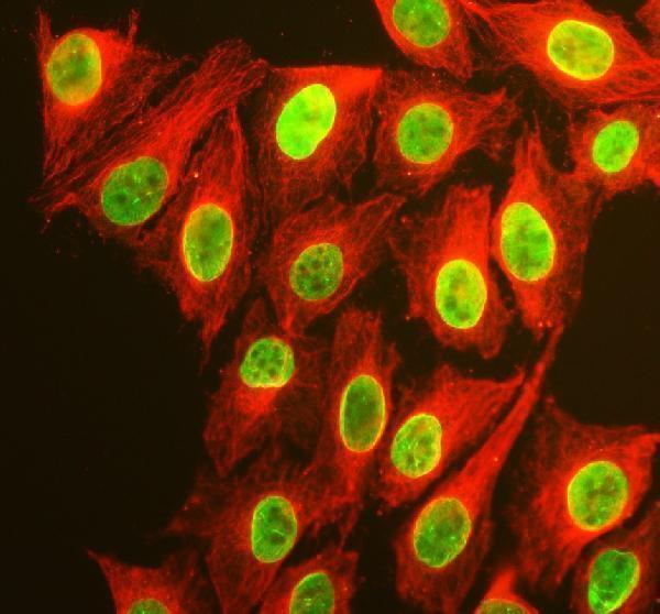
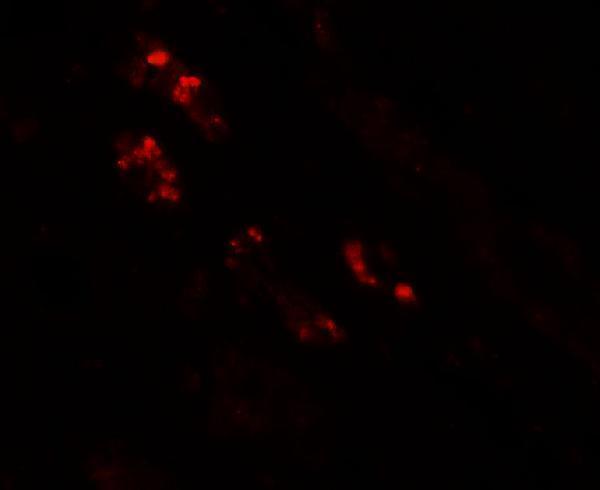
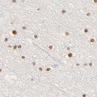
Anti-NKX2-2 IHC images cover human kidney and pancreas samples and mouse and rat brain (catalog IHC captions); IF images cover human kidney and U2OS cells (catalog IF captions).

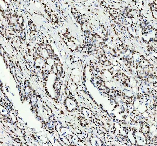
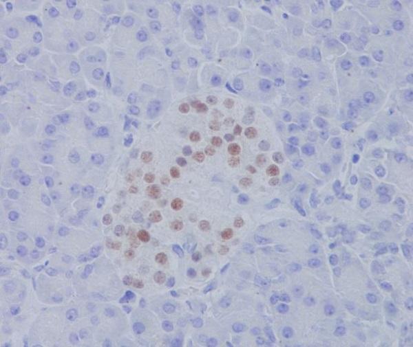
Real IHC data IHC analysis of NKX2-2 using anti-NKX2-2 antibody (A04740-2). NKX2-2 was detected in a paraffin-embedded section of human pancreas cancer tissue. Heat mediated antigen retrieval was performed in EDTA buffer (pH 8.0, epitope retrieval solution). The tissue section was blocked with 10% goat serum. The tissue section was then incubated with 2 μg/ml rabbit anti-NKX2-2 Antibody (A04740-2) overnight at 4°C. Peroxidase Conjugated Goat Anti-rabbit IgG was used as secondary antibody and incubated for 30 minutes at 37°C. The tissue section was developed using HRP Conjugated Rabbit IgG Super Vision Assay Kit (Catalog # SV0002) with DAB as the chromogen.
Anti-NKX2-2 Antibody
Cat # A04740-2
Real IHC data Immunohistochemistry of NKX2-2 in human kidney tissue with NKX2-2 antibody at 2.5 μg/mL.
Anti-Homeobox protein Nkx-2.2 NKX2-2 Antibody
Cat # A04740
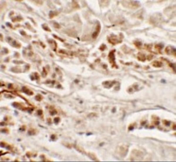
Real IHC data Immunohistochemical analysis of paraffin-embedded human pancreas, using NKX2.2 Antibody.
Anti-NKX2.2 Rabbit Monoclonal Antibody
Cat # M04740-1

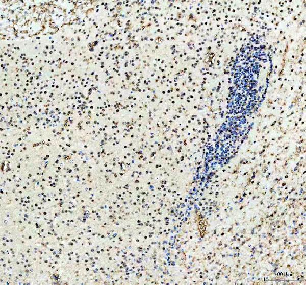
A04740 has human kidney IHC and IF images (A04740 image captions); A04740-2 has paraffin-section IHC images from human pancreas cancer and prostate cancer and mouse and rat brain, plus IF in U2OS cells (A04740-2 image captions). M04740-1 has a paraffin-section IHC image from human pancreas (M04740-1 IHC caption).

Which to pick: For tissue IHC, choose M04740-1 when a rabbit monoclonal is preferred: its human pancreas image documents paraffin sections, but does not report the fixative (M04740-1 catalog entry and IHC caption). For IF/ICC, choose A04740-2, which lists ICC/IF and shows IF in U2OS cells; A04740 also has a human kidney IF image (A04740-2 and A04740 catalog entries and IF captions). For IHC across the listed species, choose A04740-2: its own paraffin-section captions document human, mouse and rat samples, with EDTA retrieval at pH 8.0 and primary antibody at 2 μg/ml; the fixative is unreported (A04740-2 IHC captions).

Each figure is that product's own IHC / IF validation image from its datasheet.

References

  1. UniProt Consortium. UniProt entry O95096 (NKX22_HUMAN, Homeobox protein Nkx-2.2).
  2. Human Protein Atlas. NKX2-2 tissue IHC expression (reliability: Approved).
  3. Human Protein Atlas. NKX2-2 subcellular location (ICC-IF): Highest expression in HAP1: 3.8 nTPM.
  4. Human Protein Atlas. NKX2-2 antibody validation summary (1 antibodies).
  5. Role of Nkx2.2 immunohistochemistry in distinguishing Ewing sarcoma from neuroendocrine neoplasms at different sites. Journal of Taibah University Medical Sciences 2026 — PMC13325908.
  6. Primary Ewing Sarcoma of the Lung: Integrative Analysis of Clinical, Histopathological, Immunohistochemical, and Cytogenetic Features with Literature Review. Indian journal of surgical oncology 2026 — PMC13518683.
  7. Ontogenetic distribution of the transcription factor nkx2.2 in the developing forebrain of Xenopus laevis. Frontiers in neuroanatomy 2011 — PMC3049246.
  8. Adamantinoma-like Ewing Sarcoma of the Salivary Glands: A Newly Recognized Mimicker of Basaloid Salivary Carcinomas. The American journal of surgical pathology 2019 — PMC8115302.
  9. PubMed PMID:9703340 — UniProt-cited evidence.
  10. PubMed PMID:11780052 — UniProt-cited evidence.
  11. PubMed PMID:15489334 — UniProt-cited evidence.