NLRP12 / NACHT, LRR and PYD domains-containing protein 12 · IHC design guide

Design Immunohistochemistry for NLRP12

Plan NLRP12 IHC on paraffin sections around cytoplasmic staining in tissue immune cells (HPA tissue IHC). The catalog antibody has a starting IHC concentration of 5 μg/mL (datasheet); interpret staining with care because agreement with RNA expression is very low (HPA tissue IHC).

Evidence assembled Oct 2026 · For research use; verify linked source records and product datasheet before use
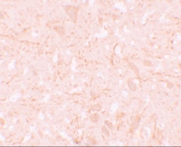
Immunohistochemistry protocol sheet for NLRP12 (IHC for NLRP12): expected localisation Cytoplasm of tissue immune cells (HPA tissue IHC), antibody A02124, validated IHC image, and IHC protocol steps
Printable NLRP12 IHC protocol sheet — expected localisation Cytoplasm of tissue immune cells (HPA tissue IHC), antibody A02124, controls and protocol steps. Open the full NLRP12 IHC guide →

NLRP12 Immunohistochemistry Experimental Design Guide

Expected localisation, validated protocols, controls and antibodies — the at-a-glance facts below, then the full design guide.

Must know before staining
Expected localisation Cytoplasm of tissue immune cells (HPA tissue IHC)
Staining pattern Cytoplasmic immune-cell staining, most abundant in GI tract (HPA tissue IHC)
Antigen retrieval Citrate pH 6.0 HIER, 95–98 °C, 20 min (rule: cytoplasmic / membrane antigen)
Positive control ⓘ Fallopian tube+4 more · see all
Negative control ⓘ Adipose tissue+4 more · see all
Important caveats
Reasons your staining may differ from the expected pattern.
Fixation Keep fixation consistent across sections (standard IHC practice; not target-specific)
Caveat Very low agreement between staining and RNA expression (HPA tissue IHC)
Regulation Leukocyte abundance affects signal (UniProt)
Isoform / epitope 7 isoforms; epitope coverage is unspecified (UniProt)
Section 1

Recommended NLRP12 IHC & IF Protocols

The catalog antibody’s IHC-P protocol (datasheet) is accompanied by published chromogenic IHC protocols for glioma (PMC12755504) and prostate tissue (PMC5491527).

Recommended immunohistochemistry (IHC-P) protocol parameters
SampleTissue sections; selected-image fixative not specified (standard IHC workflow)
FixationImage fixative and duration unreported (datasheet A02124); verify before use.
Sectioning4–5 µm sections on charged slides (standard)
DeparaffinisationXylene, graded ethanol series to water (standard)
Antigen retrievalHeat-induced epitope retrieval in citrate buffer, pH 6.0, 20 min at 95–98 °C (standard rule: cytoplasmic / membrane antigen)
Peroxidase block3% H2O2, 10 min, room temperature (standard)
Blocking10% normal serum of the secondary host, 30 min, room temperature (standard)
Primary antibodyRabbit anti-NLRP12, 5 μg/mL (datasheet A02124)
Primary incubationOvernight at 4 °C (standard)
DetectionHRP-polymer secondary, DAB chromogen 5–10 min (standard)
CounterstainHematoxylin, blue, dehydrate and mount (standard)
Expected resultNLRP12-positive staining in glandular cells of fallopian tube (HPA tissue IHC: High). HPA tissue profile: Cytoplasmic expression in immune cells of several tissues, most abundant in gastrointestinal tract. No signal in the no-primary control.
💡Decision noteStart with citrate pH 6.0 retrieval at 95–98 °C for 20 min (page retrieval rule); both published protocols also use pH 6.0 retrieval (PMC12755504; PMC5491527).
Section 2

What Is the Expected NLRP12 Staining Pattern?

NLRP12 is cytoplasmic and has no transmembrane segment (UniProt P59046). In paraffin sections, expect cytoplasmic staining in immune cells, especially in gastrointestinal tissue; bone marrow hematopoietic cells show medium staining (HPA tissue IHC). Interpret this pattern cautiously: the HPA antibody is rated Approved for IHC, but its staining has very low consistency with RNA expression and includes cells not annotated in its tissue profile (HPA tissue IHC).

What am I looking at on my slide?
Discrete cytoplasmic staining in immune cells, especially in gastrointestinal tissue; medium staining in bone marrow hematopoietic cells (HPA tissue IHC).This fits the reported tissue pattern and cytoplasmic location (HPA tissue IHC; UniProt P59046). Score the stained cells and compartment, rather than treating all cells in a positive tissue as positive (standard IHC practice).
Predominantly nuclear or sharply membrane-bound staining.That distribution conflicts with the reported cytoplasmic location and lack of a transmembrane segment (UniProt P59046). Check morphology, counterstain and detection controls before calling it NLRP12 (standard IHC practice).
Strong staining mainly in an unexpected cell population.Consider nonspecific antibody binding or endogenous detection activity (standard IHC practice). High glandular-cell staining in fallopian tube and high myocyte staining in skeletal muscle are reported observations, but the tissue profile has very low RNA concordance; these findings alone do not establish specificity (HPA tissue IHC).
Broad, diffuse colour obscures cell boundaries across the section.Treat this as possible background, not a defined cytoplasmic pattern (standard IHC practice). Review blocking, antibody concentration, washes and the no-primary control (standard IHC practice); HPA's tissue profile does not identify the cause of background in this specimen.
No staining in bone marrow hematopoietic cells.HPA reports medium staining in that population, so absence of signal merits a technical check (HPA tissue IHC). Compare morphology and a concurrent positive control, then review the catalog antibody's IHC-P conditions (standard IHC practice).
💡Expected NLRP12 appearanceCall a result positive when identifiable immune or hematopoietic cells show discrete cytoplasmic staining, with medium staining reported in bone marrow hematopoietic cells (HPA tissue IHC; UniProt P59046); widespread nuclear, membrane-bound or cell-independent colour warrants control review (standard IHC practice).
How each factor affects the staining
Cell population and tissueHPA describes cytoplasmic expression in immune cells of several tissues, most abundant in the gastrointestinal tract, and medium staining in bone marrow hematopoietic cells (HPA tissue IHC). UniProt reports detection in peripheral blood leukocytes, predominantly eosinophils and granulocytes, with lower levels in monocytes (UniProt P59046).
Antibody evidenceHPA042981 is Approved for IHC, while HPA reports very low consistency between antibody staining and RNA expression, including staining in cells or structures outside its annotated profile (HPA tissue IHC; HPA antibodies). Treat a positive stain as an observation that needs cell-level and control-based interpretation (standard IHC practice).
Negative comparisonAdipocytes in adipose tissue, smooth muscle cells, and urinary bladder urothelial cells are reported as not detected (HPA tissue IHC). These observations can help compare staining patterns, but a negative comparison alone cannot establish antibody specificity (standard IHC practice).
Protein form and compartmentUniProt lists seven isoforms, one full-length chain, no signal peptide or propeptide, and no transmembrane segment (UniProt P59046). The supplied record does not identify the antibody epitope, so it cannot establish which isoforms this stain detects or support a claim about cleavage or shedding.
IF/ICC evidenceDoes the HPA record confirm an IF/ICC pattern? No: it lists no main subcellular location or cell lines with ICC-IF images, and HPA042981 has no ICC validation entry (HPA subcellular; HPA antibodies). Use the separate IF/ICC guide for that application.
Why is my staining missing, weak or wrong?
SituationLikely causeNext action
The bone marrow positive comparison is blank.A technical failure is possible because HPA reports medium staining in bone marrow hematopoietic cells (HPA tissue IHC); the record does not identify a target-specific fixation effect.Check the concurrent positive control, section quality and detection reagents, then follow the catalog antibody's IHC-P conditions for retrieval and dilution (standard IHC practice).
Colour appears mainly in nuclei or along cell membranes.The pattern conflicts with cytoplasmic localisation and lack of a transmembrane segment (UniProt P59046); misread counterstain or nonspecific detection is possible (standard IHC practice).Compare with the no-primary control and inspect the compartment at higher magnification before scoring positive cells (standard IHC practice).
Many cell types stain uniformly.Diffuse background or endogenous detection activity can obscure cell-specific staining (standard IHC practice); HPA also reports low RNA concordance for its tissue staining (HPA tissue IHC).Review the no-primary control, blocking, antibody concentration and washes; score only distinguishable cellular staining (standard IHC practice).
Strong signal is confined to glandular cells or myocytes.HPA reports high staining in fallopian tube glandular cells and skeletal muscle myocytes, but rates overall antibody–RNA consistency as very low (HPA tissue IHC).Record the tissue and cell type without assuming specificity; compare controls and the reported immune-cell pattern (standard IHC practice; HPA tissue IHC).
A presumed negative tissue shows focal staining.A tissue-level negative label applies to the specified cell population: for example, HPA reports adipocytes and urothelial cells as not detected (HPA tissue IHC). Other cells may need separate identification (standard IHC practice).Identify the stained cells and their compartment, then compare the matching HPA cell-level entry and a no-primary control (HPA tissue IHC; standard IHC practice).
An IF/ICC image differs from the paraffin-section result.The supplied HPA record has no ICC-IF images, main ICC-IF location or ICC validation entry for HPA042981 (HPA subcellular; HPA antibodies).Do not transfer the IHC-P staining call to IF/ICC; assess the IF/ICC result with application-specific controls and its separate guide (standard IF practice).

Sample controls for NLRP12 IHC & IF

🧪Run fallopian tube first: glandular cells should stain (HPA: High in fallopian tube glandular cells). Use smooth muscle as the negative tissue (HPA: Not detected in smooth muscle cells); on the positive slide, the fallopian tube smooth muscle layer should show background-level staining, but confirm it as an internal negative rather than assuming every nonglandular cell is negative (HPA: Not detected in smooth muscle cells).
Positive control tissue: Fallopian tube (Glandular cells, HPA High)
Negative control tissue: Adipose tissue (HPA Not detected)
ICC-IF cell lines (HPA subcellular resource): HPA carries no ICC-IF cell line for NLRP12; derive a cell-line control from the positive tissue's cell type (Glandular cells) and confirm it by RNA or western blot first.
Technical controls: Include no-primary (secondary-only) and host-species- and isotype-matched nonimmune antibody controls; assess specificity with NLRP12 knockout material, if available, or a peptide block when the immunizing peptide is available (standard IHC practice). Quench endogenous peroxidase for chromogenic detection, and block endogenous biotin if using avidin–biotin detection (standard IHC practice).
⚠️Feasibility: A target-specific fixation window or fixation effect is unreported, and the selected A02124 tissue-IHC caption does not state a fixative (A02124 caption: fixative not stated). Retrieval dependence is unreported; compare retrieved and unretrieved paraffin sections in a pilot (standard IHC practice). The supplied evidence does not establish that frozen sections or IF/ICC are easier, or identify a fallopian tube-specific artefact; assess tissue background with the no-primary control (HPA subcellular: no ICC-IF image cell lines; standard IHC practice).

HPA tissue IHC evidence for NLRP12

Comprehensive Human Protein Atlas IHC scoring per tissue (reliability: Approved — Very low consistency between antibody staining and RNA expression data. Antibody staining in cells/structures not annotated, view images.). Rows are taken directly from the HPA tissue chart — click any row's HPA link to view the source.

Positive expression · recommended positive controls

TissueCell typeLevelEvidenceSource
Fallopian tube Glandular cells High Protein (IHC) HPA →
Skeletal muscle Myocytes High Protein (IHC) HPA →
Appendix Glandular cells Medium Protein (IHC) HPA →
Bone marrow Hematopoietic cells Medium Protein (IHC) HPA →
Bronchus Respiratory epithelial cells Medium Protein (IHC) HPA →

Undetected expression · recommended negative controls

TissueCell typeLevelEvidenceSource
Adipose tissue Adipocytes Not detected Protein (IHC) HPA →
Adrenal gland Glandular cells Not detected Protein (IHC) HPA →
Parathyroid gland Glandular cells Not detected Protein (IHC) HPA →
Smooth muscle Smooth muscle cells Not detected Protein (IHC) HPA →
Urinary bladder Urothelial cells Not detected Protein (IHC) HPA →
Section 3

Advanced NLRP12 IHC Tips

Use the IHC observations as a starting point, then verify NLRP12 staining by compartment, cell type and controls before interpreting tissue patterns.

Which retrieval conditions should I try first for NLRP12 in paraffin sections?
Start with citrate buffer at pH 6.0, heated to 95–98 °C for 20 min (page retrieval setting). Let sections cool in buffer, then compare staining with a matched section processed without retrieval to judge whether heat improves the signal (standard IHC practice). NLRP12 is cytoplasmic and has no transmembrane segment, so assess cytoplasmic staining in identifiable cells rather than treating membrane staining as successful retrieval (UniProt P59046). If staining remains weak, test a second retrieval condition on adjacent sections while holding antibody concentration and detection constant (standard IHC practice). Record the condition used for every scored section, since retrieval can change both signal and tissue background (standard IHC practice).
How should I troubleshoot fixation when NLRP12 staining is weak or uneven?
Target-specific fixation sensitivity is unknown from the supplied evidence; the selected antibody caption reports brain staining at 5 µg/mL but does not state a fixative (caption: A02124). Compare sections with documented fixation histories, using the same retrieval, antibody concentration and detection settings across each comparison (standard IHC practice). Check whether weak staining tracks with poorly preserved morphology or varies across a block before attributing it to NLRP12 abundance (standard IHC practice). Include a matched control tissue in each run to distinguish a processing change from a staining-run change (standard IHC practice). Do not infer an optimal fixation time from the tissue staining profile or from NLRP12 topology (HPA tissue IHC; UniProt P59046).
What localisation should count as plausible NLRP12 staining?
Score well-defined cytoplasmic staining first, because UniProt assigns NLRP12 to the cytoplasm and reports no transmembrane segment (UniProt P59046). Its reported peripheral-blood expression is strongest in eosinophils and granulocytes and lower in monocytes, so identify cell types before assigning a tissue-level result (UniProt P59046). HPA describes cytoplasmic staining in immune cells across several tissues, but also reports very low agreement between antibody staining and RNA expression (HPA tissue IHC). Treat isolated nuclear or crisp cell-surface staining as a specificity question and compare it with omission and background controls (standard IHC practice; UniProt P59046). Assess epithelial or neuronal staining cautiously, even where HPA lists positive cells, because those observations alone do not establish specificity (HPA tissue IHC).
How can isoforms or epitope placement affect NLRP12 IHC interpretation?
NLRP12 has 7 listed isoforms, so establish which sequence the catalog antibody recognizes before comparing staining between specimens (UniProt P59046). Map any disclosed immunogen against the pyrin domain at residues 1–95, FISNA at 129–201 and NACHT at 211–528 (UniProt P59046). If the immunogen sequence is unavailable, report that isoform coverage is unresolved rather than claiming that every isoform is detected (standard IHC interpretation; UniProt P59046). Compare staining from an independently targeted antibody or an appropriate loss-of-target control where available, while retaining identical tissue processing (standard IHC practice). NLRP12 has no listed signal peptide, propeptide or glycosylation sites; those annotations do not reveal this antibody’s epitope (UniProt P59046).
How should I adapt an NLRP12 tissue experiment to multiplex immunofluorescence?
For the separate IF assay, pair NLRP12 with a validated marker of the immune cell population being examined, and score overlap at the single-cell level (UniProt P59046; standard IF practice). Choose spectrally separated fluorophores and place the weaker signal in a channel with low tissue autofluorescence after checking unstained sections (standard IF practice). Because NLRP12 is cytoplasmic without a transmembrane segment, include a controlled permeabilisation step to admit antibody across the plasma membrane, then check morphology and background (UniProt P59046; standard IF practice). Run single-label and secondary-only controls to identify channel bleed-through and nonspecific fluorescence before interpreting overlap (standard IF practice). The selected 5 µg/mL brain-tissue caption documents IHC staining, not an IF concentration or IF fixation condition (caption: A02124).
What should I check when NLRP12 chromogenic staining is widespread or diffuse?
Inspect a no-primary section and a matched control section to separate detection-system background from staining that depends on the primary antibody (standard IHC practice). Block endogenous peroxidase before peroxidase-based detection, and compare a shorter chromogen development with the current run if diffuse DAB obscures cell boundaries (standard IHC practice). Titrate the primary antibody downward if background rises with concentration; the selected caption’s 5 µg/mL documents one brain-tissue image, not a universal working concentration (caption: A02124). Compare tissue edges, folds and necrotic regions with intact interior tissue, since these areas can produce misleading signal (standard IHC practice). Require plausible cytoplasmic cellular staining before counting a diffuse deposit as NLRP12 (UniProt P59046; standard IHC practice).
How should I quantify NLRP12 staining across tissue sections? ⚠ ANSWER MARKED FOR VERIFICATION
Define the scored cell population and region before reviewing stain intensity, then exclude folds, necrosis and section edges by the same rule in every specimen (standard IHC practice). For identifiable immune cells, report the percentage with cytoplasmic staining and, if intensity matters, an H-score using 0–3 intensity grades (UniProt P59046; standard IHC practice). For spatial comparisons, report positive-cell density per mm² of evaluable tissue and the total number of eligible cells assessed (standard IHC practice). Normalise comparisons to the relevant cell population or evaluable area, since immune-cell abundance can differ between sections (standard IHC practice). State the retrieval condition, staining threshold and controls alongside each score, especially given HPA’s very low staining–RNA consistency (page retrieval setting; HPA tissue IHC).
How can I distinguish credible NLRP12 staining from an artefact?
A credible result has reproducible cytoplasmic signal in identifiable cells and remains distinguishable from the no-primary control (UniProt P59046; standard IHC practice). Give particular attention to leukocytes, including granulocytes and monocytes, while recording the actual cell type present in each section (UniProt P59046). Investigate predominantly nuclear or membrane-like signal, tissue-edge gradients, necrotic deposits and staining reproduced by the detection control before calling a sample positive (UniProt P59046; standard IHC practice). HPA lists epithelial and neuronal positives but warns of very low consistency between antibody staining and RNA expression, so these patterns need independent support (HPA tissue IHC). The selected brain-tissue image at 5 µg/mL establishes an IHC observation, not the specificity of every stained cell (caption: A02124).
Boster reagents

Best NLRP12 / NACHT, LRR and PYD domains-containing protein 12 IHC Antibodies

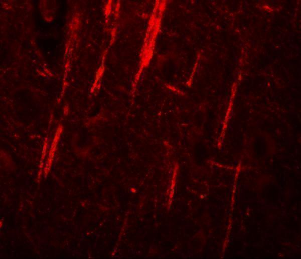
Anti-NLRP12 A02124 has human brain tissue IHC and IF images (catalog: A02124 image captions) and lists human, mouse and rat reactivity (catalog: A02124 reactivity).

Real IHC data Immunohistochemistry of NALP12 in human brain tissue with NALP12 antibody at 5 μg/mL.
Anti-NALP12 NLRP12 Antibody
Cat # A02124

A02124 lists IHC-P and IF applications (catalog: A02124 applications), with human brain tissue shown at 5 μg/mL for IHC and 20 μg/mL for IF (catalog: A02124 image captions). Its listed reactivity covers human, mouse and rat (catalog: A02124 reactivity); the images show human tissue only (catalog: A02124 image captions).

Which to pick: For paraffin-section tissue IHC, choose A02124 (catalog: A02124 IHC-P application); its IHC caption shows human brain tissue, but reports no fixative (catalog: A02124 IHC image caption). For IF, A02124 has a human brain tissue image (catalog: A02124 IF image caption); ICC-specific validation is unreported (catalog: A02124 applications and image captions). For cross-species work, A02124 lists human, mouse and rat reactivity (catalog: A02124 reactivity), although the supplied IHC and IF images show human tissue only (catalog: A02124 image captions).

Each figure is that product's own IHC / IF validation image from its datasheet.

References

  1. UniProt Consortium. UniProt entry P59046 (NAL12_HUMAN, NACHT, LRR and PYD domains-containing protein 12).
  2. Human Protein Atlas. NLRP12 tissue IHC expression (reliability: Approved).
  3. Human Protein Atlas. NLRP12 subcellular location (ICC-IF): Highest expression in THP-1: 2.5 nTPM.
  4. Human Protein Atlas. NLRP12 antibody validation summary (1 antibodies).
  5. High expression of NLRP12 predicts poor prognosis in patients with intracranial glioma. Journal of the Chinese Medical Association : JCMA 2023 — PMC12755504.
  6. NLRP12 decreases TRIM25-mediated HK2 degradation to promote glycolysis and H3K18la in gastric cancer. Cell death & disease 2025 — PMC12343871.
  7. NLRP12 Alleviated Periodontal Destruction via Suppressing Nuclear Factor Kappa-B Signalling Pathway. International dental journal 2026 — PMC12905752.
  8. Expression analysis of inflammasome sensors and implication of NLRP12 inflammasome in prostate cancer. Scientific reports 2017 — PMC5491527.
  9. PubMed PMID:12019269 — UniProt-cited evidence.
  10. PubMed PMID:12563287 — UniProt-cited evidence.
  11. PubMed PMID:11167794 — UniProt-cited evidence.