This website uses cookies to ensure you get the best experience on our website.


- Table of Contents
Plan NLRP3 paraffin IHC around the reported cytoplasmic tissue pattern (HPA tissue IHC). Compare stained sections with appropriate controls, and interpret intensity cautiously because HPA rates the tissue evidence uncertain (HPA tissue IHC).

Expected localisation, validated protocols, controls and antibodies — the at-a-glance facts below, then the full design guide.
| Expected localisation | Cytoplasmic staining in tissue (HPA tissue IHC) | |
| Staining pattern | Cytoplasmic staining in several tissues, including bone marrow (HPA tissue IHC) | |
| Antigen retrieval | Tris-EDTA pH 9.0 HIER, 95–98 °C, 20 min (rule: nuclear antigen) | |
| Positive control | Adrenal gland+4 more · see all | |
| Negative control | Adipose tissue+4 more · see all |
| Fixation | Keep fixation consistent across paraffin sections. (standard IHC practice; not target-specific) | |
| Caveat | Tissue staining has low consistency with RNA expression (HPA tissue IHC) | |
| Regulation | LPS-treated granulocytes express NLRP3 (UniProt) | |
| Isoform / epitope | 6 isoforms; map the antibody epitope to each (UniProt) |
The catalog antibody’s IHC protocol is followed by 4 published NLRP3 staining protocols (PMC6842944; PMC4807043; PMC12720632; PMC5331985).
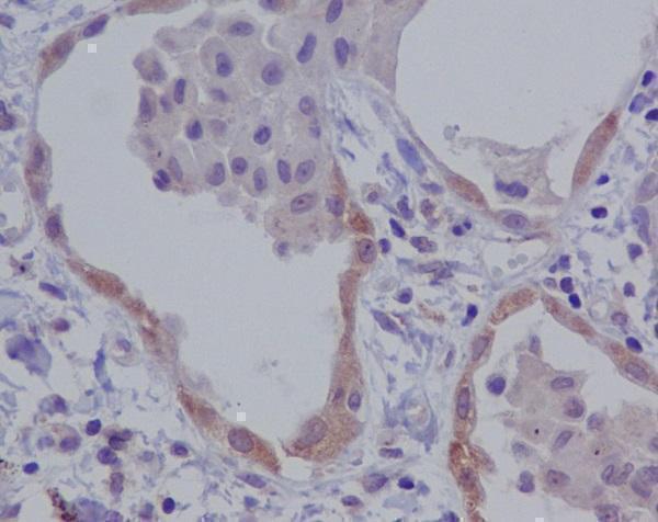
| Sample | Paraffin-embedded human lung carcinoma tissue; fixative not specified (datasheet M00034) |
| Fixation | Image fixative and duration unreported (datasheet M00034); verify before use. |
| Sectioning | 4–5 µm sections on charged slides (standard) |
| Deparaffinisation | Xylene, graded ethanol series to water (standard) |
| Antigen retrieval | Heat-induced epitope retrieval in Tris-EDTA buffer, pH 9.0, 20 min at 95–98 °C (standard rule: nuclear antigen) |
| Peroxidase block | 3% H2O2, 10 min, room temperature (standard) |
| Blocking | 10% normal serum of the secondary host, 30 min, room temperature (standard) |
| Primary antibody | Rabbit monoclonal (clone FBD-14) anti-NLRP3, 1:50-1:200 (datasheet M00034) |
| Primary incubation | Overnight at 4 °C (standard) |
| Detection | HRP-polymer secondary, DAB chromogen 5–10 min (standard) |
| Counterstain | Hematoxylin, blue, dehydrate and mount (standard) |
| Expected result | NLRP3-positive staining in glandular cells of adrenal gland (HPA tissue IHC: Medium). HPA tissue profile: Cytoplasmic expression in several tissues including bone marrow. No signal in the no-primary control. |
NLRP3 is mainly cytosolic in resting macrophages and can associate with organelle membranes (UniProt Q96P20). It has no transmembrane segment (UniProt Q96P20 topology). HPA reports cytoplasmic staining in several tissues, including bone marrow, but rates its tissue IHC pattern Uncertain because antibody staining and RNA expression have low consistency (HPA tissue IHC).
| Cytoplasmic staining in macrophages. | Fits the main resting-cell location and predominant cell type (UniProt Q96P20). |
| Strong, uniform nuclear staining in tissue cells. | Check for artefact; resting macrophage NLRP3 is mainly cytosolic (UniProt Q96P20). |
| Strong staining in adipocytes. | Check cross-reactivity or detection activity; HPA reports adipocytes Not detected (HPA tissue IHC). |
| Diffuse stain across cells and empty areas. | Suspect background; check a no-primary control (standard IHC practice). |
| No signal in bone marrow. | Check the run; HPA reports cytoplasmic expression there, with Uncertain reliability (HPA tissue IHC). |
| Activation state (UniProt Q96P20) | NLRP3 can move toward the MTOC and dispersed trans-Golgi vesicles (UniProt Q96P20). |
| Cell type (UniProt Q96P20) | Macrophages predominate; resting monocytes have very weak protein expression (UniProt Q96P20). |
| Tissue IHC confidence (HPA tissue IHC) | HPA rates the pattern Uncertain; antibody HPA012878 is also IHC Uncertain (HPA antibodies). |
| Situation | Likely cause | Next action |
|---|---|---|
| No cytoplasmic signal in bone marrow (HPA tissue IHC). | A failed run is possible; HPA tissue IHC reliability is Uncertain (HPA tissue IHC). | Check tissue preservation and a positive control (standard IHC practice). |
| Stain appears mainly nuclear in macrophages. | Unexpected for resting macrophages (UniProt Q96P20). | Review morphology and a no-primary control (standard IHC practice). |
| Adipocytes stain strongly. | HPA reports adipocytes Not detected (HPA tissue IHC). | Check cross-reactivity and endogenous detection activity (standard IHC practice). |
| Diffuse chromogen covers the section. | Nonspecific background or endogenous detection activity is possible (standard IHC practice). | Review blocking, washes and a no-primary control (standard IHC practice). |
| HPA tissue patterns disagree with this slide. | HPA rates tissue IHC reliability Uncertain (HPA tissue IHC). | Compare cell type and compartment before scoring (standard IHC practice). |
| Can IF/ICC confirm the punctate pattern? | HPA lists no ICC-IF images or main location (HPA subcellular). | Use the separate IF/ICC guide; this HPA record cannot confirm puncta (HPA subcellular). |


Comprehensive Human Protein Atlas IHC scoring per tissue (reliability: Uncertain — Low consistency between antibody staining and RNA expression data.). Rows are taken directly from the HPA tissue chart — click any row's HPA link to view the source.
| Tissue | Cell type | Level | Evidence | Source |
|---|---|---|---|---|
| Adrenal gland | Glandular cells | Medium | Protein (IHC) | HPA → |
| Appendix | Glandular cells | Medium | Protein (IHC) | HPA → |
| Bronchus | Respiratory epithelial cells | Medium | Protein (IHC) | HPA → |
| Cerebral cortex | Neuronal cells | Medium | Protein (IHC) | HPA → |
| Cervix | Squamous epithelial cells | Medium | Protein (IHC) | HPA → |
| Tissue | Cell type | Level | Evidence | Source |
|---|---|---|---|---|
| Adipose tissue | Adipocytes | Not detected | Protein (IHC) | HPA → |
| Caudate | Glial cells | Not detected | Protein (IHC) | HPA → |
| Cerebellum | Cells in granular layer | Not detected | Protein (IHC) | HPA → |
| Fallopian tube | Glandular cells | Not detected | Protein (IHC) | HPA → |
| Heart muscle | Cardiomyocytes | Not detected | Protein (IHC) | HPA → |
Troubleshoot NLRP3 staining in paraffin sections by checking retrieval, cell identity, compartment, and controls before interpreting chromogenic signal.
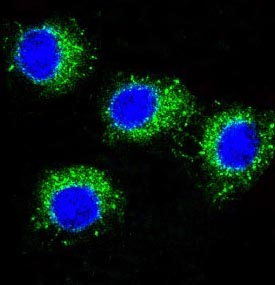
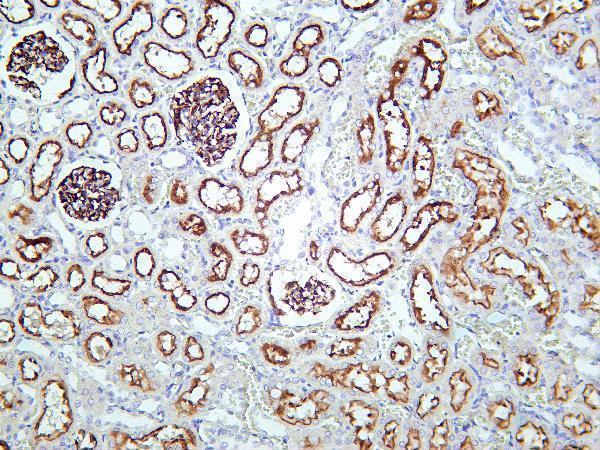
Catalog IHC images show human lung carcinoma in paraffin sections and rat kidney tissue; an IF image shows Raw264.7 cells (M00034 and M00034-1 image captions).
M00034 has an IHC image of paraffin-embedded human lung carcinoma and an IF image of Raw264.7 cells; its listed reactivity is human (M00034 image captions; catalog reactivity). M00034-1 has an IHC image of rat kidney tissue and lists IHC and IF applications with human, mouse and rat reactivity (M00034-1 IHC image caption; catalog applications and reactivity).
Which to pick: For human tissue IHC in paraffin sections, choose M00034: its own image documents that preparation, though the fixative is unreported (M00034 IHC image caption). For IF/ICC, choose M00034 when the listed human reactivity fits; it lists both applications and has an IF image, while its Raw264.7 image does not establish additional species reactivity (M00034 catalog applications and reactivity; IF image caption). For work requiring listed human, mouse and rat reactivity, choose M00034-1; its rat kidney IHC caption does not report section processing or fixative (M00034-1 catalog reactivity; IHC image caption).