This website uses cookies to ensure you get the best experience on our website.


- Table of Contents
Plan NLRP9 chromogenic IHC in paraffin sections using intestinal glandular staining as a reference (HPA tissue IHC). This guide covers fixation, controls and interpretation, including the reported mismatch between staining and RNA expression (HPA tissue IHC).

Expected localisation, validated protocols, controls and antibodies — the at-a-glance facts below, then the full design guide.
| Expected localisation | Membranous intestinal staining (HPA tissue IHC); cytoplasmic location (UniProt) | |
| Staining pattern | Membranous staining in intestinal glandular cells (HPA tissue IHC) | |
| Antigen retrieval | Citrate pH 6.0 HIER, 95–98 °C, 20 min (rule: cytoplasmic / membrane antigen) | |
| Positive control | Appendix+4 more · see all | |
| Negative control | Bone marrow+4 more · see all |
| Fixation | Keep fixation consistent across paraffin sections (standard IHC practice; not target-specific) | |
| Caveat | Staining and RNA show very low consistency (HPA tissue IHC) | |
| Regulation | Expression regulation is not established (UniProt) | |
| Isoform / epitope | 2 isoforms; assess epitope coverage (UniProt) |
The catalog antibody’s IHC-P protocol (datasheet) is accompanied by published protocols for gastric and colorectal cancers (PMC8262223) and MS brain lesions (PMC6138928).
| Sample | Tissue sections; selected-image fixative not specified (standard IHC workflow) |
| Fixation | Image fixative and duration unreported (datasheet A14119); verify before use. |
| Sectioning | 4–5 µm sections on charged slides (standard) |
| Deparaffinisation | Xylene, graded ethanol series to water (standard) |
| Antigen retrieval | Heat-induced epitope retrieval in citrate buffer, pH 6.0, 20 min at 95–98 °C (standard rule: cytoplasmic / membrane antigen) |
| Peroxidase block | 3% H2O2, 10 min, room temperature (standard) |
| Blocking | 10% normal serum of the secondary host, 30 min, room temperature (standard) |
| Primary antibody | Rabbit anti-NLRP9, 10 μg/mL (datasheet A14119) |
| Primary incubation | Overnight at 4 °C (standard) |
| Detection | HRP-polymer secondary, DAB chromogen 5–10 min (standard) |
| Counterstain | Hematoxylin, blue, dehydrate and mount (standard) |
| Expected result | NLRP9-positive staining in glandular cells of appendix (HPA tissue IHC: High). HPA tissue profile: Membranous expression in intestines. No signal in the no-primary control. |
NLRP9 is annotated as cytoplasmic, with no transmembrane segment (UniProt Q7RTR0), while tissue IHC reports membranous staining in intestines (HPA tissue IHC). Look first in intestinal glandular cells, including appendix and small intestine, and in pancreatic endocrine cells, all reported High (HPA tissue IHC). HPA rates the IHC evidence Approved but reports very low agreement between antibody staining and RNA expression (HPA tissue IHC).
| Intestinal glandular cells show a membrane-associated chromogenic signal. | This matches the reported intestinal IHC profile (HPA tissue IHC: membranous expression in intestines). Record its cellular distribution as observed; UniProt places NLRP9 in the cytoplasm and lists no transmembrane segment (UniProt Q7RTR0). The two location descriptions should remain visible when interpreting the slide. |
| Appendix or small-intestine glandular cells, or pancreatic endocrine cells, stain more strongly than surrounding cells. | These are useful positive comparators because each cell population is reported High by tissue IHC (HPA tissue IHC). High is a reported staining category, not a promised intensity for every section; assess the signal alongside tissue morphology and controls (general IHC practice). |
| A strong nuclear-only signal dominates the expected positive cells. | Nuclear-only staining does not match the cytoplasmic UniProt annotation or HPA's intestinal membrane-associated profile (UniProt Q7RTR0; HPA tissue IHC). Treat it as suspect and inspect background and detection controls before assigning it to NLRP9 (general IHC practice). |
| Cells reported as unstained show widespread chromogenic signal. | For example, HPA reports bone-marrow hematopoietic cells and lymph-node germinal-center cells as Not detected (HPA tissue IHC). Signal there calls for control review; cross-reactivity or endogenous detection activity are possible explanations, not conclusions from tissue identity alone (general IHC practice). |
| The whole section looks hazy, or a known-positive cell population has no signal. | Haze without cellular definition is difficult to score (general IHC practice). Absence of staining in appendix or small-intestine glandular cells needs a run-level check because HPA reports those cells High (HPA tissue IHC); a negative slide by itself does not establish absent NLRP9. |
| Compartment interpretation | UniProt lists cytoplasm and inflammasome and no transmembrane segment, whereas HPA describes membranous intestinal IHC (UniProt Q7RTR0; HPA tissue IHC). Do not infer a membrane-spanning protein or use either annotation alone to dismiss a well-controlled slide. |
| Choice of tissue and cell population | Appendix and small-intestine glandular cells and pancreatic endocrine cells are reported High; colon and duodenal glandular cells are Medium (HPA tissue IHC). Score the named cells rather than averaging signal across an entire organ. |
| Strength of antibody evidence | The tissue IHC result is Approved, with very low antibody-staining versus RNA consistency; HPA042623 is Approved for IHC (HPA tissue IHC; HPA antibodies). These labels support cautious comparison with controls, not an independently confirmed compartment assignment. |
| Isoforms and epitope scope | UniProt lists two isoforms and pyrin and NACHT domains but supplies no antibody epitope in this record (UniProt Q7RTR0). An isoform-specific staining prediction cannot be made from the supplied evidence. |
| Processing and retrieval limits | UniProt lists no signal peptide, propeptide, glycosylation sites, or processed chain beyond residues 1–991 (UniProt Q7RTR0). Target-specific effects of fixation or antigen retrieval are unreported here; choose and evaluate retrieval using the antibody's validated IHC method (general IHC practice). |
| Situation | Likely cause | Next action |
|---|---|---|
| No stain appears in appendix or small-intestine glandular cells. | These cells are reported High, so a blank result may reflect a failed staining run or sample issue; tissue identity alone cannot distinguish them (HPA tissue IHC; general IHC practice). | Check morphology and a positive control processed in the same run, then verify the catalog antibody's IHC-P conditions and detection reagents (general IHC practice). |
| A strong nuclear-only signal appears. | That location conflicts with UniProt's cytoplasmic annotation and HPA's intestinal membrane-associated profile (UniProt Q7RTR0; HPA tissue IHC). | Compare with a detection-only control and review whether the signal follows nuclei, tissue edges, or cellular morphology before scoring it (general IHC practice). |
| Staining is widespread in cells expected to be unstained. | HPA reports bone-marrow hematopoietic cells and lymph-node germinal-center cells as Not detected; cross-reactivity or endogenous detection activity may contribute (HPA tissue IHC; general IHC practice). | Check primary-omission and detection controls, then assess whether staining remains restricted to defined cells in a reported positive tissue (general IHC practice; HPA tissue IHC). |
| Diffuse brown background obscures cell boundaries. | Nonspecific reagent binding or endogenous chromogenic detection activity can obscure cellular staining (general IHC practice). | Inspect primary-omission controls; review blocking, antibody dilution, washing, and endogenous-activity control for the detection system (general IHC practice). |
| Intestinal membrane-associated signal seems inconsistent with a cytoplasmic annotation. | The descriptions differ: HPA reports membranous intestinal IHC, while UniProt annotates cytoplasm and no transmembrane segment (HPA tissue IHC; UniProt Q7RTR0). | Document the actual distribution and compare named positive cells and controls; avoid treating membrane-associated chromogen as proof of membrane topology (general IHC practice; UniProt Q7RTR0). |
| Can IF/ICC establish the same subcellular pattern? | HPA provides no main ICC-IF location and lists no cell lines with ICC-IF images for NLRP9 (HPA subcellular). | Use the separate IF/ICC guide for that application; this IHC result cannot validate an IF compartment pattern, though cytoplasm is UniProt's annotated location (HPA subcellular; UniProt Q7RTR0). |


Comprehensive Human Protein Atlas IHC scoring per tissue (reliability: Approved — Very low consistency between antibody staining and RNA expression data. External characterization data supports antibody staining.). Rows are taken directly from the HPA tissue chart — click any row's HPA link to view the source.
| Tissue | Cell type | Level | Evidence | Source |
|---|---|---|---|---|
| Appendix | Glandular cells | High | Protein (IHC) | HPA → |
| Pancreas | Pancreatic endocrine cells | High | Protein (IHC) | HPA → |
| Small intestine | Glandular cells | High | Protein (IHC) | HPA → |
| Colon | Glandular cells | Medium | Protein (IHC) | HPA → |
| Duodenum | Glandular cells | Medium | Protein (IHC) | HPA → |
| Tissue | Cell type | Level | Evidence | Source |
|---|---|---|---|---|
| Bone marrow | Hematopoietic cells | Not detected | Protein (IHC) | HPA → |
| Caudate | Glial cells | Not detected | Protein (IHC) | HPA → |
| Cerebellum | Cells in granular layer | Not detected | Protein (IHC) | HPA → |
| Endometrium | Cells in endometrial stroma | Not detected | Protein (IHC) | HPA → |
| Fallopian tube | Glandular cells | Not detected | Protein (IHC) | HPA → |
Troubleshoot NLRP9 staining in paraffin sections by checking retrieval, compartment, cell identity, controls and scoring before interpreting signal (UniProt Q7RTR0; HPA tissue IHC).
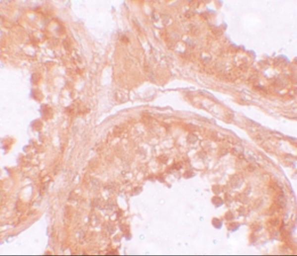
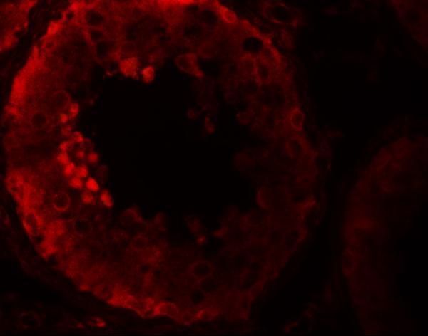
A14119 has human testis IHC and IF images (catalog image captions) and lists Human and Mouse reactivity (catalog reactivity).
A14119 is listed for IHC-P and shows human testis IHC at 10 μg/mL (catalog applications; IHC image caption). The same SKU is listed for IF and shows human testis IF at 20 μg/mL (catalog applications; IF image caption).
Which to pick: Choose A14119 for paraffin-section tissue IHC: IHC-P is listed, and its own image shows human testis staining; the fixative is unreported (catalog applications; IHC image caption). For IF, choose A14119 on the strength of its human testis image; ICC validation is unreported (catalog applications; IF image caption). A14119 also lists Mouse reactivity, but the supplied IHC and IF images show Human tissue only; it is rabbit-hosted, with clonality unreported (catalog reactivity, image captions and host/clone fields).