This website uses cookies to ensure you get the best experience on our website.


- Table of Contents
Plan NNMT IHC-P around cytoplasmic staining in hepatocytes and kidney tubule cells (HPA tissue IHC). The catalog antibody has an IHC-P dilution of 1:10–1:50 (datasheet: A02676-1 IHC-P); compare sections with consistent fixation and appropriate controls (standard IHC practice).

Expected localisation, validated protocols, controls and antibodies — the at-a-glance facts below, then the full design guide.
| Expected localisation | Cytoplasmic staining in a few tissues (HPA tissue IHC) | |
| Staining pattern | Cytoplasmic signal in hepatocytes and kidney tubule cells (HPA tissue IHC) | |
| Antigen retrieval | Citrate pH 6.0 HIER, 95–98 °C, 20 min (rule: cytoplasmic / membrane antigen) | |
| Positive control | Kidney+4 more · see all | |
| Negative control | Adipose tissue+4 more · see all |
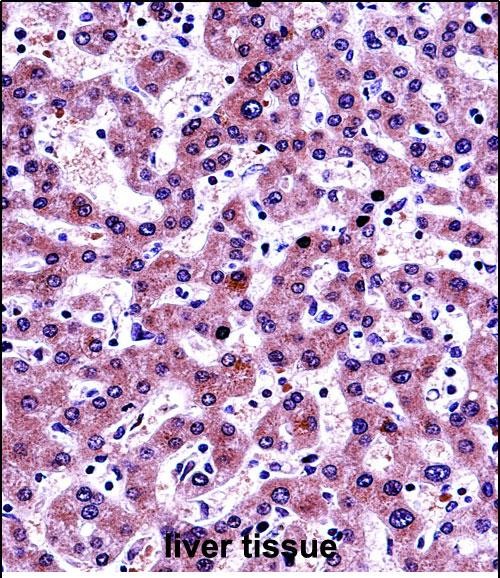
| Fixation | Formalin-fixed paraffin liver is shown (selected-SKU IHC image A02676-1); Matched tissue-IHC evidence does not establish the fixation claim. Validate the specimen-specific method before use. (selected-SKU IHC image A02676-1) | |
| Caveat | Antibody staining has low consistency with RNA expression (HPA tissue IHC) | |
| Regulation | Predominantly expressed in liver (UniProt) | |
| Isoform / epitope | 0 annotated isoforms or cleavage events; no mapped epitope shift (UniProt) |
The catalog antibody’s IHC-P protocol is accompanied by published NNMT staining methods for prostate tissue, ovarian clear cell carcinoma, and paraffin sections (PMC4114624; PMC12378295; PMC12098763).
| Sample | FFPE human liver tissue (datasheet A02676-1) |
| Fixation | Image formalin-fixed; duration unreported (datasheet A02676-1); verify before use. |
| Sectioning | 4–5 µm sections on charged slides (standard) |
| Deparaffinisation | Xylene, graded ethanol series to water (standard) |
| Antigen retrieval | Heat-induced epitope retrieval in citrate buffer, pH 6.0, 20 min at 95–98 °C (standard rule: cytoplasmic / membrane antigen) |
| Peroxidase block | 3% H2O2, 10 min, room temperature (standard) |
| Blocking | 10% normal serum of the secondary host, 30 min, room temperature (standard) |
| Primary antibody | Rabbit anti-NNMT, 1:10-1:50 (datasheet A02676-1) |
| Primary incubation | Overnight at 4 °C (standard) |
| Detection | HRP-polymer secondary, DAB chromogen 5–10 min (standard) |
| Counterstain | Hematoxylin, blue, dehydrate and mount (standard) |
| Expected result | NNMT-positive staining in cells in tubules of kidney (HPA tissue IHC: High). HPA tissue profile: Cytoplasmic expression in a few tissues. No signal in the no-primary control. |
NNMT is a cytoplasmic protein without a transmembrane segment (UniProt P40261). In paraffin-section IHC, expect strong cytoplasmic staining in hepatocytes, kidney tubular cells and pancreatic exocrine glandular cells (HPA tissue IHC: High). Interpret tissue comparisons cautiously: HPA rates its antibody staining Approved but reports low consistency with RNA expression (HPA tissue IHC: reliability).
| Strong cytoplasmic signal in hepatocytes or kidney tubular cells, with cellular boundaries still readable. | This fits the reported compartment and high-staining cell populations (UniProt P40261: Cytoplasm; HPA tissue IHC: High in hepatocytes and kidney tubular cells). Judge the cells stained as well as the overall intensity; a positive-looking region alone does not establish the expected pattern (general IHC practice). |
| Predominantly nuclear, membranous or extracellular signal in an otherwise positive-looking area. | These compartments do not match the reported cytoplasmic localization or lack of a transmembrane segment (UniProt P40261). Review morphology and controls before assigning NNMT positivity; misplaced chromogen can reflect nonspecific staining or detection artefact (general IHC practice). |
| Strong staining chiefly in adipocytes or hematopoietic cells. | HPA reports NNMT as not detected in those cell populations (HPA tissue IHC: adipocytes; bone marrow hematopoietic cells). Consider cross-reactivity or endogenous detection activity, then inspect matched controls (general IHC practice). A reported negative population is a comparison, not proof that every specimen must be negative. |
| Diffuse chromogen across several tissue compartments, including spaces without recognizable cells. | That distribution does not resemble the reported cell-associated cytoplasmic pattern (HPA tissue IHC: cytoplasmic expression in a few tissues). Check the primary-omitted control and background from the detection system; widespread haze makes intensity scoring unreliable (general IHC practice). |
| No convincing signal in hepatocytes on a liver section. | HPA reports high hepatocyte staining and liver-enriched RNA, while UniProt describes predominant liver expression (HPA tissue IHC; UniProt P40261: tissue specificity). First confirm tissue morphology and control performance, then review the IHC-validated antibody's documented conditions (general IHC practice). |
| Tissue and cell selection | Hepatocytes, kidney tubular cells and pancreatic exocrine glandular cells are reported High; cervix, endometrium and epididymis glandular cells are Medium (HPA tissue IHC). Compare the named cell populations, since adjacent cells need not share the same score (general IHC practice). |
| Tissue evidence conflict | Pancreatic exocrine cells stain High by HPA IHC, whereas UniProt says NNMT was not detected in pancreas (HPA tissue IHC; UniProt P40261: tissue specificity). Treat pancreas as an observed IHC pattern with this explicit caveat, rather than a settled expression control. |
| Antibody validation | HPA059180 is Approved for IHC, but HPA reports low consistency between antibody staining and RNA expression (HPA antibodies; HPA tissue IHC: reliability). The supplied status is Approved, so do not infer an Enhanced validation claim from it. |
| Topology and processing | NNMT is cytoplasmic, lacks a transmembrane segment and signal peptide, and is annotated as one chain spanning residues 1–264 (UniProt P40261). These annotations support a cellular cytoplasmic expectation; they do not identify an antibody epitope or predict antigen retrieval performance. |
| IF/ICC question: should Golgi staining match tissue IHC? | HPA ICC-IF places NNMT mainly at the Golgi apparatus and additionally in the cytosol, whereas its tissue IHC profile is cytoplasmic (HPA subcellular; HPA tissue IHC). Compare each observation with its own assay and specimen context; the IF/ICC workflow belongs to its separate guide. |
| Situation | Likely cause | Next action |
|---|---|---|
| Liver section has no hepatocyte signal. | This conflicts with the reported High hepatocyte pattern (HPA tissue IHC); the image alone does not identify the technical cause. | Confirm the expected cells are present, inspect run controls, then review the catalog antibody's IHC-P instructions for retrieval, dilution and detection conditions (general IHC practice). Do not infer a target-specific fixation effect from this result. |
| Kidney staining is scored from the whole section rather than tubules. | The reported High signal is assigned specifically to cells in tubules (HPA tissue IHC), so a region-wide score can obscure the relevant cell population. | Recheck morphology and score tubular cells separately from other compartments (general IHC practice). Compare localization with the cytoplasmic expectation (UniProt P40261). |
| Pancreatic exocrine cells stain strongly, but the result seems inconsistent with the expression summary. | HPA reports High exocrine-cell IHC staining, while UniProt reports no pancreatic detection (HPA tissue IHC; UniProt P40261: tissue specificity). | Record both observations and examine antibody and detection controls before interpreting the signal (general IHC practice). Avoid using pancreas alone to settle the discrepancy. |
| Adipocytes, hematopoietic cells or acellular areas dominate the positive signal. | The named cell populations are reported Not detected (HPA tissue IHC); acellular staining also departs from a cellular cytoplasmic pattern (HPA tissue IHC). | Examine a primary-omitted control and detection background, then reassess whether signal follows cell morphology (general IHC practice). Treat unexplained staining as unresolved specificity, not confirmed NNMT. |
| Signal is predominantly nuclear or outlines cell membranes. | That localization differs from cytoplasm, and NNMT has no annotated transmembrane segment (UniProt P40261). | Check section morphology, counterstain and control slides; adjust interpretation only after confirming where the chromogen sits within cells (general IHC practice). Do not score compartment-mismatched signal as an expected positive. |
| Tissue IHC looks diffuse while a separate IF/ICC image appears Golgi enriched. | HPA describes cytoplasmic tissue IHC and mainly Golgi, additionally cytosolic, ICC-IF localization (HPA tissue IHC; HPA subcellular). | Document the assay and specimen for each image, then compare each with its corresponding HPA pattern. If the IHC signal lacks recognizable cellular localization, revisit IHC controls before scoring (general IHC practice). |


Comprehensive Human Protein Atlas IHC scoring per tissue (reliability: Approved — Low consistency between antibody staining and RNA expression data.). Rows are taken directly from the HPA tissue chart — click any row's HPA link to view the source.
| Tissue | Cell type | Level | Evidence | Source |
|---|---|---|---|---|
| Kidney | Cells in tubules | High | Protein (IHC) | HPA → |
| Liver | Hepatocytes | High | Protein (IHC) | HPA → |
| Pancreas | Exocrine glandular cells | High | Protein (IHC) | HPA → |
| Cervix | Glandular cells | Medium | Protein (IHC) | HPA → |
| Endometrium | Glandular cells | Medium | Protein (IHC) | HPA → |
| Tissue | Cell type | Level | Evidence | Source |
|---|---|---|---|---|
| Adipose tissue | Adipocytes | Not detected | Protein (IHC) | HPA → |
| Appendix | Glandular cells | Not detected | Protein (IHC) | HPA → |
| Bone marrow | Hematopoietic cells | Not detected | Protein (IHC) | HPA → |
| Breast | Adipocytes | Not detected | Protein (IHC) | HPA → |
| Bronchus | Respiratory epithelial cells | Not detected | Protein (IHC) | HPA → |
These NNMT troubleshooting questions focus on chromogenic IHC in paraffin sections, with one entry for IF/ICC.
The catalog shows human liver IHC data for A02676-1 (image caption) and human ICC/IF validation for A02676-2 (applications and reactivity); A02676-2 also lists mouse and rat reactivity (catalog reactivity).
The rendered A02676-1 card shows IHC-P staining of formalin-fixed, paraffin-embedded human liver with peroxidase/DAB detection (A02676-1 image caption). A02676-2 is listed for human ICC/IF, but has no IF figure in the supplied payload (A02676-2 applications, reactivity and image alts).
Which to pick: Choose A02676-1 for human tissue IHC-P: it is polyclonal, has a 1:10–1:50 IHC-P dilution, and its own image documents formalin-fixed, paraffin-embedded human liver (A02676-1 catalog and image caption). Choose A02676-2 for human ICC/IF at a listed 5 μg/ml; its applications do not include IHC-P (A02676-2 catalog). A02676-2 lists human, mouse and rat reactivity, but the supplied ICC/IF validation is human only (A02676-2 catalog applications, reactivity and dilution data).