This website uses cookies to ensure you get the best experience on our website.


- Table of Contents
Plan chromogenic NOS1AP IHC in paraffin sections using the catalog antibody’s 2 μg/ml example (datasheet A03060-3). Compare neuronal cytoplasmic staining with the HPA tissue pattern and assess suspected off-target signal before scoring (HPA tissue IHC).

Expected localisation, validated protocols, controls and antibodies — the at-a-glance facts below, then the full design guide.
| Expected localisation | Neuronal cytoplasm observed (HPA tissue IHC); filopodia and podosomes annotated (UniProt) | |
| Staining pattern | Cytoplasmic staining in neuronal cells (HPA tissue IHC) | |
| Antigen retrieval | EDTA pH 8.0 HIER, heat-mediated (datasheet A03060-3) | |
| Positive control | Caudate+4 more · see all | |
| Negative control | Adipose tissue+4 more · see all |
| Fixation | Keep fixation consistent across sections (standard IHC practice; not target-specific) | |
| Caveat | Presumed off-target binding may confound scoring (HPA tissue IHC) | |
| Regulation | Expression varies by tissue (HPA tissue IHC) | |
| Isoform / epitope | 3 isoforms; epitope coverage is unspecified (UniProt) |
Compare the catalog antibody’s IHC-P protocol with the published breast cancer IHC protocol (PMC3419983).
| Sample | Paraffin-embedded mouse brain tissue; fixative not specified (datasheet A03060-3) |
| Fixation | Image fixative and duration unreported (datasheet A03060-3); verify before use. |
| Sectioning | 4–5 µm sections on charged slides (standard) |
| Deparaffinisation | Xylene, graded ethanol series to water (standard) |
| Antigen retrieval | Heat retrieval: EDTA pH 8.0 (datasheet A03060-3); 20 min, 95–100 °C (standard) |
| Peroxidase block | 3% H2O2, 10 min, room temperature (standard) |
| Blocking | 10% goat serum (datasheet A03060-3) |
| Primary antibody | Rabbit anti-NOS1AP, 2-5 μg/ml (datasheet A03060-3) |
| Primary incubation | Overnight at 4 °C (datasheet A03060-3) |
| Detection | HRP-conjugated secondary, DAB chromogen (datasheet A03060-3) |
| Counterstain | Hematoxylin, blue, dehydrate and mount (standard) |
| Expected result | NOS1AP-positive staining in neuronal cells of caudate (HPA tissue IHC: Medium). HPA tissue profile: Cytoplasmic expression in neuronal cells. No signal in the no-primary control. |
In paraffin-section IHC, expect NOS1AP mainly in neuronal cytoplasm, with Medium staining reported in caudate, cerebral cortex, and hippocampus (HPA tissue IHC: Approved). UniProt also places NOS1AP in filopodia and podosomes and reports no transmembrane segment (UniProt O75052). Interpret this pattern cautiously: HPA reports low agreement between antibody staining and RNA expression and disregarded presumed off-target staining (HPA tissue IHC: reliability description).
| Neuronal cell bodies show cytoplasmic chromogen in caudate, cortex, or hippocampus. | This fits HPA's observed Medium neuronal staining and cytoplasmic profile (HPA tissue IHC). Judge the labeled cells and compartment together; intensity alone cannot establish specificity, given HPA's reported RNA–staining inconsistency and presumed off-target binding (HPA tissue IHC: reliability description). |
| Brain sections show only nuclear staining, or a sharp outline confined to the plasma membrane. | That differs from HPA's neuronal cytoplasmic IHC profile (HPA tissue IHC). A membrane-only interpretation is also unsupported by the absence of a transmembrane segment (UniProt O75052). Nuclear staining needs an assay-specific check: nucleoplasm is an approved ICC-IF location, so it is not automatically an artefact in IF (HPA subcellular). |
| Strong staining appears across cells or tissues listed as undetected. | Check whether the staining is confined to the cell type HPA actually scored: adipocytes in adipose tissue and glandular cells in adrenal gland were Not detected (HPA tissue IHC). Widespread signal in those scored cells raises concern for nonspecific binding or endogenous chromogen activity (general IHC practice); the HPA reliability note strengthens the need for controls. |
| Chromogen forms a diffuse haze over tissue, including areas without identifiable labeled cells. | An indistinct field cannot support the expected neuronal cytoplasmic call (HPA tissue IHC: profile). Consider excess primary or detection reagent, inadequate washing, or endogenous enzyme activity, and assess a matched no-primary section (general chromogenic IHC practice). Do not score haze as NOS1AP-positive cells. |
| No neuronal signal is visible in a caudate, cortex, or hippocampus section. | Those tissues contain HPA-scored Medium neuronal staining (HPA tissue IHC), so first check that recognizable neuronal cells and an interpretable counterstain are present. A failed run, unsuitable retrieval conditions, or weak detection can also erase an expected pattern (general IHC practice); one negative section does not establish biological absence. |
| Reference tissue and scored cell type | Caudate, cortex, and hippocampus provide observed Medium neuronal examples; HPA also reports Medium staining in fallopian-tube ciliated cell bodies and testis Leydig cells (HPA tissue IHC). Compare like cell types rather than treating every cell in a positive tissue as positive. |
| Location depends on assay | HPA describes neuronal cytoplasm by tissue IHC, but approves nucleoplasm and vesicles by ICC-IF (HPA tissue IHC; HPA subcellular). UniProt adds filopodia and podosomes (UniProt O75052). Keep those observations tied to their assay and context when judging an apparent compartment mismatch. |
| Antibody evidence | Three listed rabbit antibodies have IHC status Approved; only HPA030066 also has ICC status Approved (HPA antibodies). HPA still reports low staining–RNA consistency and presumed off-target binding for the tissue profile (HPA tissue IHC). An Approved status does not replace slide controls. |
| Isoforms and epitope coverage | UniProt lists 3 NOS1AP isoforms and a PID domain at residues 26–196 (UniProt O75052). The supplied record does not map an antibody epitope or show which isoforms it detects, so isoform-specific staining predictions would be unsupported. |
| Processing and topology | The annotated chain spans residues 1–506, with no signal peptide, propeptide, or transmembrane segment (UniProt O75052). These annotations support caution about a membrane-only or shed-protein explanation; they do not establish an antigen-retrieval condition or fixation sensitivity. |
| Situation | Likely cause | Next action |
|---|---|---|
| Brain reference tissue has little or no neuronal staining. | The run may have inadequate detection, or the chosen retrieval conditions may perform poorly (general IHC practice); HPA reports Medium neuronal staining in caudate, cortex, and hippocampus (HPA tissue IHC). | Confirm the section contains identifiable neurons, check the run's positive control and reagent steps, then optimize retrieval and antibody concentration within the laboratory's IHC workflow (general IHC practice). Reassess the same cell type rather than increasing exposure until background appears. |
| The chromogen is predominantly nuclear in neuronal cells. | A nuclear-only tissue IHC pattern conflicts with HPA's neuronal cytoplasmic profile (HPA tissue IHC), although nucleoplasm is an approved ICC-IF location (HPA subcellular). The two assays should not be assigned one universal compartment rule. | Check the counterstain and focus plane, compare a matched no-primary control, and repeat with a validated IHC antibody if the pattern persists (general IHC practice; HPA antibodies: IHC Approved). Report the compartment and assay explicitly. |
| Adipose adipocytes or adrenal glandular cells appear broadly positive. | HPA scores those particular cells Not detected and notes presumed off-target binding in its tissue assessment (HPA tissue IHC). Endogenous detection activity can also produce chromogen without specific primary binding (general chromogenic IHC practice). | Run no-primary and detection-only controls, block the endogenous activity relevant to the chosen chromogen system, and compare staining with the scored cell types on HPA-positive sections (general IHC practice; HPA tissue IHC). |
| Signal is diffuse, granular, or present beyond recognizable cells. | The expected IHC call requires identifiable neuronal cytoplasm (HPA tissue IHC: profile). Excess reagent, incomplete washing, or endogenous enzyme activity may create nonspecific background (general chromogenic IHC practice); appearance alone cannot identify which cause applies. | Inspect no-primary controls, review washing and blocking, and titrate the primary and detection reagents under the same staining conditions (general IHC practice). Score only cellular staining that remains distinguishable from background. |
| A ciliated-cell or Leydig-cell signal seems inconsistent with brain-enriched RNA. | HPA reports Medium staining in fallopian-tube ciliated cell bodies and testis Leydig cells while describing RNA as brain-enriched and staining–RNA agreement as low (HPA tissue IHC). Neither observation alone resolves antibody specificity. | Record the labeled cell type and compartment, compare the pattern with HPA's scored cells, and include specificity controls before treating the signal as confirmed NOS1AP (HPA tissue IHC; general IHC practice). |
| ICC-IF images show nucleoplasm or vesicles while the IHC slide shows neuronal cytoplasm. | HPA approves nucleoplasm and vesicles for ICC-IF and describes cytoplasmic neuronal tissue IHC staining (HPA subcellular; HPA tissue IHC). Those are observations from different assays and cellular settings. | Interpret each image against its own HPA reference and controls (HPA subcellular; HPA tissue IHC; general IF/IHC practice). Keep IF optimization on its separate guide page; this IHC decision concerns cellular staining in paraffin sections. |


Comprehensive Human Protein Atlas IHC scoring per tissue (reliability: Approved — Low consistency between antibody staining and RNA expression data. Presumed off target binding observed and disregarded.). Rows are taken directly from the HPA tissue chart — click any row's HPA link to view the source.
| Tissue | Cell type | Level | Evidence | Source |
|---|---|---|---|---|
| Caudate | Neuronal cells | Medium | Protein (IHC) | HPA → |
| Cerebral cortex | Neuronal cells | Medium | Protein (IHC) | HPA → |
| Fallopian tube | Ciliated cells (cell body) | Medium | Protein (IHC) | HPA → |
| Hippocampus | Neuronal cells | Medium | Protein (IHC) | HPA → |
| Testis | Leydig cells | Medium | Protein (IHC) | HPA → |
| Tissue | Cell type | Level | Evidence | Source |
|---|---|---|---|---|
| Adipose tissue | Adipocytes | Not detected | Protein (IHC) | HPA → |
| Adrenal gland | Glandular cells | Not detected | Protein (IHC) | HPA → |
| Appendix | Glandular cells | Not detected | Protein (IHC) | HPA → |
| Bone marrow | Hematopoietic cells | Not detected | Protein (IHC) | HPA → |
| Bronchus | Basal cells | Not detected | Protein (IHC) | HPA → |
Troubleshoot NOS1AP staining in paraffin sections by separating retrieval and detection problems from cell type, compartment, and antibody specificity.
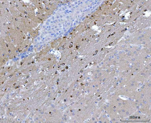
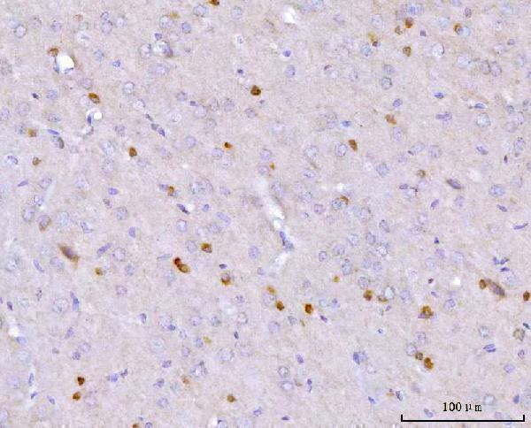
Anti-NOS1AP A03060-3 has real IHC images from paraffin-embedded mouse, rat, and human brain sections (A03060-3 IHC image captions). No IF/ICC images are provided (A03060-3 IF image alts).
The sole card is A03060-3, a rabbit antibody listed for IHC and human, mouse, and rat reactivity (A03060-3 catalog). Its IHC captions document staining in paraffin-embedded brain sections from all three species (A03060-3 IHC image captions).
Which to pick: For tissue IHC and cross-species studies, choose A03060-3: its own captions show paraffin-section staining in mouse, rat, and human brain using EDTA retrieval at pH 8.0 and 2 μg/ml primary antibody (A03060-3 IHC image captions). The section fixative is unreported (A03060-3 IHC image captions). For IF/ICC, this payload offers no validated choice: A03060-3 has no IF/ICC application listing or IF images (A03060-3 catalog applications; IF image alts).