This website uses cookies to ensure you get the best experience on our website.


- Table of Contents
Plan NOVA1 staining in paraffin sections using the IHC-validated antibody (datasheet A06087-1). Compare the nuclear expectation with the reported nuclear and cytoplasmic tissue staining, while accounting for uncertain agreement between staining and RNA expression (UniProt; HPA tissue IHC).

Expected localisation, validated protocols, controls and antibodies — the at-a-glance facts below, then the full design guide.
| Expected localisation | Nuclear expected (UniProt); nuclear and cytoplasmic observed (HPA tissue IHC) | |
| Staining pattern | Nuclear and cytoplasmic staining in most tissues (HPA tissue IHC) | |
| Antigen retrieval | EDTA pH 8.0 HIER, heat-mediated (datasheet A06087-1) | |
| Positive control | Cerebellum+4 more · see all | |
| Negative control | Adipose tissue+4 more · see all |
| Fixation | Keep fixation consistent across paraffin sections (standard IHC practice; not target-specific) | |
| Caveat | Staining and RNA expression show low consistency (HPA tissue IHC) | |
| Regulation | Brain-enhanced RNA expression (HPA tissue IHC: RNA specificity) | |
| Isoform / epitope | 3 isoforms; verify epitope coverage (UniProt) |
Compare the catalog antibody’s IHC-P protocol (datasheet A06087-1) with four published NOVA1 tissue IHC methods (PMC8799250; PMC4502388; PMC8798774; PMC4804152).
| Sample | Paraffin-embedded human breast cancer tissue; fixative not specified (datasheet A06087-1) |
| Fixation | Image fixative and duration unreported (datasheet A06087-1); verify before use. |
| Sectioning | 4–5 µm sections on charged slides (standard) |
| Deparaffinisation | Xylene, graded ethanol series to water (standard) |
| Antigen retrieval | Heat retrieval: EDTA pH 8.0 (datasheet A06087-1); 20 min, 95–100 °C (standard) |
| Peroxidase block | 3% H2O2, 10 min, room temperature (standard) |
| Blocking | 10% goat serum (datasheet A06087-1) |
| Primary antibody | Rabbit anti-NOVA1, 2-5μg/ml (datasheet A06087-1) |
| Primary incubation | Overnight at 4 °C (datasheet A06087-1) |
| Detection | HRP-conjugated secondary, DAB chromogen (datasheet A06087-1) |
| Counterstain | Hematoxylin, blue, dehydrate and mount (standard) |
| Expected result | NOVA1-positive staining in cells in granular layer of cerebellum (HPA tissue IHC: Medium). HPA tissue profile: Cytoplasmic and nuclear expression in most tissues. No signal in the no-primary control. |
NOVA1 is a nuclear RNA-binding protein with no transmembrane segment (UniProt P51513). In paraffin-section IHC, look first for staining in cerebellar granular-layer cells and hippocampal neuronal cells, both reported at medium level (HPA tissue IHC). HPA also reports cytoplasmic and nuclear staining across most tissues, but rates the tissue IHC profile Uncertain because antibody staining and RNA expression have low consistency (HPA tissue IHC).
| Medium staining in cerebellar granular-layer cells or hippocampal neuronal cells, with a nuclear component (HPA tissue IHC; UniProt P51513). | This is a useful positive-reference pattern: those exact cell populations have medium HPA tissue IHC staining, and UniProt places NOVA1 in the nucleus (HPA tissue IHC; UniProt P51513). Judge the named cells rather than treating every cell in a brain section as an expected positive (HPA tissue IHC). |
| Signal is confined to cell membranes, extracellular space, or an unrelated compartment, without convincing cellular nuclear staining (UniProt P51513). | Treat this as a possible staining artefact and inspect controls: NOVA1 has no transmembrane segment and is annotated as nuclear (UniProt P51513). Cytoplasmic signal alone needs caution, because HPA describes cytoplasmic as well as nuclear tissue staining, while its tissue IHC reliability is Uncertain (HPA tissue IHC). |
| Strong signal appears in adipocytes, cardiomyocytes, or skeletal myocytes, which HPA reports as not detected (HPA tissue IHC). | Consider antibody cross-reactivity or endogenous chromogenic detection activity, especially if a no-primary control also stains (general IHC practice). These HPA negatives refer to the specified cell types in sampled tissues; they do not establish that every cell in those organs lacks NOVA1 (HPA tissue IHC). |
| Diffuse colour covers the section, including tissue spaces, and obscures nuclei and cell boundaries (general IHC practice). | This is background rather than an interpretable NOVA1 distribution (general IHC practice). Check the no-primary control and review blocking, antibody concentration, washing, and chromogen development before assigning cell-level staining (general IHC practice). HPA's Uncertain tissue IHC rating warrants particular restraint in calling a weak diffuse signal positive (HPA tissue IHC). |
| No signal is visible in cerebellar granular-layer cells or hippocampal neuronal cells (HPA tissue IHC). | The run lacks staining in HPA-reported medium positive cell populations, so assess the assay before interpreting study negatives (HPA tissue IHC; general IHC practice). Check tissue identity, the catalog antibody's IHC-P instructions, detection reagents, and a positive control processed alongside the samples (general IHC practice). |
| Tissue and cell selection (HPA tissue IHC; UniProt P51513) | UniProt reports NOVA1 expression in cerebellum, brain stem, hippocampus, and frontal cortex; HPA records medium staining in cerebellar granular-layer cells and hippocampal neuronal cells (UniProt P51513; HPA tissue IHC). HPA's cerebral-cortex example is endothelial cells, so its positive result should not be recast as a neuronal result (HPA tissue IHC). |
| Tissue IHC evidence quality (HPA tissue IHC; HPA antibody validation) | HPA rates the tissue profile Uncertain, citing low consistency with RNA expression and a splice or transcript discrepancy pending external verification (HPA tissue IHC). The listed antibody, HPA004155, has Uncertain IHC validation; a plausible slide pattern alone therefore cannot establish antibody specificity (HPA antibody validation). |
| Subcellular evidence (UniProt P51513; HPA tissue IHC) | UniProt annotates the nucleus, whereas HPA tissue IHC describes cytoplasmic and nuclear expression in most tissues (UniProt P51513; HPA tissue IHC). Record nuclear and cytoplasmic staining separately when scoring; do not require every IHC-positive cell to show exclusively nuclear colour (HPA tissue IHC; general IHC practice). |
| Isoforms and epitope coverage (UniProt P51513) | UniProt lists 3 NOVA1 isoforms and three KH domains, but the supplied evidence gives no catalog-antibody epitope or isoform coverage (UniProt P51513). A difference between sections cannot be attributed to a particular isoform from these records alone (UniProt P51513). |
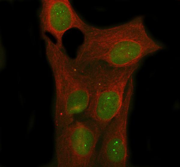
| IF/ICC Q: Where should signal appear? (HPA subcellular ICC-IF) | A: HPA ICC-IF reports NOVA1 mainly in nucleoli, with additional nucleoplasmic localisation, both supported (HPA subcellular ICC-IF). This is cell-imaging evidence; use the tissue IHC record to interpret paraffin-section chromogenic staining (HPA subcellular ICC-IF; HPA tissue IHC). |
| Situation | Likely cause | Next action |
|---|---|---|
| Expected cerebellar or hippocampal cells are blank (HPA tissue IHC). | A failed staining run or a missed target cell population is possible; HPA reports those populations at medium level (HPA tissue IHC; general IHC practice). | Confirm the section and cell identity, then inspect the parallel positive control and follow the catalog antibody's IHC-P instructions for retrieval, dilution, and detection (general IHC practice). Do not infer NOVA1-specific retrieval sensitivity from the supplied records. |
| The whole section has diffuse chromogen colour (general IHC practice). | Non-specific antibody binding, insufficient washing, or endogenous detection activity can produce widespread background (general IHC practice). | Compare a no-primary control; review blocking, washing, antibody dilution, and chromogen development as general IHC checks (general IHC practice). Score NOVA1 only where individual cells remain interpretable. |
| A proposed negative cell type stains strongly (HPA tissue IHC). | Cross-reactivity or endogenous detection activity is possible if adipocytes, cardiomyocytes, or skeletal myocytes stain despite HPA's not detected entries (HPA tissue IHC; general IHC practice). | Check a no-primary control and compare the exact cell type with an expected-positive section in the same run (general IHC practice; HPA tissue IHC). Treat discordance as unresolved because HPA tissue IHC reliability is Uncertain (HPA tissue IHC). |
| Colour appears only along membranes or outside cells (UniProt P51513). | That distribution conflicts with UniProt's nuclear localisation and absence of a transmembrane segment (UniProt P51513). | Review counterstain, cell boundaries, and no-primary staining before assigning a positive call (general IHC practice). Report a distinct cytoplasmic component separately if present, given HPA's broader tissue IHC description (HPA tissue IHC). |
| Cytoplasmic staining accompanies nuclear staining (HPA tissue IHC; UniProt P51513). | HPA describes both compartments in tissue IHC, while UniProt annotates the nucleus; the supplied evidence does not resolve every tissue-level difference (HPA tissue IHC; UniProt P51513). | Score the two compartments separately and compare the named cell population with HPA's tissue observations (HPA tissue IHC; general IHC practice). Keep the interpretation provisional because the HPA tissue profile is Uncertain (HPA tissue IHC). |
| Two sections show different cell patterns despite apparently similar staining quality (general IHC practice). | Cell composition may differ; HPA reports positives and negatives for specified cells, and its tissue IHC profile has low consistency with RNA expression (HPA tissue IHC). | Identify and score comparable cell populations, include the same expected-positive control across runs, and document the discrepancy (HPA tissue IHC; general IHC practice). The supplied records do not establish a NOVA1-specific fixation effect. |
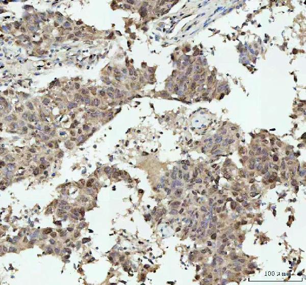
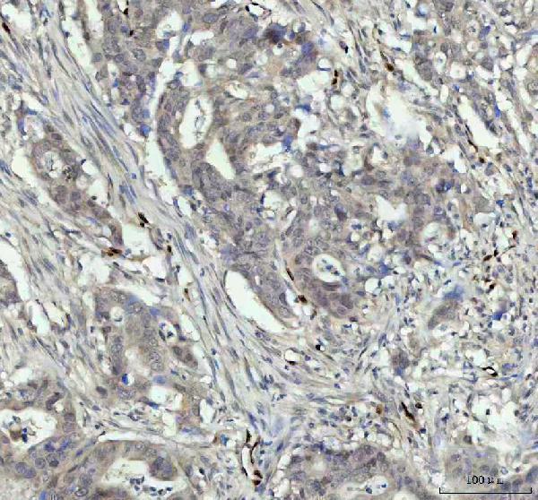


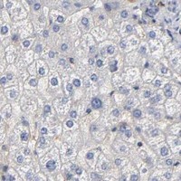
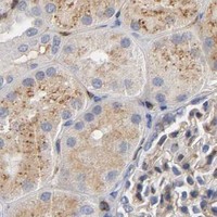


Comprehensive Human Protein Atlas IHC scoring per tissue (reliability: Uncertain — Low consistency between antibody staining and RNA expression data. Caution, Splice and/or transcript discrepancy exists. Pending external verification.). Rows are taken directly from the HPA tissue chart — click any row's HPA link to view the source.
| Tissue | Cell type | Level | Evidence | Source |
|---|---|---|---|---|
| Cerebellum | Cells in granular layer | Medium | Protein (IHC) | HPA → |
| Cerebral cortex | Endothelial cells | Medium | Protein (IHC) | HPA → |
| Colon | Glandular cells | Medium | Protein (IHC) | HPA → |
| Hippocampus | Neuronal cells | Medium | Protein (IHC) | HPA → |
| Kidney | Cells in glomeruli | Medium | Protein (IHC) | HPA → |
| Tissue | Cell type | Level | Evidence | Source |
|---|---|---|---|---|
| Adipose tissue | Adipocytes | Not detected | Protein (IHC) | HPA → |
| Bone marrow | Hematopoietic cells | Not detected | Protein (IHC) | HPA → |
| Esophagus | Squamous epithelial cells | Not detected | Protein (IHC) | HPA → |
| Heart muscle | Cardiomyocytes | Not detected | Protein (IHC) | HPA → |
| Liver | Cholangiocytes | Not detected | Protein (IHC) | HPA → |
Troubleshoot NOVA1 staining in paraffin sections by checking retrieval, nuclear localisation, controls and cell-specific scoring before interpreting chromogenic signal.
A06087-1 has paraffin-section IHC images from human tumors and mouse brain (catalog IHC captions), and IF/ICC images from U2OS and A549 cells (catalog IF captions). Rat reactivity is listed (catalog reactivity).
A06087-1 will render with its own IHC figure of a human breast cancer paraffin section (A06087-1 figure caption). Other IHC captions show human colorectal adenocarcinoma, human liver cancer, and mouse brain paraffin sections; IF captions show U2OS and A549 cells (A06087-1 catalog captions).
Which to pick: For tissue IHC, choose A06087-1: its own figure shows a human breast cancer paraffin section stained after EDTA pH 8 retrieval with 2 μg/ml antibody; the fixative is unreported (A06087-1 IHC figure caption). For IF/ICC, the same SKU has U2OS and A549 cell images at 2 μg/ml (A06087-1 IF captions). For cross-species work, A06087-1 lists human, mouse, and rat reactivity, with a mouse brain IHC image; clonality is unreported (A06087-1 catalog reactivity, IHC captions, and clone field).