This website uses cookies to ensure you get the best experience on our website.


- Table of Contents
Plan NOVA2 chromogenic IHC in paraffin sections around the nuclear staining observed in neuronal cells (HPA tissue IHC). This guide covers fixation consistency, antibody concentration, controls and interpretation of the differing cell-type annotations (datasheet A10622-2; UniProt; HPA tissue IHC).

Expected localisation, validated protocols, controls and antibodies — the at-a-glance facts below, then the full design guide.
| Expected localisation | Nuclear staining in neuronal cells (HPA tissue IHC) | |
| Staining pattern | Neuronal nuclei stain in brain tissue (HPA tissue IHC) | |
| Antigen retrieval | EDTA pH 8.0 HIER, heat-mediated (datasheet A10622-2) | |
| Positive control | Caudate+4 more · see all | |
| Negative control | Adipose tissue+4 more · see all |
| Fixation | Keep fixation consistent across sections (standard IHC practice; not target-specific) | |
| Caveat | Astrocyte annotation differs from neuronal IHC staining (UniProt tissue specificity; HPA tissue IHC) | |
| Regulation | Brain-enhanced RNA expression (HPA tissue RNA) | |
| Isoform / epitope | No isoforms or processing reported; one 1–492 chain (UniProt) |
Compare the catalog antibody’s IHC-P protocol (datasheet A10622-2) with three published NOVA2 paraffin-section IHC protocols (PMC12185593; PMC6157992; PMC6398979).
| Sample | Paraffin-embedded human appendicitis tissue; fixative not specified (datasheet A10622-2) |
| Fixation | Image fixative and duration unreported (datasheet A10622-2); verify before use. |
| Sectioning | 4–5 µm sections on charged slides (standard) |
| Deparaffinisation | Xylene, graded ethanol series to water (standard) |
| Antigen retrieval | Heat retrieval: EDTA pH 8.0 (datasheet A10622-2); 20 min, 95–100 °C (standard) |
| Peroxidase block | 3% H2O2, 10 min, room temperature (standard) |
| Blocking | 10% goat serum (datasheet A10622-2) |
| Primary antibody | Rabbit anti-NOVA2, 0.5-1μg/ml (datasheet A10622-2) |
| Primary incubation | Overnight at 4 °C (datasheet A10622-2) |
| Detection | Streptavidin-biotin complex (SABC), DAB chromogen (datasheet A10622-2) |
| Counterstain | Hematoxylin, blue, dehydrate and mount (standard) |
| Expected result | NOVA2-positive staining in neuronal cells of caudate (HPA tissue IHC: Medium). HPA tissue profile: Nuclear expression in neuronal cells. No signal in the no-primary control. |
In paraffin-section IHC, expect predominantly nuclear NOVA2 staining in neuronal cells of the brain (HPA tissue IHC: nuclear neuronal profile; Enhanced reliability). HPA reports medium consistency between antibody staining and RNA expression (HPA tissue IHC: reliability description). UniProt places NOVA2 in the nucleus and reports no transmembrane segment (UniProt Q9UNW9: subcellular location and topology). UniProt also reports brain expression restricted to astrocytes, a cell-type assignment that differs from HPA's IHC profile (UniProt Q9UNW9: tissue specificity; HPA tissue IHC: profile).
| Distinct nuclear staining in neuronal cells of caudate, cerebral cortex, or hippocampus. | This matches the reported compartment and cell type; HPA records medium staining in neuronal cells at each of these sites (HPA tissue IHC: profile and positive tissues). Score the nuclei of identifiable cells rather than the overall darkness of the section, and compare them with the counterstain (standard IHC practice). |
| Prominent cytoplasmic or membranous staining in brain cells, with little nuclear signal. | That distribution conflicts with the nuclear tissue-IHC profile and UniProt location (HPA tissue IHC: profile; UniProt Q9UNW9: subcellular location). Treat it as a questionable IHC result and examine control sections and detection background before assigning it to NOVA2 (standard IHC practice). HPA's separate ICC-IF Golgi observation does not establish a cytoplasmic paraffin-IHC pattern (HPA subcellular ICC-IF: main location). |
| Strong staining in an unexpected cell population, such as adipocytes in adipose tissue. | HPA reports NOVA2 as undetected in adipocytes there (HPA tissue IHC: adipose tissue). Consider nonspecific antibody binding or endogenous detection activity, then check a section processed without primary antibody (standard IHC practice). Do not label every nonneuronal cell positive by default: HPA also reports medium staining in lung endothelial cells (HPA tissue IHC: lung). |
| Diffuse chromogen across tissue, including areas without clear cell boundaries or nuclear enrichment. | A widespread haze obscures the reported nuclear neuronal pattern (HPA tissue IHC: profile). Possible workflow causes include excess primary or detection reagent, inadequate washing, or endogenous detection activity (standard IHC practice). Judge cell-specific staining only after background is low enough to resolve nuclei and tissue structures (standard IHC practice). |
| No staining in neuronal cells of a caudate, cerebral-cortex, or hippocampal control section. | These are reported medium-positive populations, so an entirely blank run needs investigation (HPA tissue IHC: positive tissues). Check control-section integrity, reagent delivery, retrieval conditions, and detection performance using the validated IHC workflow (standard IHC practice). HPA's Enhanced rating supports the reported pattern but does not guarantee signal in every preparation (HPA tissue IHC: reliability). |
| Tissue and cell selection | Caudate, cerebral cortex, and hippocampus provide reported medium-positive neuronal populations; cerebellar molecular-layer cells are also medium positive (HPA tissue IHC: positive tissues). Adipose-tissue adipocytes and colon endothelial cells are reported undetected and can help assess cell-specific background (HPA tissue IHC: negative tissues). |
| Cell-type evidence | HPA describes nuclear expression in neuronal cells, while UniProt states brain expression is restricted to astrocytes (HPA tissue IHC: profile; UniProt Q9UNW9: tissue specificity). Record the morphology of stained cells and avoid resolving that disagreement from chromogen color alone (standard IHC practice). HPA rates tissue-IHC reliability Enhanced, with medium staining-to-RNA consistency (HPA tissue IHC: reliability). |
| Antibody evidence and target structure | HPA045607 has an Enhanced IHC validation status (HPA antibodies: HPA045607). NOVA2 is recorded as one chain, with no signal peptide, transmembrane segment, or listed isoforms (UniProt Q9UNW9: processing, topology, isoforms). These annotations support a nuclear interpretation but do not identify the antibody epitope or predict retrieval sensitivity (UniProt Q9UNW9: subcellular location; supplied record: no epitope or fixation-effect data). |
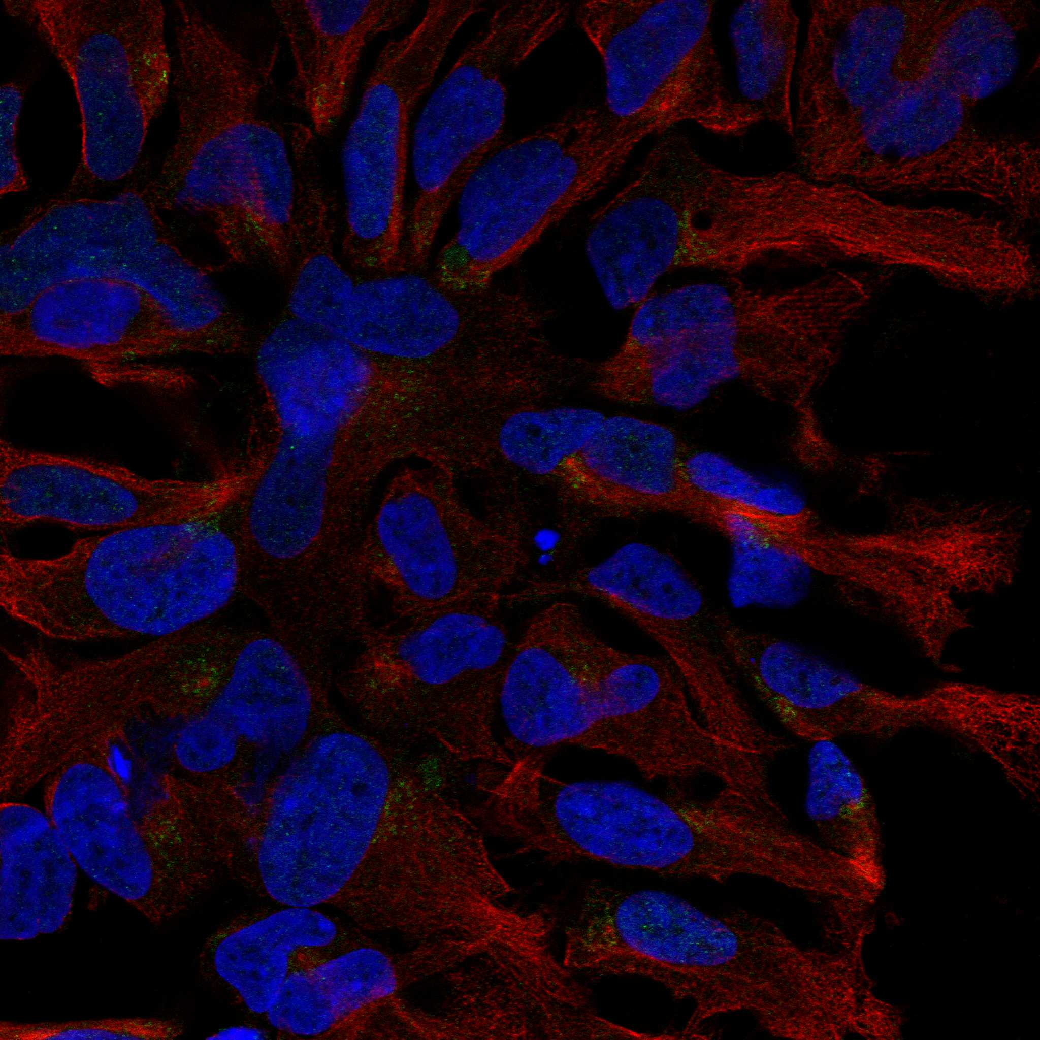
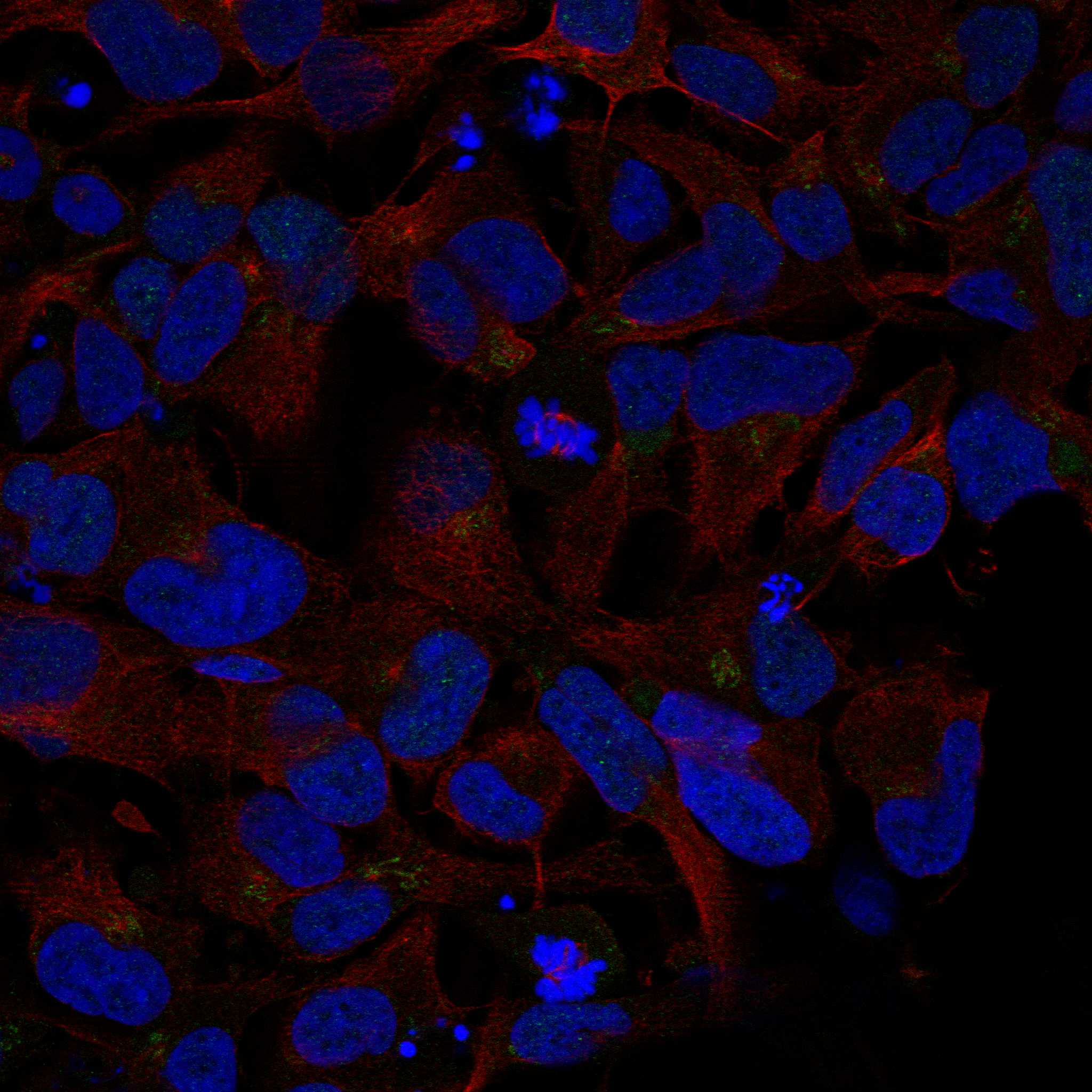
| IF/ICC Q&A: should fluorescence show only nuclei? | No single compartment can be carried over from paraffin IHC: HPA's ICC-IF summary places NOVA2 mainly at the Golgi apparatus, with additional nucleoplasm and nuclear-body signal, and rates HPA045607 ICC Approved (HPA subcellular ICC-IF; HPA antibodies: HPA045607). Interpret fluorescence on its own guide page while retaining the nuclear neuronal profile for tissue IHC (HPA tissue IHC: profile). |
| Situation | Likely cause | Next action |
|---|---|---|
| The expected brain control is blank. | The selected neuronal population is reported medium positive, so absent signal may reflect a failed IHC run or unsuitable section rather than a valid negative call (HPA tissue IHC: caudate, cerebral cortex, hippocampus; standard IHC practice). | Repeat with an intact reported-positive brain section; verify the IHC-validated antibody, retrieval, primary incubation, detection reagents, and counterstain against the validated workflow (HPA antibodies: HPA045607 IHC Enhanced; standard IHC practice). |
| Every compartment is brown, including reported-negative cells. | Broad staining can arise from nonspecific binding or endogenous detection activity (standard IHC practice). It is inconsistent with HPA's cell-resolved nuclear neuronal profile and undetected adipose adipocytes (HPA tissue IHC: profile; adipose tissue). | Run a no-primary section, inspect the detection system's endogenous-activity control, and optimize blocking, washing, and reagent concentration as appropriate for that system (standard IHC practice). |
| Nuclei appear weak against a strong counterstain. | The counterstain can reduce visual contrast, while the reported positive populations are only medium intensity (standard IHC practice; HPA tissue IHC: positive tissues). | Compare with the known-positive section in the same run and adjust counterstain strength and imaging exposure before changing the interpretation threshold (standard IHC practice; HPA tissue IHC: positive tissues). |
| Cytoplasm dominates the signal in brain tissue. | That pattern differs from HPA's nuclear tissue-IHC profile (HPA tissue IHC: profile). ICC-IF Golgi localization is a separate assay observation and cannot by itself validate this paraffin-IHC result (HPA subcellular ICC-IF: main location). | Inspect morphology and a no-primary control; compare nuclear and cytoplasmic signal in the same identifiable cells, then reassess antibody and detection conditions (standard IHC practice). |
| Staining appears in lung endothelial cells. | This is a reported medium IHC finding, even though HPA summarizes the overall profile as nuclear expression in neuronal cells (HPA tissue IHC: lung and profile). | Assess whether the signal is cell-resolved and nuclear, and compare it with the no-primary control; do not reject lung endothelial staining solely because the brain profile is neuronal (HPA tissue IHC: lung and profile; standard IHC practice). |
| A change in retrieval conditions changes staining intensity. | Target-specific fixation effects are not established by the supplied assay evidence. Verify with a matched IHC source before attributing a result to fixation. | Compare conditions on matched positive and negative control sections, keeping detection and scoring consistent; report the observed effect as an assay result without attributing it to a documented NOVA2 epitope mechanism (standard IHC practice). |


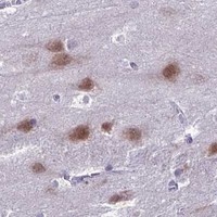
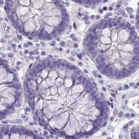
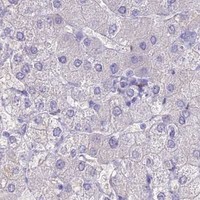
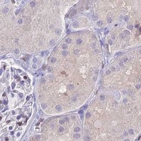
Comprehensive Human Protein Atlas IHC scoring per tissue (reliability: Enhanced — Medium consistency between antibody staining and RNA expression data.). Rows are taken directly from the HPA tissue chart — click any row's HPA link to view the source.
| Tissue | Cell type | Level | Evidence | Source |
|---|---|---|---|---|
| Caudate | Neuronal cells | Medium | Protein (IHC) | HPA → |
| Cerebellum | Cells in molecular layer | Medium | Protein (IHC) | HPA → |
| Cerebral cortex | Neuronal cells | Medium | Protein (IHC) | HPA → |
| Hippocampus | Neuronal cells | Medium | Protein (IHC) | HPA → |
| Lung | Endothelial cells | Medium | Protein (IHC) | HPA → |
| Tissue | Cell type | Level | Evidence | Source |
|---|---|---|---|---|
| Adipose tissue | Adipocytes | Not detected | Protein (IHC) | HPA → |
| Adrenal gland | Glandular cells | Not detected | Protein (IHC) | HPA → |
| Appendix | Glandular cells | Not detected | Protein (IHC) | HPA → |
| Bone marrow | Hematopoietic cells | Not detected | Protein (IHC) | HPA → |
| Breast | Adipocytes | Not detected | Protein (IHC) | HPA → |
Troubleshoot NOVA2 staining in paraffin sections by checking retrieval, cell identity, subcellular pattern, and assay controls before assigning biological meaning.
The IHC-validated anti-NOVA2 antibody has paraffin-section images from human appendicitis tissue and mouse and rat brain (catalog IHC captions). No IF/ICC data are supplied (catalog applications and images).
A10622-2 has IHC images from human appendicitis tissue and mouse and rat brain paraffin sections (A10622-2 IHC captions). Its listed applications include IHC, and its stated reactivity covers human, mouse and rat (catalog applications and reactivity).
Which to pick: For tissue IHC, choose A10622-2: its captions document paraffin sections, EDTA retrieval at pH 8.0 and DAB detection; the fixative is unreported (A10622-2 IHC captions). No IF/ICC-validated option is listed, and A10622-2 has no IF/ICC figure; its clonality is unreported (catalog applications, IF images and clone field). For cross-species IHC, A10622-2 has human, mouse and rat reactivity and a paraffin-section caption for each species (catalog reactivity; A10622-2 IHC captions).