This website uses cookies to ensure you get the best experience on our website.


- Table of Contents
Plan chromogenic NPAS4 IHC in paraffin brain sections with the catalog antibody at 1:100–1:300 (datasheet). Compare neuronal staining with controls, since the reported cytoplasmic CNS pattern has uncertain reliability (HPA tissue IHC).

Expected localisation, validated protocols, controls and antibodies — the at-a-glance facts below, then the full design guide.
| Expected localisation | CNS cytoplasm (HPA tissue IHC); nucleus (UniProt) | |
| Staining pattern | Cytoplasmic staining in CNS neurons and molecular-layer cells (HPA tissue IHC) | |
| Antigen retrieval | Tris-EDTA pH 9.0 HIER, 95–98 °C, 20 min (rule: nuclear antigen) | |
| Positive control | Caudate+3 more · see all | |
| Negative control | Adipose tissue+4 more · see all |
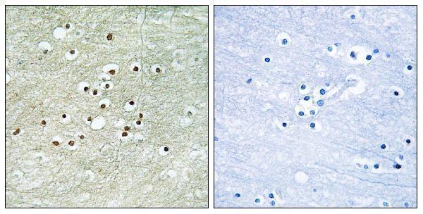
| Fixation | Matched tissue-IHC evidence does not establish the fixation claim. Validate the specimen-specific method before use. (selected-SKU IHC image A02045-1) | |
| Caveat | Presumed off-target binding; tissue staining is uncertain (HPA tissue IHC) | |
| Regulation | Activity-responsive in neurons (UniProt) | |
| Isoform / epitope | 2 isoforms; epitope coverage is unresolved (UniProt) |
The catalog antibody’s IHC-P protocol is accompanied by published NPAS4 staining protocols for rat paraffin sections, mouse free-floating sections, and mounted sections (PMC4275263; PMC3460929; PMC13172362).
| Sample | Paraffin-embedded human brain tissue; fixative not specified (datasheet A02045-1) |
| Fixation | Image fixative and duration unreported (datasheet A02045-1); verify before use. |
| Sectioning | 4–5 µm sections on charged slides (standard) |
| Deparaffinisation | Xylene, graded ethanol series to water (standard) |
| Antigen retrieval | Heat-induced epitope retrieval in Tris-EDTA buffer, pH 9.0, 20 min at 95–98 °C (standard rule: nuclear antigen) |
| Peroxidase block | 3% H2O2, 10 min, room temperature (standard) |
| Blocking | 10% normal serum of the secondary host, 30 min, room temperature (standard) |
| Primary antibody | Rabbit anti-NPAS4, 1:100-1:300 (datasheet A02045-1) |
| Primary incubation | Overnight at 4 °C (standard) |
| Detection | HRP-polymer secondary, DAB chromogen 5–10 min (standard) |
| Counterstain | Hematoxylin, blue, dehydrate and mount (standard) |
| Expected result | NPAS4-positive staining in neuronal cells of caudate (HPA tissue IHC: Medium). HPA tissue profile: Cytoplasmic expression in CNS. No signal in the no-primary control. |
NPAS4 is a nuclear, non-membrane neuronal transcription factor with brain tissue specificity (UniProt Q8IUM7: nucleus, topology, tissue specificity). In paraffin sections, examine neuronal cells in brain regions reported positive by HPA (HPA tissue IHC: medium staining). Treat the compartment call as provisional: HPA describes cytoplasmic CNS staining, rates its tissue IHC reliability Uncertain, and notes presumed off-target binding (HPA tissue IHC: reliability and profile).
| Defined nuclear staining in neuronal cells of cerebral cortex, hippocampus, or caudate; some cells in the cerebellar molecular layer stain. | This fits the nuclear localization of NPAS4 and the reported positive cell populations (UniProt Q8IUM7: nucleus; HPA tissue IHC: medium staining in these regions). HPA does not verify a nuclear paraffin-section pattern, so assess specificity with controls. |
| Strong cytoplasmic staining with little or no nuclear signal. | Interpret cautiously. HPA reports cytoplasmic CNS tissue staining but rates that result Uncertain and notes presumed off-target binding; UniProt places NPAS4 in the nucleus (HPA tissue IHC: profile and reliability; UniProt Q8IUM7: nucleus). |
| Prominent staining in adipocytes, glandular cells, or hematopoietic cells. | This conflicts with HPA's undetected results for those assessed cell types (HPA tissue IHC: adipose tissue, appendix and bone marrow). Check whether signal follows a plausible cell pattern and whether control sections reveal cross-reactivity or endogenous detection activity (general IHC practice). |
| Diffuse color across cells and surrounding tissue, without recognizable nuclear boundaries. | An indistinct pattern cannot support a compartment call (general IHC interpretation). Assess the primary-antibody omission control and background from the detection system, then reassess blocking, washing and antibody concentration (general IHC practice). |
| No staining in neuronal cells of a cerebral cortex, hippocampus or caudate section. | HPA reports medium staining in these neuronal populations, but its tissue IHC assessment is Uncertain (HPA tissue IHC: positive tissues and reliability). Confirm tissue integrity and assay controls before calling NPAS4 absent; a negative slide alone cannot distinguish biology from assay failure (general IHC practice). |
| Compartment evidence | UniProt assigns NPAS4 to the nucleus, and HPA ICC-IF approves nucleoplasm and nuclear bodies as main locations (UniProt Q8IUM7: subcellular location; HPA subcellular: approved main locations). HPA's cytoplasmic tissue IHC profile has Uncertain reliability, so reconcile any cytoplasmic chromogenic pattern with independent controls (HPA tissue IHC: profile and reliability). |
| Choice of tissue and cells | HPA reports medium staining in neuronal cells of caudate, cerebral cortex and hippocampus, and in cells of the cerebellar molecular layer (HPA tissue IHC: positive tissues). Select a documented positive population for interpretation; HPA's Uncertain reliability limits its use as a definitive specificity standard (HPA tissue IHC: reliability). |
| Antibody validation | The listed tissue IHC antibody HPA039255 is rated Uncertain, while HPA039248 has Approved ICC status and no listed IHC status (HPA antibodies: HPA039255, HPA039248). Approval in ICC does not establish paraffin IHC performance; interpret the catalog antibody's staining using its own IHC controls (general assay interpretation). |
| Isoforms and epitope coverage | UniProt lists two NPAS4 isoforms, but the supplied record gives no antibody epitope or isoform recognition data (UniProt Q8IUM7: isoforms; supplied antibody records). An unexpected negative result cannot be assigned to isoform exclusion from these sources alone. |
| IF/ICC Q: Where should NPAS4 appear? | A: HPA's approved ICC-IF main locations are nucleoplasm and nuclear bodies; nuclear membrane and vesicles are additional approved locations (HPA subcellular: ICC-IF). Those cell-image findings inform localization interpretation but do not supply a paraffin IHC protocol or resolve the Uncertain tissue IHC profile (HPA tissue IHC: reliability). |
| Situation | Likely cause | Next action |
|---|---|---|
| Cytoplasmic signal dominates the brain section. | HPA records cytoplasmic CNS staining but flags tissue IHC as Uncertain, with presumed off-target binding (HPA tissue IHC: profile and reliability). | Compare neuronal nuclear staining with control sections and an independently validated antibody when available; keep a cytoplasmic-only result provisional (UniProt Q8IUM7: nucleus; general IHC practice). |
| Color appears in HPA-negative cell populations. | Adipocytes, selected glandular cells and hematopoietic cells were not detected in the listed HPA tissues (HPA tissue IHC: negative tissues). | Check primary-antibody omission and detection controls for background or endogenous activity; then reassess antibody specificity in the affected cell type (general IHC practice). |
| Positive brain tissue shows no interpretable neuronal signal. | A negative section may reflect an assay problem; HPA reports medium staining in selected brain cells but rates the tissue result Uncertain (HPA tissue IHC: positives and reliability; general IHC practice). | Verify section quality, control performance, retrieval conditions and antibody application using the catalog antibody's IHC instructions (general IHC practice). |
| Diffuse chromogen obscures nuclear boundaries. | Excess nonspecific signal or detection background can prevent compartment assessment (general IHC practice). | Review the omission control, blocking and washes; adjust antibody concentration or detection development within the assay's validated range (general IHC practice). |
| Nuclear signal is visible but weak or uneven. | HPA reports medium tissue staining, not uniform staining of every neuronal nucleus (HPA tissue IHC: positive tissues and levels). | Score identifiable cells and compartments, compare a documented positive region processed alongside the section, and avoid treating weak fields alone as proof of absence (HPA tissue IHC: positives; general IHC practice). |
| ICC-IF localization seems clearer than chromogenic IHC. | HPA approves nuclear ICC-IF locations, whereas its tissue IHC profile remains Uncertain (HPA subcellular: main locations; HPA tissue IHC: reliability). | Record the two assay observations separately and use IHC-specific controls to judge the paraffin-section result; ICC approval alone cannot validate that result (HPA antibodies: ICC and IHC statuses; general assay interpretation). |
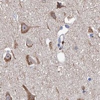
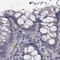
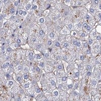


Comprehensive Human Protein Atlas IHC scoring per tissue (reliability: Uncertain — Pending external verification. Presumed off target binding observed and disregarded.). Rows are taken directly from the HPA tissue chart — click any row's HPA link to view the source.
| Tissue | Cell type | Level | Evidence | Source |
|---|---|---|---|---|
| Caudate | Neuronal cells | Medium | Protein (IHC) | HPA → |
| Cerebellum | Cells in molecular layer | Medium | Protein (IHC) | HPA → |
| Cerebral cortex | Neuronal cells | Medium | Protein (IHC) | HPA → |
| Hippocampus | Neuronal cells | Medium | Protein (IHC) | HPA → |
| Tissue | Cell type | Level | Evidence | Source |
|---|---|---|---|---|
| Adipose tissue | Adipocytes | Not detected | Protein (IHC) | HPA → |
| Appendix | Glandular cells | Not detected | Protein (IHC) | HPA → |
| Bone marrow | Hematopoietic cells | Not detected | Protein (IHC) | HPA → |
| Breast | Adipocytes | Not detected | Protein (IHC) | HPA → |
| Bronchus | Respiratory epithelial cells | Not detected | Protein (IHC) | HPA → |
Use nuclear staining, cell identity and matched controls to evaluate NPAS4 in paraffin section IHC; treat tissue staining patterns cautiously because HPA rates its IHC evidence uncertain.
A02045-1 has real IHC data from paraffin-embedded human brain tissue (IHC image caption: human brain) and lists IF plus Human, Mouse and Rat reactivity (catalog: applications and reactivity).
A02045-1 will render with an IHC figure from paraffin-embedded human brain tissue and a peptide-blocked comparison (IHC image caption: human brain and peptide block). It lists IHC and IF applications and Human, Mouse and Rat reactivity; no IF figure is supplied (catalog: applications, reactivity and image captions).
Which to pick: For tissue IHC, choose A02045-1: its own image shows paraffin-embedded human brain tissue, while the fixative is unreported (IHC image caption: paraffin-embedded human brain). For IF, A02045-1 is listed as an application, but ICC validation and an IF image are unreported (catalog: applications and image captions). For Mouse or Rat work, A02045-1 lists those species as reactive, although its shown IHC result is from Human tissue (catalog: reactivity; IHC image caption: human brain).