This website uses cookies to ensure you get the best experience on our website.


- Table of Contents
Plan chromogenic NPC1 tissue IHC with catalog antibody A00428, starting at 2.5 μg/mL (datasheet: A00428). Expect generally cytoplasmic tissue staining (HPA tissue IHC), while NPC1 resides in late endosome and lysosome membranes (UniProt).

Expected localisation, validated protocols, controls and antibodies — the at-a-glance facts below, then the full design guide.
| Expected localisation | Cytoplasmic tissue IHC (HPA tissue IHC); late endosome/lysosome membrane (UniProt) | |
| Staining pattern | High staining in kidney tubules; generally cytoplasmic (HPA tissue IHC) | |
| Antigen retrieval | Citrate pH 6.0 HIER, 95–98 °C, 20 min (rule: cytoplasmic / membrane antigen) | |
| Positive control | Adrenal gland+4 more · see all | |
| Negative control | None in HPA (detected in all 45 tissues); use no-primary + isotype controls |
| Fixation | Keep fixation consistent across sections. (standard IHC practice; not target-specific) | |
| Caveat | IHC staining has medium consistency with RNA expression (HPA tissue IHC) | |
| Regulation | Expression regulation not annotated (UniProt) | |
| Isoform / epitope | 2 isoforms; check whether the epitope is lumenal or cytoplasmic (UniProt) |
The catalog antibody's IHC-P protocol is accompanied by two published NPC1 IHC protocols (PMC11961396; PMC12292827).
| Sample | Tissue sections; selected-image fixative not specified (standard IHC workflow) |
| Fixation | Image fixative and duration unreported (datasheet A00428); verify before use. |
| Sectioning | 4–5 µm sections on charged slides (standard) |
| Deparaffinisation | Xylene, graded ethanol series to water (standard) |
| Antigen retrieval | Heat-induced epitope retrieval in citrate buffer, pH 6.0, 20 min at 95–98 °C (standard rule: cytoplasmic / membrane antigen) |
| Peroxidase block | 3% H2O2, 10 min, room temperature (standard) |
| Blocking | 10% normal serum of the secondary host, 30 min, room temperature (standard) |
| Primary antibody | Rabbit anti-NPC1, 2.5 μg/mL (datasheet A00428) |
| Primary incubation | Overnight at 4 °C (standard) |
| Detection | HRP-polymer secondary, DAB chromogen 5–10 min (standard) |
| Counterstain | Hematoxylin, blue, dehydrate and mount (standard) |
| Expected result | NPC1-positive staining in glandular cells of adrenal gland (HPA tissue IHC: High). HPA tissue profile: General cytoplasmic expression. No signal in the no-primary control. |
NPC1 is a 13-pass protein of late-endosome and lysosome membranes, so positive cells should show cytoplasmic staining consistent with those compartments (UniProt O15118 topology and subcellular location). HPA reports general cytoplasmic IHC expression, with high staining in adrenal glandular cells, kidney tubular cells, ovarian follicle cells, seminal-vesicle glandular cells, and cells in testicular seminiferous ducts (HPA tissue IHC). Its tissue IHC reliability is Approved, with medium agreement between staining and RNA data (HPA tissue IHC).
| Cytoplasmic staining in kidney tubular cells or adrenal glandular cells, with little nuclear signal. | This fits HPA's high staining in those cell types and its general cytoplasmic profile (HPA tissue IHC). A punctate or granular appearance would be consistent with late-endosome and lysosome membranes, but HPA does not establish a required granule pattern in these sections (UniProt O15118 subcellular location; HPA tissue IHC). |
| Strong staining confined mainly to nuclei, plasma membranes, or extracellular material. | That distribution conflicts with the expected late-endosome and lysosome membrane location and HPA's cytoplasmic IHC profile (UniProt O15118 subcellular location; HPA tissue IHC). Treat it as a possible staining artefact; assess section morphology and controls before assigning NPC1 positivity (general IHC practice). |
| Intense staining in cells outside the expected stained population, while the expected cells remain faint. | For a chosen tissue, compare the stained cells with HPA's cell-level observations; for example, kidney staining is reported as high in tubular cells (HPA tissue IHC). An unexpected cell distribution can reflect cross-reactivity or endogenous detection activity (general IHC practice). It is not definitive evidence against NPC1 because HPA reports low tissue specificity and no negative tissues (HPA tissue IHC). |
| A diffuse stain covers many cell types, lumina, or section-free areas without a clear cytoplasmic pattern. | This obscures the general cytoplasmic profile reported by HPA (HPA tissue IHC). Background outside cells suggests nonspecific reagent or detection signal; compare a no-primary control and inspect wash and blocking conditions (general IHC practice). Uniform brown color alone does not establish NPC1-positive cells (general IHC practice). |
| No detectable staining in kidney tubular cells or adrenal glandular cells. | Both are reported as high-staining cell populations, so an absent result warrants a technical check (HPA tissue IHC). Check section integrity, retrieval, antibody conditions, and detection controls using the IHC-validated antibody's instructions (general IHC practice). HPA's Approved rating has medium staining–RNA agreement and does not guarantee positivity in every specimen (HPA tissue IHC). |
| Cell population and tissue | HPA reports high staining in five specified cell populations, medium staining in appendix glandular, bone-marrow hematopoietic, and bronchial respiratory epithelial cells, and low staining in several others (HPA tissue IHC). Choose a documented high-staining population for a positive control; low staining alone is a weak technical benchmark (HPA tissue IHC; general IHC practice). |
| Antibody validation | HPA lists HPA026618 and CAB070132 as IHC Approved; neither is listed as IHC Enhanced in the supplied record (HPA antibodies). HPA describes overall tissue IHC reliability as Approved with medium staining–RNA consistency, so interpret unexpected patterns with controls (HPA tissue IHC; general IHC practice). |
| Membrane topology and epitope position | NPC1 has 13 transmembrane segments and lumenal and cytoplasmic regions (UniProt O15118 topology). The antibody epitope is not supplied, so topology cannot identify which region the antibody detects or predict a target-specific retrieval requirement (UniProt O15118 topology; supplied antibody record). |
| Processing, glycosylation, and isoforms | UniProt lists a signal peptide at residues 1–22, a mature chain beginning at residue 23, 19 glycosylation sites, and two isoforms (UniProt O15118 processing, glycosylation, isoforms). Without an epitope or isoform-specific antibody record, these features do not justify predicting different tissue staining or fixation sensitivity (supplied antibody record). |
| Situation | Likely cause | Next action |
|---|---|---|
| Known high-staining cells are blank. | The assay may have failed, although HPA's reported high level does not guarantee every specimen will stain (HPA tissue IHC; general IHC practice). | Run a documented high-staining tissue alongside the sample; check section preservation, the antibody's IHC-P instructions, retrieval and detection steps, and reagent controls (HPA tissue IHC; general IHC practice). |
| Color appears mainly in nuclei or extracellular spaces. | This conflicts with NPC1's late-endosome and lysosome membrane location and HPA's cytoplasmic profile (UniProt O15118 subcellular location; HPA tissue IHC). | Review morphology and a no-primary control; adjust nonspecific staining controls before scoring the compartment as NPC1 positive (general IHC practice). |
| Unexpected cells stain more strongly than the documented population. | Cross-reactivity or endogenous detection activity is possible, but NPC1 also has low tissue specificity and HPA supplies no negative-tissue list (general IHC practice; HPA tissue IHC). | Compare cell identities with the HPA tissue profile; include no-primary and detection controls, and use an independently validated antibody if the assignment remains uncertain (HPA tissue IHC; HPA antibodies; general IHC practice). |
| The whole section has weak, diffuse color. | Background from antibody or detection reagents may obscure cellular staining (general IHC practice). | Inspect a no-primary control, washing and blocking; score cells only where staining can be distinguished from section-wide background (general IHC practice). |
| A low-staining tissue gives little or no signal. | HPA reports low staining in several specified cell populations; weak signal there is a poor standalone test of assay performance (HPA tissue IHC). | Evaluate a documented high-staining population in parallel before changing assay conditions, and avoid calling the low-staining sample NPC1 negative on that result alone (HPA tissue IHC; general IHC practice). |
| IF/ICC: what pattern should be expected? | UniProt places NPC1 on late-endosome and lysosome membranes, but HPA provides only a Membrane summary, no main ICC-IF location, and no cell-line images (UniProt O15118 subcellular location; HPA subcellular ICC-IF). | Assess intracellular membrane-associated signal against appropriate IF controls; treat a specific punctate pattern as a hypothesis, not an HPA-verified ICC-IF finding (UniProt O15118 subcellular location; HPA subcellular ICC-IF; general IF practice). |


Comprehensive Human Protein Atlas IHC scoring per tissue (reliability: Approved — Medium consistency between antibody staining and RNA expression data.). Rows are taken directly from the HPA tissue chart — click any row's HPA link to view the source.
| Tissue | Cell type | Level | Evidence | Source |
|---|---|---|---|---|
| Adrenal gland | Glandular cells | High | Protein (IHC) | HPA → |
| Kidney | Cells in tubules | High | Protein (IHC) | HPA → |
| Ovary | Follicle cells | High | Protein (IHC) | HPA → |
| Seminal vesicle | Glandular cells | High | Protein (IHC) | HPA → |
| Testis | Cells in seminiferous ducts | High | Protein (IHC) | HPA → |
| Tissue | Cell type | Level | Evidence | Source |
|---|---|---|---|---|
| None in HPA: NPC1 is detected in all 45 scored tissues. Use a no-primary (secondary-only) and an isotype control instead. | ||||
Troubleshoot NPC1 staining in paraffin sections by checking retrieval, compartmental pattern, controls, and scoring before interpreting biological differences.
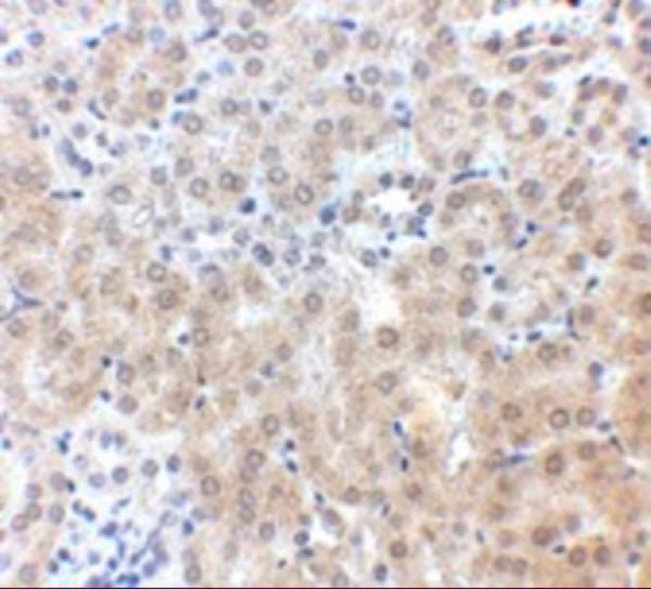
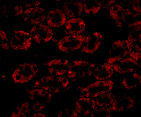
A00428 has IHC data from mouse kidney tissue and IF data from mouse kidney cells; its listed reactivity covers human and mouse (catalog image captions; catalog reactivity).
Only A00428 will render a card: its IHC figure shows mouse kidney tissue stained at 2.5 μg/mL (catalog IHC image caption). Its IF figure shows mouse kidney cells at 20 μg/mL (catalog IF image caption).
Which to pick: For paraffin-section IHC, choose A00428: IHC-P is listed, and its own IHC figure shows mouse kidney tissue staining; the caption does not report the fixative (catalog applications; catalog IHC image caption). For IF with figure evidence, choose A00428; for ICC, M00428-1 lists ICC/IF and is a rabbit monoclonal, but has no supplied figure (catalog IF image caption; catalog applications; catalog host/clone; catalog image alts). For rat samples, M00428-1 lists rat reactivity, while A00428 lists human and mouse (catalog reactivity).