This website uses cookies to ensure you get the best experience on our website.
- Table of Contents
8 Citations 1 Q&As
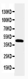
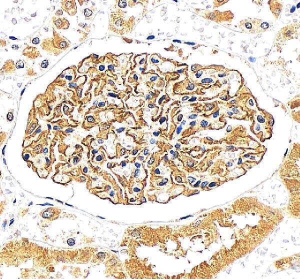
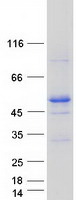
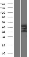
Facts about Podocin.
Human | |
---|---|
Gene Name: | NPHS2 |
Uniprot: | Q9NP85 |
Entrez: | 7827 |
Belongs to: |
---|
band 7/mec-2 family |
Nephrosis 2; nephrosis 2, idiopathic, steroid-resistant (podocin); NPHS2; PDCN; PDCNSRN1podocin; Podocin; SRN1
Mass (kDA):
42.201 kDA
Human | |
---|---|
Location: | 1q25.2 |
Sequence: | 1; NC_000001.11 (179550539..179575987, complement) |
Almost exclusively expressed in the podocytes of fetal and mature kidney glomeruli.
[Isoform 1]: Cell membrane; Peripheral membrane protein.; [Isoform 2]: Endoplasmic reticulum.
The glomerular protein NPHS2 is encoded by the NPHS2 gene. Boster has created antisense NPHS2 Riboprobes to detect this glomerular protein. These antibodies have been validated for high specificity and affinity in a series of known positive and negative samples. Boster rewards the first reviewers with product credits and other benefits. The product credits are awarded to scientists across the world.
Recent research found that NPHS2 mutations are linked to late-onset focal segmental glomerulosclerosis and steroid-resistant nephrotic disorder. The study also revealed an NPHS2 mutation NPHS2, R229Q. This mutation is the cause of steroid resistant nephrotic disorder. The mutation is also associated with mutation-dependent recessive inheritance.
Both sporadic and familial cases have been affected by the nephrotic syndrome that is caused by the defect in NPHS2. The mice with NPHS2-/ were proteinuric at birth , but produced urine during embryogenesis. Podocin-deficient mice have normal kidney function, however, podocyte foot processes don't develop normally in the. Podocin is required for the assembly and maintenance of the glomerular barrier.
NPHS1 and NPHS2 are two glomerular proteins that play crucial roles in regulating glomerular filtration. Mutations in either of these proteins can cause the nephrotic disorder. This study highlights the role of these proteins in regulating glomerular filtering. This study doesn't address the precise mechanism behind NPHS2 mutations in nephrotic syndrome.
One of the most well-known causes of SRNS is a mutation in the NPHS2 gene. The mutation causes steroid resistance kidney disease. It is unclear which gene is responsible this mutation. The glomerular protein podocin is coded by the NPHS2 gene. In sporadic cases, the mutation of NPHS2 causes steroid-resistant renal syndrome. This is why it is vital to detect a genetic defect in NPHS2 in patients.
The phenotype of the nephrotic syndrome has been implicated in CD2AP haploinsufficiency. CD2AP haploinsufficiency is associated with an increased susceptibility to glomerular alteration. The mouse model of NPHS2 mice shares a genetic background to that of B6/129 Nphs2 mice. However the kidneys of B6/129 Nphs2mice suffered from an excessive amount of proteinuria by the time they reached day 25.
The NPHS2 is a vital glomerular protein in nephrotric disorder. It regulates the function of the GFB which causes the oedema and the proteinuria. Nephrotic syndrome is the most common glomerular disorder in children . It is often classified into two types: steroid-sensitive and steroid-resistant NS. Ninety percent of cases in children respond to corticosteroid treatment.
The Anti-Podocin antibody NPHS2-Boster can be used as a potent test for kidney disease diagnosis. This antibody detects podocin in the glomerular capillary wall, that is closely related to the distribution of podocin in the human kidney. It is visible using double immunofluorescence and is in sync with the human A3b1 integrin.
The four antibodies were raised against different regions of podocin. P18 and P21 targeted the complete N-terminal region , which is located prior to the membrane-associated domain, whereas P35 and P39 were raised against the C-terminal region. Cells transiently transfected by the expression construct pNPHS2 displayed the similar pattern. The antibodies P18 and P39 identified a single band of 49kD in transfected cells. None was detected in cells that were untreated.
In a mouse model of PAN the Anti-Podocin-NPHS2-Boster antibody against PAN was increased. PAN + AC1903 stimulated podocytes via decreasing TRPC5 channel activation. The podocytes of the rat treated with PAN were significantly protected by the presence of SYNPO and Nphs2 marker proteins.
A riboprobe and antisense probe may be used to determine the RNA-based NPHS2 transcription in postnatal kidneys. The antisense probe exhibits strong labeling in podocytes and in the peripheral region of the glomerular tuft. This protein is also found heavily in the proximal glomerular tuft of an S-shaped foetus exhibiting a frameshift mutation.
In situ hybridization allows the detection of mRNA through analysis of the levels of mRNA. It is performed with paraffin-embedded tissue sections. Six millimeter thick sections were deparaffinized and heated in sodium citrate buffer (pH 6.0). The riboprobes that were used in this procedure were synthesized from a 10-65-bp product of PCR that covers the range of bases spanning from 728 to 1792 in NPHS2 DNA. These probes were subcloned to an PGEM Teasy vector, followed by SacII digestion and SalI digestion to obtain sense and antisense NPHS2 riboprobes.
The NPHS2 gene is expressed in the podocytes and is highly regulated by nephrin. We have studied the distribution of podocin mRNA by immunohistology and in situ hybridization. These techniques were employed in the past to detect podocin. These riboprobes enabled us to detect the levels of the gene within the extracellular glomerular mat.
In-situ hybridization can be carried out with various samples. In contrast to conventional methods, in situ hybridization is a relatively simple method that can be executed without compromising the quality of the sample. We can visualize gene expression in intact tissue samples with a riboprobe with an antisense NPHS2 tag. We recommend using digoxigenin-labeled antisense DNA for in situ hybridization.
In-situ hybridization with antisense DNA probes labelled with RNA is a great way to examine the expression levels of genes in the body. It is beneficial for studies of mRNA expression and localization levels in different organs and tissues. It's not inexpensive, but it's extremely efficient and can be utilized to serve a variety of research objectives.
It is now time to begin the actual process of hybridization. Hybridization is performed at temperatures between 55 and 62 degrees Celsius. The sections should be placed in the incubator for a night to allow the probe to work. The probe mixture is held in contact with the tissue with surface tension. The slides are then washed in 0.5x SSC.
In-situ hybridization with anti-sense or sense NPHS2 the riboprobes are made using inserts of cDNA which have specific sequences. During hybridization, the probes bind with the target mRNA. This way, researchers can control whether the probes are forming a hybrid to the targeted mRNA. Antisense riboprobes can be more specific than sense riboprobes but they are still useful when studies require quantitative expression of RNA.
Before incubating the probe the sample needs to be prehybridized to 42°C. Then, the probe mixture (two ul of 35S riboprobe in 1xTE, one ul of tRNA in dH2O, and seventeen micrograms of rHB2) is pipetted into the bubble of prehybridization solution. The mixture is then heated to 95degC for three minutes. The probe and tRNA then are added to a microfuge tub.
The NPHS2 gene was previously found to be only present in the kidneys. Although the fetal kidneys don't contain any podocyte signals but there was a distinct signal in the lower half the body with the shape of an S. This signal is in the area of future podocytes. It was also observed in mature glomeruli as well as immature glomeruli from the deep cortex. These findings are in agreement with pathology, and the protein encoded by the NPHS2 gene has been identified as podocin.
The NPHS2 gene is composed of eight exons. Each one contains a nucleotide sequence of SEQ ID NO. 1. Exons 2 3 6, 8 and 10, all contain SEQ ID No. 2 (A) of SEQ ID NO.2 (see Figure 1).
A mutation in the NPHS2 gene may result in frameshifts or premature stop codons that help to verify the gene's identity. Missense mutations are found in regions of the gene that are conserved. These mutations are common within families that suffer from the disease, but mRNautes. Thce ofkaemililtion.
PMID: 10742096 by Boute N., et al. NPHS2, encoding the glomerular protein podocin, is mutated in autosomal recessive steroid-resistant nephrotic syndrome.
PMID: 11562357 by Huber T.B., et al. Interaction with podocin facilitates nephrin signaling.
*More publications can be found for each product on its corresponding product page