Click image to see more details
-

-
-
-
-
+4
Product Info Summary
| SKU: | PA1322-1 |
|---|---|
| Size: | 100 μg/vial |
| Reactive Species: | Human, Mouse, Rat |
| Host: | Rabbit |
| Application: | IHC, WB |
Customers Who Bought This Also Bought

Product info
Product Name
Anti-Podocin NPHS2 Antibody Picoband®
SKU/Catalog Number
PA1322-1
BA1688 is an alternative SKU for this antibody, used in previous lots.
Size
100 μg/vial
Form
Lyophilized
Description
Boster Bio Anti-Podocin NPHS2 Antibody catalog # PA1322-1. Tested in IHC, WB applications. This antibody reacts with Human, Mouse, Rat. The brand Picoband indicates this is a premium antibody that guarantees superior quality, high affinity, and strong signals with minimal background in Western blot applications. Only our best-performing antibodies are designated as Picoband, ensuring unmatched performance.
Storage & Handling
Store at -20˚C for one year from date of receipt. After reconstitution, at 4˚C for one month. It can also be aliquotted and stored frozen at -20˚C for six months. Avoid repeated freeze-thaw cycles.
Cite This Product
Anti-Podocin NPHS2 Antibody Picoband® (Boster Biological Technology, Pleasanton CA, USA, Catalog # PA1322-1)
Host
Rabbit
Contents
Each vial contains antibody formulated with stabilizing components, 0.9mg NaCl, 0.2mg Na2HPO4, 0.05mg Thimerosal, 0.05mg NaN3.
*This antibody is supplied in a stabilized formulation.
Compatibility with conjugation reactions depends on the chemistry of the conjugation method used.
For conjugation methods that are not compatible with the stabilizing components present in this formulation, a carrier-free antibody format is required.
Clonality
Polyclonal
Isotype
Rabbit IgG
Immunogen
A synthetic peptide corresponding to a sequence at the C-terminus of human NPHS2, identical to the related mouse sequence, and different from the related rat sequence by one amino acid.
Cross-reactivity
No cross-reactivity with other proteins
Reactive Species
PA1322-1 is reactive to NPHS2 in Human, Mouse, Rat
Observed Molecular Weight
45 kDa
Calculated molecular weight
42.2 kDa
Background of NPHS2
Podocin (PDCN) is a protein which lines the podocytes and assists in maintaining the barrier at the glomerular basement membrane. NPHS2 is a causative gene for Familial idiopathic nephrotic syndromes, which represents a heterogeneous group of kidney disorders, and include autosomal recessive steroid-resistant nephrotic syndrome, which is characterized by early childhood onset of proteinuria, rapid progression to end-stage renal disease and focal segmental glomerulosclerosis. By positional cloning, NPHS2 was mapped to 1q25-31. It is almost exclusively expressed in the podocytes of fetal and mature kidney glomeruli, and encodes a new integral membrane protein, podocin, belonging to the stomatin protein family. Ten different NPHS2 mutations were found, comprising nonsense, frameshift and missense mutations, to segregate with the disease, demonstrating a crucial role for podocin in the function of the glomerular filtration barrier.
Antibody Validation
Boster validates all antibodies on WB, IHC, ICC, Immunofluorescence, and ELISA with known positive control and negative samples to ensure specificity and high affinity, including thorough antibody incubations.
Application & Images
Applications
PA1322-1 is guaranteed for IHC, WB Boster Guarantee
Recommend Dilution
| Application | Dilution | Species |
|---|---|---|
| Immunohistochemistry (Frozen Section) | 0.5-1μg/ml | Rat, Human, Mouse |
| Immunohistochemistry (Paraffin-embedded Section) | 0.5-1μg/ml | Rat, Human, Mouse |
| Western blot | 0.1-0.5μg/ml | Rat, Human, Mouse |
Tested application
Suggested blocking solution with 5% non-fat milk or BSA; (*)Recommended protein loading: 20-40 µg per lane
Use TE buffer pH 9.0 for antigen retrieval; (*) citrate buffer pH 6.0 is an alternative.
Validation Images & Assay Conditions
Click image to see more details
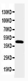
Western blot analysis of NPHS2 using anti-NPHS2 antibody (PA1322-1).
Electrophoresis was performed on a 5-20% SDS-PAGE gel at 70V (Stacking gel) / 90V (Resolving gel) for 2-3 hours. The sample well of each lane was loaded with 50ug of sample under reducing conditions.
Lane 1: rat kidney tissue lysate.
After Electrophoresis, proteins were transferred to a Nitrocellulose membrane at 150mA for 50-90 minutes. Blocked the membrane with 5% Non-fat Milk/ TBS for 1.5 hour at RT. The membrane was incubated with rabbit anti-NPHS2 antigen affinity purified polyclonal antibody (Catalog # PA1322-1) at 0.5 μg/mL overnight at 4°C, then washed with TBS-0.1%Tween 3 times with 5 minutes each and probed with a goat anti-rabbit IgG-HRP secondary antibody at a dilution of 1:10000 for 1.5 hour at RT. The signal is developed using an Enhanced Chemiluminescent detection (ECL) kit (Catalog # EK1002) with Tanon 5200 system. A specific band was detected for NPHS2 at approximately 45KD. The expected band size for NPHS2 is at 42KD.

Click image to see more details
IHC analysis of NPHS2 using anti-NPHS2 antibody (PA1322-1).
NPHS2 was detected in frozen section of rat kidney tissues. The tissue section was blocked with 10% goat serum. The tissue section was then incubated with 1μg/ml rabbit anti-NPHS2 Antibody (PA1322-1) overnight at 4°C. Biotinylated goat anti-rabbit IgG was used as secondary antibody and incubated for 30 minutes at 37°C. The tissue section was developed using Strepavidin-Biotin-Complex (SABC)(Catalog # SA1022) with DAB as the chromogen.

Click image to see more details
IHC analysis of NPHS2 using anti-NPHS2 antibody (PA1322-1).
NPHS2 was detected in a paraffin-embedded section of human kidney tissue. Heat mediated antigen retrieval was performed in EDTA buffer (pH 8.0, epitope retrieval solution). The tissue section was blocked with 10% goat serum. The tissue section was then incubated with 2 μg/ml rabbit anti-NPHS2 Antibody (PA1322-1) overnight at 4°C. Peroxidase Conjugated Goat Anti-rabbit IgG was used as secondary antibody and incubated for 30 minutes at 37°C. The tissue section was developed using HRP Conjugated Rabbit IgG Super Vision Assay Kit (Catalog # SV0002) with DAB as the chromogen.

Click image to see more details
Western blot analysis of NPHS2 using anti-NPHS2 antibody (PA1322-1).
Electrophoresis was performed on a 5-20% SDS-PAGE gel at 70V (Stacking gel) / 90V (Resolving gel) for 2-3 hours. The sample well of each lane was loaded with 50ug of sample under reducing conditions.
Lane 1: human 293T whole cell lysates.
After Electrophoresis, proteins were transferred to a Nitrocellulose membrane at 150mA for 70 minutes. Blocked the membrane with 5% Non-fat Milk/ TBS for 1.5 hour at RT. The membrane was incubated with rabbit anti-NPHS2 antigen affinity purified polyclonal antibody (Catalog # PA1322-1) at 0.5 μg/mL overnight at 4°C, then washed with TBS-0.1%Tween 3 times with 5 minutes each and probed with a goat anti-rabbit IgG-HRP secondary antibody at a dilution of 1:10000 for 1.5 hour at RT. The signal is developed using an Enhanced Chemiluminescent detection (ECL) kit (Catalog # EK1002) with Tanon 5200 system. A specific band was detected for NPHS2 at approximately 45KD. The expected band size for NPHS2 is at 42KD.

Click image to see more details
IHC analysis of NPHS2 using anti-NPHS2 antibody (PA1322-1).
NPHS2 was detected in a paraffin-embedded section of human kidney tissue. Heat mediated antigen retrieval was performed in EDTA buffer (pH 8.0, epitope retrieval solution). The tissue section was blocked with 10% goat serum. The tissue section was then incubated with 2 μg/ml rabbit anti-NPHS2 Antibody (PA1322-1) overnight at 4°C. Peroxidase Conjugated Goat Anti-rabbit IgG was used as secondary antibody and incubated for 30 minutes at 37°C. The tissue section was developed using HRP Conjugated Rabbit IgG Super Vision Assay Kit (Catalog # SV0002) with DAB as the chromogen.

Click image to see more details
IHC analysis of NPHS2 using anti-NPHS2 antibody (PA1322-1).
NPHS2 was detected in paraffin-embedded section of mouse kidney tissues. Heat mediated antigen retrieval was performed in citrate buffer (pH6, epitope retrieval solution) for 20 mins. The tissue section was blocked with 10% goat serum. The tissue section was then incubated with 1μg/ml rabbit anti-NPHS2 Antibody (PA1322-1) overnight at 4°C. Biotinylated goat anti-rabbit IgG was used as secondary antibody and incubated for 30 minutes at 37°C. The tissue section was developed using Strepavidin-Biotin-Complex (SABC)(Catalog # SA1022) with DAB as the chromogen.

Click image to see more details
IHC analysis of NPHS2 using anti-NPHS2 antibody (PA1322-1).
NPHS2 was detected in paraffin-embedded section of rat kidney tissues. Heat mediated antigen retrieval was performed in citrate buffer (pH6, epitope retrieval solution) for 20 mins. The tissue section was blocked with 10% goat serum. The tissue section was then incubated with 1μg/ml rabbit anti-NPHS2 Antibody (PA1322-1) overnight at 4°C. Biotinylated goat anti-rabbit IgG was used as secondary antibody and incubated for 30 minutes at 37°C. The tissue section was developed using Strepavidin-Biotin-Complex (SABC)(Catalog # SA1022) with DAB as the chromogen.

Click image to see more details
Scutellarin Restored Podocyte Injury of the DN Mice. a Representative images of immunohistochemistry for NPHS1 and NPHS2 of the mice treated with vehicle, scutellarin or empagliflozin (× 200; scale bar = 50 µm). b Representative images of Western-blotting for NPHS1, NPHS2. c Quantitative plot of the expression of NPHS1 of the mice. d Quantitatification of NPHS1 expression of the mice. e Representative images of Western-blotting for β-catenin, Axin2, snail and DKK1 of the mice. f – i Quantifications of the protein levels for β-catenin, Axin2, snail and DKK1 from E. All data are presented as the mean ± S.D.; n = 4–6 for each group, “n” stands for the number of animals; p vs. the model group (STZ)
Index in PubMed under a CC BY license. PMID: 38656633
Specific Publications For Anti-Podocin NPHS2 Antibody Picoband® (PA1322-1)
Loading publications
Recommended Resources
Here are featured tools and databases that you might find useful.
- Boster's Pathways Library
- Protein Databases
- Bioscience Research Protocol Resources
- Data Processing & Analysis Software
- Photo Editing Software
- Scientific Literature Resources
- Research Paper Management Tools
- Molecular Biology Software
- Primer Design Tools
- Bioinformatics Tools
- Phylogenetic Tree Analysis
Customer Reviews
Have you used Anti-Podocin NPHS2 Antibody Picoband®?
Share your experimental results or join a short interview to earn up to $1,000 in product credits or other rewards.
0 Reviews For Anti-Podocin NPHS2 Antibody Picoband®
Customer Q&As
Have a question?
Find answers in Q&As, reviews.
Can't find your answer?
Submit your question
1 Customer Q&As for Anti-Podocin NPHS2 Antibody Picoband®
Question
We are currently using anti-NPHS2 antibody PA1322-1 for mouse tissue, and we are happy with the IHC results. The species of reactivity given in the datasheet says human, mouse, rat. Is it likely that the antibody can work on feline tissues as well?
Verified Customer
Verified customer
Asked: 2020-03-06
Answer
The anti-NPHS2 antibody (PA1322-1) has not been tested for cross reactivity specifically with feline tissues, but there is a good chance of cross reactivity. We have an innovator award program that if you test this antibody and show it works in feline you can get your next antibody for free. Please contact me if I can help you with anything.
Boster Scientific Support
Answered: 2020-03-06
