This website uses cookies to ensure you get the best experience on our website.


- Table of Contents
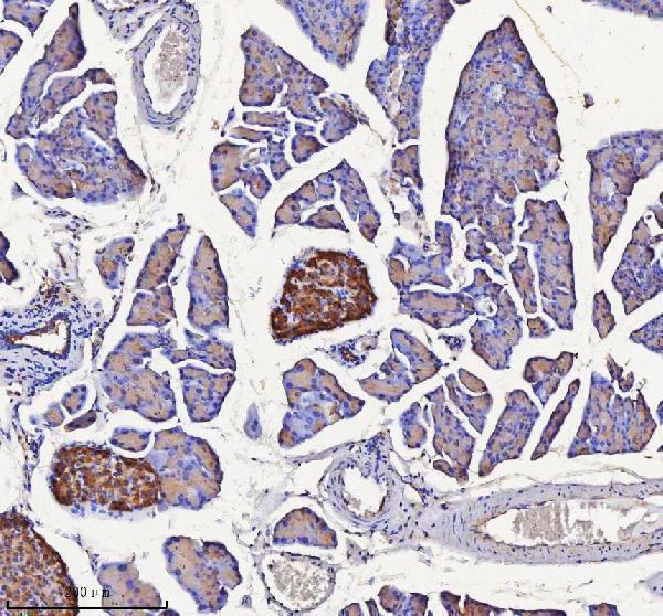
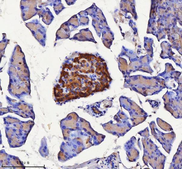
Plan chromogenic NPTX2 IHC on paraffin sections using pancreatic endocrine cells as a positive reference (HPA tissue IHC). This guide covers fixation, retrieval and interpretation of cytoplasmic staining, with a catalog antibody IHC range of 2–5 μg/mL (HPA tissue IHC; datasheet A09667-1).

Expected localisation, validated protocols, controls and antibodies — the at-a-glance facts below, then the full design guide.
| Expected localisation | Cytoplasmic tissue staining (HPA tissue IHC) | |
| Staining pattern | Cytoplasm: islets, cortical neurons, pituitary, adrenal medulla (HPA tissue IHC) | |
| Antigen retrieval | EDTA pH 8.0 HIER, heat-mediated (datasheet A09667-1) | |
| Positive control | Pancreas+2 more · see all | |
| Negative control | Adipose tissue+4 more · see all |
| Fixation | Keep paraffin-section fixation consistent. (standard IHC practice; not target-specific) | |
| Caveat | Secreted protein may stain away from its RNA source (HPA tissue IHC) | |
| Regulation | Pituitary-enriched RNA (HPA tissue RNA) | |
| Isoform / epitope | No listed isoforms; mature chain lacks signal peptide 1–15 (UniProt) |
The catalog antibody protocol is followed by three published chromogenic IHC protocols for NPTX2, covering paraffin sections and brain sections (PMC7985451; PMC6046040; PMC10813937).
| Sample | Paraffin-embedded mouse pancreas tissue; fixative not specified (datasheet A09667-1) |
| Fixation | Image fixative and duration unreported (datasheet A09667-1); verify before use. |
| Sectioning | 4–5 µm sections on charged slides (standard) |
| Deparaffinisation | Xylene, graded ethanol series to water (standard) |
| Antigen retrieval | Heat retrieval: EDTA pH 8.0 (datasheet A09667-1); 20 min, 95–100 °C (standard) |
| Peroxidase block | 3% H2O2, 10 min, room temperature (standard) |
| Blocking | 10% goat serum (datasheet A09667-1) |
| Primary antibody | Rabbit anti-NPTX2, 2-5 μg/ml (datasheet A09667-1) |
| Primary incubation | Overnight at 4 °C (datasheet A09667-1) |
| Detection | HRP-conjugated secondary, DAB chromogen (datasheet A09667-1) |
| Counterstain | Hematoxylin, blue, dehydrate and mount (standard) |
| Expected result | NPTX2-positive staining in pancreatic endocrine cells of pancreas (HPA tissue IHC: High). HPA tissue profile: Cytoplasmic expression in islets of Langerhans, neuronal cells in cerebral cortex, and in pituitary gland and adrenal medulla. No signal in the no-primary control. |
NPTX2 is secreted and has no transmembrane segment (UniProt P47972 topology). In paraffin-section IHC, expect chiefly cytoplasmic staining in pancreatic endocrine cells and anterior pituitary cells, with weaker staining in adrenal medullary cells and cerebral cortical neurons (HPA tissue IHC). HPA rates the tissue profile Enhanced; because NPTX2 is secreted, its protein location need not match the cells with the most RNA (HPA tissue IHC).
| Strong cytoplasmic staining in pancreatic islets or anterior pituitary cells. | This matches the reported high staining in pancreatic endocrine and anterior pituitary cells (HPA tissue IHC). Assess the named cells and their compartment, rather than treating any staining in the organ as confirmation. |
| Predominantly nuclear staining in cells expected to be positive. | A nuclear-dominant pattern conflicts with the reported cytoplasmic tissue profile (HPA tissue IHC). Treat it as suspect; compare with a known-positive section and the negative control before interpreting it as NPTX2. |
| Strong staining in adipocytes or another cell type reported as unstained. | HPA reports adipocytes as not detected in adipose tissue and breast (HPA tissue IHC). Consider antibody cross-reactivity or endogenous chromogenic detection activity; check staining in a no-primary control (general IHC practice). |
| Diffuse color across cells and surrounding tissue, obscuring cell boundaries. | This does not establish the cell-restricted cytoplasmic pattern reported by HPA (HPA tissue IHC). General IHC practice: review background controls, detection activity, blocking, washes and primary-antibody concentration before scoring. |
| No staining in pancreatic endocrine cells or anterior pituitary cells. | Both are reported high-staining cell populations (HPA tissue IHC). An absent signal makes a negative result elsewhere difficult to interpret; inspect section quality and verify the antibody, retrieval and detection workflow (general IHC practice). |
| Secreted protein and processing | NPTX2 has a signal peptide at residues 1–15, a mature chain at 16–431 and no transmembrane segment (UniProt P47972). Interpret the observed cytoplasmic IHC profile without assuming a membrane-bound target (HPA tissue IHC; UniProt P47972 topology). |
| Tissue and cell choice | Pancreatic endocrine and anterior pituitary cells stain High; adrenal medullary cells stain Medium, while cortical neurons stain Low (HPA tissue IHC). Choose the high-staining cells to judge assay sensitivity. |
| RNA–protein location | HPA calls pituitary RNA tissue enriched but cautions that RNA and protein locations can differ for a secreted protein (HPA tissue IHC). Do not score an IHC result solely by RNA abundance. |
| Testis comparison | UniProt reports the highest tissue levels in testis (UniProt P47972 tissue specificity), while HPA records Low staining in pachytene spermatocytes (HPA tissue IHC). Keep assay-specific evidence separate when selecting an IHC control. |
| IHC antibody evidence | HPA049799 and CAB020801 have Enhanced IHC status (HPA antibodies). HPA defines this as a pattern supported by independent antibodies or orthogonal data; it does not establish the performance of every antibody. |
| IF/ICC Q: Should Golgi or filament staining define a tissue IHC positive? | No. HPA reports Golgi apparatus and actin filaments as main ICC-IF locations, with centrosome additional (HPA subcellular). Judge paraffin-section IHC against its tissue cytoplasmic profile (HPA tissue IHC). |
| Situation | Likely cause | Next action |
|---|---|---|
| Pancreatic islets and anterior pituitary both lack signal. | The absence conflicts with their reported High staining (HPA tissue IHC); the failed step is undetermined. | General IHC practice: verify primary-antibody identity and working dilution, antigen retrieval, detection reagents and a known-positive section before scoring negatives. |
| Only adrenal medulla stains faintly. | Adrenal medullary cells have a Medium rating, below the High ratings in pancreatic endocrine and anterior pituitary cells (HPA tissue IHC). | Evaluate a High-rated cell population alongside the section (HPA tissue IHC); use the positive control to decide whether faint staining reflects the assay (general IHC practice). |
| Color appears in adipocytes and in the no-primary control. | Adipocytes are reported Not detected (HPA tissue IHC); color without primary antibody suggests endogenous detection activity (general IHC practice). | General IHC practice: address endogenous chromogenic activity and repeat the no-primary control before attributing adipocyte color to NPTX2. |
| Nuclei stain more strongly than cytoplasm. | This conflicts with the cytoplasmic tissue profile (HPA tissue IHC); its specific cause is unresolved. | General IHC practice: compare a known-positive section and no-primary control, then review antibody concentration and detection conditions before interpretation. |
| The section has diffuse background that hides islet cells. | The reported cell-specific pattern cannot be assessed through widespread color (HPA tissue IHC); several workflow steps can contribute (general IHC practice). | General IHC practice: inspect the no-primary control and review blocking, washes, primary-antibody dilution and chromogen development. |
| Testis is weak despite an expectation of high staining. | UniProt reports highest tissue levels in testis, while HPA rates pachytene spermatocyte IHC staining Low (UniProt P47972; HPA tissue IHC). | Use pancreatic endocrine or anterior pituitary cells, both rated High, to assess the IHC run (HPA tissue IHC); do not infer assay failure from testis alone. |
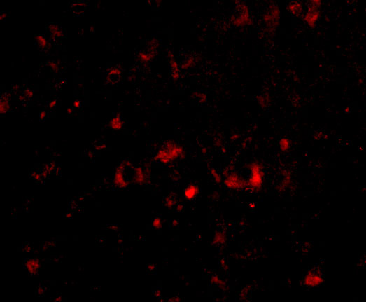


![HPA ICC-IF BJ [Human fibroblast] — NPTX2 (HPA058320)](https://images.proteinatlas.org/58320/1742_F8_18_cr5804950376bc9_blue_red_green.jpg)
![HPA ICC-IF BJ [Human fibroblast] — NPTX2 (HPA058320)](https://images.proteinatlas.org/58320/1742_F8_22_cr5804950d083f7_blue_red_green.jpg)
Comprehensive Human Protein Atlas IHC scoring per tissue (reliability: Enhanced — High consistency between antibody staining and RNA expression data. Secreted protein, tissue location of RNA and protein is expected to differ. External characterization data supports antibody staining.). Rows are taken directly from the HPA tissue chart — click any row's HPA link to view the source.
| Tissue | Cell type | Level | Evidence | Source |
|---|---|---|---|---|
| Pancreas | Pancreatic endocrine cells | High | Protein (IHC) | HPA → |
| Pituitary gland | Cells in anterior | High | Protein (IHC) | HPA → |
| Adrenal gland | Medullary cells | Medium | Protein (IHC) | HPA → |
| Tissue | Cell type | Level | Evidence | Source |
|---|---|---|---|---|
| Adipose tissue | Adipocytes | Not detected | Protein (IHC) | HPA → |
| Appendix | Endocrine cells | Not detected | Protein (IHC) | HPA → |
| Bone marrow | Hematopoietic cells | Not detected | Protein (IHC) | HPA → |
| Breast | Adipocytes | Not detected | Protein (IHC) | HPA → |
| Bronchus | Respiratory epithelial cells | Not detected | Protein (IHC) | HPA → |
For NPTX2 chromogenic IHC in paraffin sections, anchor troubleshooting to the catalog antibody’s pancreas example and interpret staining by cell type and compartment.
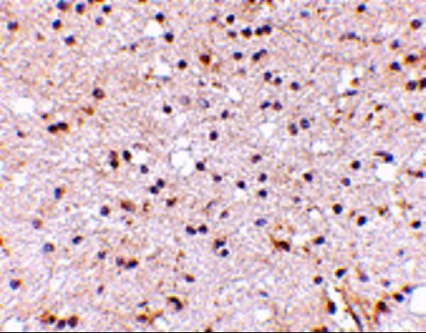
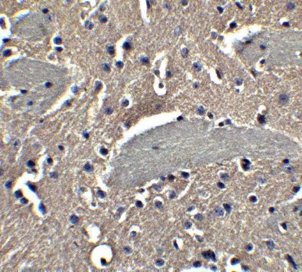
Anti-NPTX2 antibodies have IHC images from human and mouse brain and mouse and rat pancreas; IF images cover human and mouse brain (catalog image captions).
A09667 has paraffin-section IHC images from formaldehyde-fixed human and mouse brain and IF images from paraformaldehyde-fixed human and mouse brain (A09667 image captions). A09667-1 has paraffin-section IHC images from mouse and rat pancreas; its captions do not report a fixative (A09667-1 image captions).
Which to pick: For brain IHC, choose A09667 for human or mouse sections (A09667 IHC captions); for mouse or rat pancreas IHC, choose A09667-1, whose paraffin-section captions leave the fixative unreported (A09667-1 IHC captions). For IF, choose A09667 for human or mouse brain; ICC validation is not reported (A09667 IF captions and application list). For work across species, both list human, mouse and rat reactivity, but the documented IHC examples cover human and mouse for A09667 and mouse and rat for A09667-1; clonality is unreported for both (catalog reactivity, image captions and clone fields).