This website uses cookies to ensure you get the best experience on our website.


- Table of Contents
Plan NREP chromogenic IHC in paraffin sections using cytoplasmic staining as the expected pattern (HPA tissue IHC). Compare high-staining bronchial respiratory epithelium with adipocytes where staining was not detected (HPA tissue IHC), and start the IHC-validated antibody at 1:100–1:300 (datasheet).

Expected localisation, validated protocols, controls and antibodies — the at-a-glance facts below, then the full design guide.
| Expected localisation | Cytoplasmic staining (HPA tissue IHC) | |
| Staining pattern | Cytoplasmic staining in most tissues (HPA tissue IHC) | |
| Antigen retrieval | Citrate pH 6.0 HIER, 95–98 °C, 20 min (rule: cytoplasmic / membrane antigen) | |
| Positive control | Appendix+4 more · see all | |
| Negative control | Adipose tissue+4 more · see all |
| Fixation | Keep fixation conditions consistent. (standard IHC practice; not target-specific) | |
| Caveat | Staining has low consistency with RNA expression (HPA tissue IHC) | |
| Regulation | Staining-linked regulation unreported (UniProt) | |
| Isoform / epitope | 2 isoforms; epitope coverage needs checking (UniProt) |
Compare the catalog antibody’s IHC-P protocol with published NREP staining of tissue sections and cartilage (PMC11066005; PMC10765488).
| Sample | Paraffin-embedded human heart tissue; fixative not specified (datasheet A09054-1) |
| Fixation | Image fixative and duration unreported (datasheet A09054-1); verify before use. |
| Sectioning | 4–5 µm sections on charged slides (standard) |
| Deparaffinisation | Xylene, graded ethanol series to water (standard) |
| Antigen retrieval | Heat-induced epitope retrieval in citrate buffer, pH 6.0, 20 min at 95–98 °C (standard rule: cytoplasmic / membrane antigen) |
| Peroxidase block | 3% H2O2, 10 min, room temperature (standard) |
| Blocking | 10% normal serum of the secondary host, 30 min, room temperature (standard) |
| Primary antibody | Rabbit anti-NREP, 1:100 - 1:300 (datasheet A09054-1) |
| Primary incubation | Overnight at 4 °C (standard) |
| Detection | HRP-polymer secondary, DAB chromogen 5–10 min (standard) |
| Counterstain | Hematoxylin, blue, dehydrate and mount (standard) |
| Expected result | NREP-positive staining in glandular cells of appendix (HPA tissue IHC: High). HPA tissue profile: Cytoplasmic expression in most tissues. No signal in the no-primary control. |
NREP is annotated as cytoplasmic, with no transmembrane segment (UniProt Q16612). In tissue IHC, expect cytoplasmic staining in many cell types, including strongly stained glandular cells in appendix and respiratory epithelial cells in bronchus (HPA tissue IHC). HPA rates the tissue pattern Approved but reports low consistency between antibody staining and RNA expression; treat the pattern as a guide for interpretation, with external verification still pending (HPA tissue IHC).
| Cytoplasmic signal in bronchial respiratory epithelium or appendix glands, with recognizable cell boundaries (HPA tissue IHC). | This fits the reported high staining in those cells and UniProt's cytoplasmic location (HPA tissue IHC; UniProt Q16612). Compare signal with adjacent cells and a no-primary control before scoring; HPA's tissue pattern has low antibody–RNA consistency (standard IHC practice; HPA tissue IHC). |
| Predominantly nuclear or sharply cell-surface staining, with little cytoplasmic signal. | Neither compartment is the expected tissue pattern; NREP has no annotated transmembrane segment (HPA tissue IHC; UniProt Q16612). Treat a compartment-restricted result as possible artefact until controls and an independent stain support it (standard IHC practice). |
| Strong staining in adipocytes, skeletal myocytes, or splenic red-pulp cells (HPA tissue IHC). | Those cells were reported as not detected (HPA tissue IHC). Check for antibody cross-reactivity or endogenous detection activity before calling them positive; the HPA assessment remains pending external verification (standard IHC practice; HPA tissue IHC). |
| Uniform color over tissue and empty spaces, obscuring cell outlines. | This is background rather than a scoreable cytoplasmic pattern (standard IHC practice; HPA tissue IHC). Review blocking, antibody concentration, washing, and chromogen development with appropriate controls (standard IHC practice). |
| No cytoplasmic signal in bronchial epithelium or appendix glands (HPA tissue IHC). | These are reported high-staining cells, so first assess the staining run and tissue preservation (HPA tissue IHC; standard IHC practice). A blank slide alone does not establish NREP absence, especially given HPA's low antibody–RNA consistency (standard IHC practice; HPA tissue IHC). |
| Cell and tissue choice | HPA reports high staining in appendix glands and bronchial epithelium, low staining in colon glands, and no detection in adipocytes (HPA tissue IHC). Choose a reported high-staining tissue for a run control, but do not treat an HPA-negative cell as a universal negative control because antibody–RNA consistency is low (HPA tissue IHC; standard IHC practice). |
| Compartment and protein features | NREP is cytoplasmic, has no transmembrane segment or signal peptide, and is annotated as one chain spanning residues 1–68 (UniProt Q16612). Those annotations support a cytoplasmic reading of tissue IHC; they do not establish a fixation or antigen-retrieval effect (UniProt Q16612). |
| Antibody evidence | HPA000545 is listed as IHC Approved, while the tissue profile has low antibody–RNA consistency and awaits external verification (HPA antibodies; HPA tissue IHC). Record the antibody used and judge unexpected staining with controls or independent evidence rather than treating Approved as definitive specificity proof (standard IHC practice). |
| IF/ICC Q&A: why might staining look punctate? | HPA reports vesicular localization in ICC-IF, while its tissue IHC profile is cytoplasmic (HPA subcellular; HPA tissue IHC). Cytoplasmic puncta can fit that IF observation, but the ICC-IF result does not require puncta in chromogenic paraffin IHC (HPA subcellular; HPA tissue IHC). |
| Isoforms and modification | Target-specific fixation effects are not established by the supplied assay evidence. Verify with a matched IHC source before attributing a result to fixation. |
| Situation | Likely cause | Next action |
|---|---|---|
| No signal in a reported high-staining tissue (HPA tissue IHC). | A staining-run failure or poorly preserved section is possible; a blank result alone does not identify the cause (standard IHC practice). | Check the run control, reagent sequence, detection chemistry, and tissue morphology; review antigen retrieval against the antibody's validated IHC instructions if available (standard IHC practice). |
| Widespread weak brown haze masks cytoplasm. | Concentrated primary antibody, insufficient washing, or extended chromogen development can raise background (standard IHC practice). | Compare a no-primary control, then optimize primary concentration, washes, and development time one variable at a time (standard IHC practice). |
| Color persists without primary antibody. | Endogenous detection activity can contribute; tissue peroxidase is relevant when an HRP chromogen is used (standard IHC practice). | Confirm the detection system and assess its appropriate endogenous-activity block alongside the no-primary control (standard IHC practice). |
| Nuclear-only or surface-only signal dominates. | That distribution conflicts with the reported cytoplasmic tissue pattern and no-transmembrane annotation (HPA tissue IHC; UniProt Q16612). | Review morphology and controls, then seek independent support before scoring the compartment as NREP (standard IHC practice; HPA tissue IHC). |
| Adipocytes or skeletal myocytes stain strongly (HPA tissue IHC). | Those cells were reported as not detected; cross-reactivity or background is possible, though HPA's pattern awaits external verification (HPA tissue IHC; standard IHC practice). | Compare a reported high-staining tissue in the same run and use detection controls; avoid interpreting the unexpected cells from intensity alone (HPA tissue IHC; standard IHC practice). |
| IF/ICC shows puncta while tissue IHC looks broadly cytoplasmic. | HPA reports vesicles in ICC-IF and cytoplasmic expression in tissue IHC; the observations come from different assay contexts (HPA subcellular; HPA tissue IHC). | Score each assay against its reported compartment and controls without imposing an IF punctate pattern on paraffin IHC (HPA subcellular; HPA tissue IHC; standard IHC practice). |
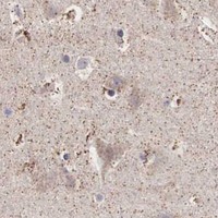
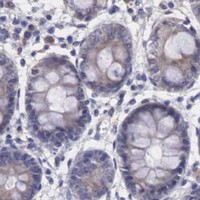
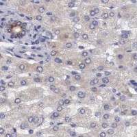
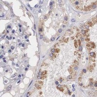


Comprehensive Human Protein Atlas IHC scoring per tissue (reliability: Approved — Low consistency between antibody staining and RNA expression data. Pending external verification.). Rows are taken directly from the HPA tissue chart — click any row's HPA link to view the source.
| Tissue | Cell type | Level | Evidence | Source |
|---|---|---|---|---|
| Appendix | Glandular cells | High | Protein (IHC) | HPA → |
| Breast | Glandular cells | High | Protein (IHC) | HPA → |
| Bronchus | Respiratory epithelial cells | High | Protein (IHC) | HPA → |
| Cerebellum | Cells in granular layer | High | Protein (IHC) | HPA → |
| Epididymis | Glandular cells | High | Protein (IHC) | HPA → |
| Tissue | Cell type | Level | Evidence | Source |
|---|---|---|---|---|
| Adipose tissue | Adipocytes | Not detected | Protein (IHC) | HPA → |
| Ovary | Ovarian stroma cells | Not detected | Protein (IHC) | HPA → |
| Skeletal muscle | Myocytes | Not detected | Protein (IHC) | HPA → |
| Smooth muscle | Smooth muscle cells | Not detected | Protein (IHC) | HPA → |
| Spleen | Cells in red pulp | Not detected | Protein (IHC) | HPA → |
Troubleshoot NREP staining in paraffin sections by checking retrieval, cell type, compartment, controls and scoring before interpreting chromogenic signal.
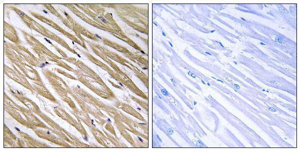
The anti-NREP catalog antibody A09054-1 has an IHC image from paraffin-embedded human heart tissue (catalog IHC caption); IF/ICC and human, mouse, and rat reactivity are listed (catalog applications/reactivity).
A09054-1 shows IHC staining of paraffin-embedded human heart tissue alongside a peptide-blocked comparison (catalog IHC caption). A09054-1 also lists IF/ICC applications and human, mouse, and rat reactivity; no IF image is supplied (catalog applications/reactivity; catalog IF image alts).
Which to pick: For tissue IHC, choose A09054-1 at the listed 1:100–1:300 dilution; its own image documents paraffin-embedded human heart tissue, but the fixative is unreported (datasheet: 1:100–1:300; catalog IHC caption). For IF/ICC, A09054-1 is the listed option, with an IF dilution of 1:50 and no supplied IF image (catalog applications; catalog IF dilution; catalog IF image alts). For cross-species work, A09054-1 is a rabbit polyclonal antibody listed as reactive with human, mouse, and rat; its IHC image documents human tissue only (catalog host/clonality/reactivity; catalog IHC caption).