This website uses cookies to ensure you get the best experience on our website.


- Table of Contents
Plan chromogenic IHC on paraffin sections with the catalog antibody at 1:100–1:300 (datasheet A17131). Use Leydig cells as a positive reference and assess follicle and adrenal cortical cell staining (HPA tissue IHC).

Expected localisation, validated protocols, controls and antibodies — the at-a-glance facts below, then the full design guide.
| Expected localisation | Tissue subcellular pattern not established (HPA tissue IHC) | |
| Staining pattern | High in Leydig cells; medium in follicle and zona fasciculata cells (HPA tissue IHC) | |
| Antigen retrieval | Tris-EDTA pH 9.0 HIER, heat-mediated (datasheet A17131) | |
| Positive control | Adrenal gland+2 more · see all | |
| Negative control | Adipose tissue+4 more · see all |
| Fixation | Keep fixation consistent across sections (standard IHC practice; not target-specific). Selected-image fixative and duration unreported (datasheet A17131); verify before use. | |
| Caveat | Catalog text mentions BIVM; verify NRIP3 specificity (datasheet A17131) | |
| Regulation | No expression regulator reported (UniProt) | |
| Isoform / epitope | No isoforms or cleavage; no TM-defined epitope side (UniProt) |
The catalog antibody’s IHC-P protocol (datasheet: A17131) is accompanied by one published protocol for human ESCC tissue (PMC7445249).
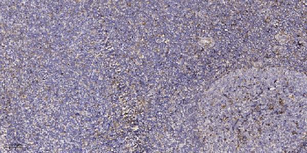
| Sample | Paraffin-embedded human tonsil tissue; fixative not specified (datasheet A17131) |
| Fixation | Image fixative and duration unreported (datasheet A17131); verify before use. |
| Sectioning | 4–5 µm sections on charged slides (standard) |
| Deparaffinisation | Xylene, graded ethanol series to water (standard) |
| Antigen retrieval | Heat retrieval: Tris-EDTA pH 9.0 (datasheet A17131); 20 min, 95–100 °C (standard) |
| Peroxidase block | 3% H2O2, 10 min, room temperature (standard) |
| Blocking | 10% normal serum of the secondary host, 30 min, room temperature (standard) |
| Primary antibody | Rabbit anti-NRIP3, 1:100-1:300 (datasheet A17131) |
| Primary incubation | Overnight at 4 °C (standard) |
| Detection | HRP-polymer secondary, DAB chromogen 5–10 min (standard) |
| Counterstain | Hematoxylin, blue, dehydrate and mount (standard) |
| Expected result | NRIP3-positive staining in cells in zona fasciculata of adrenal gland (HPA tissue IHC: Medium). HPA tissue profile: Expression in Leydig cells, ovarian follicles and adrenal cortical cells. No signal in the no-primary control. |
NRIP3 is reported in the cytosol by ICC-IF (HPA: approved cytosol); UniProt does not annotate its subcellular location and reports no transmembrane segment (UniProt Q9NQ35 topology). In tissue IHC, expect staining in Leydig cells, ovarian follicle cells and adrenal zona fasciculata cells (HPA: tissue IHC). The tissue profile is Approved, pending external verification (HPA: reliability).
| Leydig cells stain strongly; follicle and zona fasciculata cells stain moderately. | This matches the reported tissue distribution (HPA: High in Leydig cells; Medium in follicle and zona fasciculata cells). Judge the named cell populations within each section; a whole-section average can obscure their staining (standard IHC practice). HPA labels the tissue profile Approved but pending external verification (HPA: reliability). |
| Signal is mainly nuclear, membranous or extracellular in an otherwise plausible positive tissue. | These compartments differ from the approved cytosolic ICC-IF localization (HPA: subcellular). Treat the pattern as suspect and check morphology, counterstain and detection controls (standard IHC practice). The ICC-IF localization is a useful comparison, but it does not establish compartment-level validation for tissue IHC; UniProt lists no location (UniProt Q9NQ35). |
| Strong staining appears in adipocytes or appendix glandular cells while expected positive cells also stain. | HPA reports NRIP3 as Not detected in those cell populations (HPA: tissue IHC). Consider antibody cross-reactivity or endogenous detection activity, then compare with a reagent-omission control (standard IHC practice). A Not detected entry describes the reported HPA observation; it is not proof that every specimen must be completely blank. |
| Brown signal spreads through stroma or across many unrelated cell types without clear cell boundaries. | This is less convincing than cell-associated staining in the reported positive populations (HPA: tissue IHC). Diffuse staining may reflect nonspecific reagent binding, residual detection activity or section artefact (standard IHC practice). Assess an adjacent control section before assigning the signal to NRIP3. |
| No signal appears in a well-preserved testis section, including identifiable Leydig cells. | That conflicts with HPA's High Leydig-cell observation (HPA: tissue IHC). First confirm that the relevant cells are present, then review the catalog antibody's IHC-P instructions and the run's positive and negative controls (standard IHC practice). One failed section cannot establish that NRIP3 is absent. |
| Tissue and cell selection | Testis Leydig cells provide the strongest reported tissue IHC signal; ovarian follicle and adrenal zona fasciculata cells provide Medium examples (HPA: tissue IHC). Choose a section with identifiable target cells before treating a blank field as a negative result (standard IHC practice). |
| Assay-specific antibody validation | HPA072961 is Approved for IHC, while HPA058827 is Approved for ICC and has no listed IHC status (HPA: antibodies). Approval in one assay does not transfer to the other. The tissue IHC profile remains pending external verification (HPA: reliability). |
| Compartment evidence | Cytosol is the approved ICC-IF location, with images listed for SiHa and U2OS (HPA: subcellular). UniProt does not annotate localization (UniProt Q9NQ35). Use cytosolic distribution as a consistency check in IHC, with that assay distinction visible in the interpretation. |
| Topology and processing | UniProt reports no transmembrane segment, signal peptide, propeptide, annotated glycosylation sites or isoforms; it lists a single 1–241 chain (UniProt Q9NQ35). These annotations offer no basis to predict a membrane pattern, shedding-dependent staining or isoform-specific differences. They also provide no target-specific antigen-retrieval or fixation guidance. |
| Situation | Likely cause | Next action |
|---|---|---|
| Testis control is blank in the Leydig-cell region. | The section may lack identifiable Leydig cells, or the IHC run may have failed (HPA: High in Leydig cells; standard IHC practice). | Confirm cell identity and section quality, then check the catalog antibody's IHC-P instructions, detection reagents and run controls (standard IHC practice). Do not infer NRIP3 absence from this run. |
| Follicle or adrenal staining looks weaker than the testis control. | HPA reports Medium staining in follicle and zona fasciculata cells, versus High in Leydig cells (HPA: tissue IHC). | Score each named cell population against its own reported level and section morphology (HPA: tissue IHC; standard IHC practice). Check run controls before treating a weaker positive as a failure. |
| Signal is dominant in nuclei or along membranes. | That distribution conflicts with approved cytosolic ICC-IF localization (HPA: subcellular); its cause is unresolved from the supplied evidence. | Review cell boundaries and counterstain, then compare reagent-omission and positive controls (standard IHC practice). Report that the compartment reference comes from ICC-IF, not tissue IHC (HPA: subcellular). |
| Adipocytes or appendix glandular cells stain strongly. | Both are reported Not detected by HPA tissue IHC; cross-reactivity or endogenous detection activity is possible (HPA: tissue IHC; standard IHC practice). | Inspect a reagent-omission control and assess whether the signal follows tissue structures or pools outside cells (standard IHC practice). Interpret these populations relative to the reported HPA observations. |
| Diffuse brown background obscures positive cells. | Nonspecific binding or residual endogenous detection activity can obscure chromogenic IHC (standard IHC practice). | Compare control sections, review blocking and detection steps, and assess whether clear cell-associated signal remains in a reported positive population (standard IHC practice; HPA: tissue IHC). |
| IF/ICC question: should a cytosolic signal match this IHC result? | HPA approves cytosolic localization in ICC-IF, but its listed ICC-approved and IHC-approved antibodies are different (HPA: subcellular; HPA: antibodies). | A cytosolic IF/ICC signal is consistent with HPA's localization; assess it under the separate IF/ICC guide and its own antibody validation (HPA: subcellular; HPA: antibodies). Do not transfer the tissue IHC intensity ratings directly to cultured cells. |


Comprehensive Human Protein Atlas IHC scoring per tissue (reliability: Approved — Pending external verification.). Rows are taken directly from the HPA tissue chart — click any row's HPA link to view the source.
| Tissue | Cell type | Level | Evidence | Source |
|---|---|---|---|---|
| Adrenal gland | Cells in zona fasciculata | Medium | Protein (IHC) | HPA → |
| Testis | Leydig cells | High | Protein (IHC) | HPA → |
| Ovary | Follicle cells | Medium | Protein (IHC) | HPA → |
| Tissue | Cell type | Level | Evidence | Source |
|---|---|---|---|---|
| Adipose tissue | Adipocytes | Not detected | Protein (IHC) | HPA → |
| Appendix | Glandular cells | Not detected | Protein (IHC) | HPA → |
| Bone marrow | Hematopoietic cells | Not detected | Protein (IHC) | HPA → |
| Breast | Adipocytes | Not detected | Protein (IHC) | HPA → |
| Bronchus | Respiratory epithelial cells | Not detected | Protein (IHC) | HPA → |
Troubleshoot NRIP3 staining in paraffin sections by checking retrieval, cell identity and cytosolic localisation against the available tissue evidence.
A17131 is listed for human and mouse IHC and IF (catalog: applications and reactivity), with a real IHC image from a paraffin-embedded human tonsil section (IHC image caption).
A17131 will render with an IHC image of paraffin-embedded human tonsil (IHC image caption). IF is a listed application, but no IF image is supplied (catalog: applications and IF image alts).
Which to pick: For tissue IHC, choose A17131, a rabbit polyclonal antibody with a paraffin-section IHC example (catalog: host; IHC image caption); the fixative is unreported (IHC image caption). For IF/ICC planning, A17131 lists IF but supplies no IF image or ICC validation (catalog: applications and IF image alts). For cross-species work, A17131 lists human and mouse reactivity, while its IHC image documents human tissue only (catalog: reactivity; IHC image caption).