This website uses cookies to ensure you get the best experience on our website.


- Table of Contents
Plan paraffin-section NSUN2 IHC around mainly nuclear tissue staining (HPA tissue IHC). Start with the IHC-validated antibody at 1–2 μg/ml (datasheet A03878-1), using high-staining colon glandular cells and adipocytes with no detected staining as comparison controls (HPA tissue IHC).

Expected localisation, validated protocols, controls and antibodies — the at-a-glance facts below, then the full design guide.
| Expected localisation | Mainly nuclear tissue staining (HPA tissue IHC) | |
| Staining pattern | Many cell types show mainly nuclear staining (HPA tissue IHC) | |
| Antigen retrieval | EDTA pH 8.0 HIER, heat-mediated (datasheet A03878-1) | |
| Positive control | Appendix+4 more · see all | |
| Negative control | Adipose tissue+2 more · see all |
| Fixation | Matched tissue-IHC evidence does not establish the fixation claim. Validate the specimen-specific method before use. (selected-SKU IHC image A03878-1) | |
| Caveat | Mitotic cells may show spindle localization (UniProt) | |
| Regulation | Staining intensity varies by cell type (HPA tissue IHC) | |
| Isoform / epitope | 3 isoforms; epitope differences unspecified (UniProt) |
The catalog antibody protocol uses EDTA pH 8.0 heat retrieval (datasheet A03878-1). The four published NSUN2 IHC protocols below provide sample specific conditions (PMC9807547; PMC11057147; PMC8429414; PMC12997395).
| Sample | Paraffin-embedded human acinic cell carcinoma tissue; fixative not specified (datasheet A03878-1) |
| Fixation | Image fixative and duration unreported (datasheet A03878-1); verify before use. |
| Sectioning | 4–5 µm sections on charged slides (standard) |
| Deparaffinisation | Xylene, graded ethanol series to water (standard) |
| Antigen retrieval | Heat retrieval: EDTA pH 8.0 (datasheet A03878-1); 20 min, 95–100 °C (standard) |
| Peroxidase block | 3% H2O2, 10 min, room temperature (standard) |
| Blocking | 10% goat serum (datasheet A03878-1) |
| Primary antibody | Rabbit anti-NSUN2, 1-2 μg/ml (datasheet A03878-1) |
| Primary incubation | Overnight at 4 °C (datasheet A03878-1) |
| Detection | HRP-conjugated secondary, DAB chromogen (datasheet A03878-1) |
| Counterstain | Hematoxylin, blue, dehydrate and mount (standard) |
| Expected result | NSUN2-positive staining in glandular cells of appendix (HPA tissue IHC: High). HPA tissue profile: Ubiquitous mainly nuclear expression. No signal in the no-primary control. |
NSUN2 is mainly nuclear in tissue IHC, with nucleolar enrichment during interphase and possible cytoplasmic or spindle localization (HPA: ubiquitous mainly nuclear expression; UniProt Q08J23: subcellular location). Expect staining in epithelial and other cell types reported by HPA, especially glandular cells in appendix, colon, and duodenum (HPA: High). Interpret compartment and cell type together: NSUN2 has no transmembrane segment, and the tissue IHC profile has Supported reliability with medium consistency against RNA expression (UniProt Q08J23: topology; HPA: tissue IHC reliability).
| Clear nuclear staining in colon or duodenum glandular cells, with little diffuse background. | This fits a positive section: HPA rates these cells High and describes NSUN2 tissue staining as mainly nuclear (HPA: tissue IHC). Nucleolar concentration can be plausible in interphase cells (UniProt Q08J23: subcellular location). Judge staining within the relevant cells, because an apparently strong field may reflect the number of positive cells as well as their staining intensity (general IHC practice). |
| Predominantly membrane or extracellular staining, with nuclei largely unstained. | Treat this as discordant with the expected tissue pattern (HPA: ubiquitous mainly nuclear expression). UniProt lists no transmembrane segment, although it also lists extracellular exosomes (UniProt Q08J23: topology and subcellular location). Review the negative control, tissue morphology, and detection background before assigning the signal to NSUN2 (general IHC practice). Cytoplasmic or spindle signal alone needs context because both locations are annotated (UniProt Q08J23: subcellular location). |
| Strong signal in adipocytes or ovarian stroma cells while an expected positive cell type stains weakly. | That cell type contrast conflicts with HPA's Not detected calls for adipocytes and ovarian stroma cells and its High calls for several glandular cell populations (HPA: tissue IHC). Consider antibody cross-reactivity or endogenous detection activity; inspect a no-primary control and compare staining with cell morphology (general IHC practice). A Not detected HPA call is a reference pattern, not proof that every specimen must be blank (HPA: tissue IHC, Supported reliability). |
| Uniform haze across cells, stroma, and the slide background obscures nuclear detail. | The haze cannot establish NSUN2 localization because the expected tissue pattern is mainly nuclear (HPA: tissue IHC). Check the no-primary control and the distribution of chromogen; nonspecific binding, endogenous detection activity, or excessive detection signal can produce background in chromogenic IHC (general IHC practice). Score only cellular staining that can be separated from that background (general IHC practice). |
| No staining in a well-preserved colon glandular cell section. | This is unexpected for a HPA High reference cell type (HPA: colon glandular cells, High), but one negative section does not establish absent NSUN2. Confirm that the intended glands are present and that the run's positive control developed; then review antigen retrieval, primary antibody conditions, and detection reagents as general IHC checks (general IHC practice). Do not infer a target-specific fixation effect from this result (HPA: tissue IHC supplies no fixation-effect assessment). |
| Cell compartment | HPA's tissue summary is mainly nuclear, while its ICC-IF summary calls nucleoplasm uncertain; UniProt additionally annotates nucleolus, cytoplasm, mitochondrion, and spindle (HPA: tissue IHC and subcellular; UniProt Q08J23: subcellular location). Use the nuclear tissue pattern as the main IHC expectation without declaring every nonnuclear signal false. |
| Reference cell type | High calls include kidney tubule cells, hepatocytes, esophageal squamous epithelial cells, and several glandular populations; adipocytes, ovarian stroma cells, and parathyroid glandular cells are Not detected (HPA: tissue IHC). Compare like cell types when choosing a positive control or assessing unexpected staining; HPA also describes low tissue specificity at the RNA level (HPA: tissue IHC). |
| Cell-cycle context | UniProt reports nucleolar concentration in interphase and movement to the spindle during mitosis (UniProt Q08J23: subcellular location). A mitotic cell can therefore differ from neighboring interphase cells. Examine morphology before calling a localized spindle-associated signal an artifact; the dominant tissue IHC expectation remains mainly nuclear (HPA: tissue IHC). |
| Antibody evidence | The listed antibody HPA037896 is Supported for IHC and Uncertain for ICC (HPA: antibody validation). Supported tissue IHC also means medium consistency between antibody staining and RNA expression, so use the HPA pattern as a reference with that limitation (HPA: tissue IHC reliability). These ratings do not establish a target-specific retrieval condition or fixation response. |
| Isoforms and processing | UniProt lists three isoforms, no signal peptide or propeptide, and a chain spanning residues 1–767 (UniProt Q08J23: isoforms and processing). The supplied record gives no epitope position, so it cannot predict which isoforms the IHC antibody recognizes or justify a compartment change from processing (UniProt Q08J23: record scope). |
| IF/ICC Q&A | Can the IHC result define the IF/ICC pattern? Use caution: HPA lists nucleoplasm as uncertain in ICC-IF and rates HPA037896 Uncertain for ICC, whereas tissue IHC is Supported (HPA: subcellular and antibody validation). Interpret IF/ICC with its own controls and guide; this IHC section supplies no IF/ICC protocol. |
| Situation | Likely cause | Next action |
|---|---|---|
| Expected nuclear signal is absent in colon glandular cells. | The section may lack evaluable glands, or a general IHC workflow step may have failed; HPA calls these cells High but gives no NSUN2-specific fixation sensitivity (HPA: colon tissue IHC; general IHC practice). | Verify cell identity and the run's positive control, then check retrieval, primary incubation, and detection according to the IHC antibody's validated instructions (general IHC practice). Record the negative result if controls pass. |
| Staining is strongest at membranes or in extracellular deposits. | The pattern differs from HPA's mainly nuclear tissue profile; non-target staining or detection background is possible (HPA: tissue IHC; general IHC practice). UniProt's exosome annotation alone cannot validate a deposit in this section (UniProt Q08J23: subcellular location). | Compare nuclear signal and a no-primary control, then check whether the deposits follow tissue edges or damaged areas (general IHC practice). Reassess with another appropriate control before scoring them as NSUN2. |
| Adipocytes or ovarian stroma cells show strong chromogen. | These cells are Not detected in the HPA reference; cross-reactivity or endogenous detection activity could explain the signal (HPA: tissue IHC; general IHC practice). | Identify the stained cells on the counterstain, inspect a no-primary control, and compare an HPA High cell type from the same run (HPA: tissue IHC; general IHC practice). Report the discordance if controls do not resolve it. |
| Diffuse brown background hides nuclei throughout the section. | This does not match the mainly nuclear reference pattern (HPA: tissue IHC). General IHC causes include nonspecific binding or endogenous detection activity (general IHC practice). | Check a no-primary control, blocking and detection steps, and whether the chromogen reaction was overdeveloped; adjust the general workflow only as the controls indicate (general IHC practice). |
| All cells appear equally dark, including HPA Not detected cell types. | A saturated detection signal can obscure real cell type differences (general IHC practice); HPA reports both High and Not detected reference populations (HPA: tissue IHC). | Compare controls and review antibody dilution and chromogen development under the validated IHC instructions (general IHC practice). Score only where nuclei, cell boundaries, and relative intensity remain readable. |
| IF/ICC looks cytoplasmic while tissue IHC looks nuclear. | The assays have different evidence strength: tissue IHC is Supported, while HPA's nucleoplasm call and the listed antibody's ICC validation are Uncertain (HPA: tissue IHC, subcellular, and antibody validation). UniProt also annotates cytoplasmic locations (UniProt Q08J23: subcellular location). | Review IF/ICC controls and cell-cycle morphology in its separate guide; do not transfer an IHC interpretation or protocol to IF/ICC solely from this comparison (HPA: antibody validation; general assay practice). |
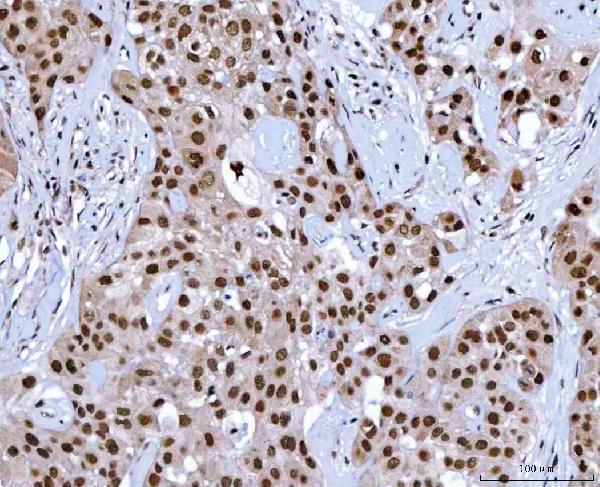
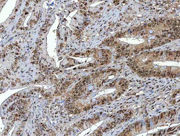
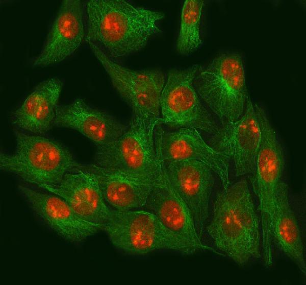


Comprehensive Human Protein Atlas IHC scoring per tissue (reliability: Supported — Medium consistency between antibody staining and RNA expression data.). Rows are taken directly from the HPA tissue chart — click any row's HPA link to view the source.
| Tissue | Cell type | Level | Evidence | Source |
|---|---|---|---|---|
| Appendix | Glandular cells | High | Protein (IHC) | HPA → |
| Colon | Glandular cells | High | Protein (IHC) | HPA → |
| Duodenum | Glandular cells | High | Protein (IHC) | HPA → |
| Epididymis | Glandular cells | High | Protein (IHC) | HPA → |
| Esophagus | Squamous epithelial cells | High | Protein (IHC) | HPA → |
Troubleshoot NSUN2 staining in paraffin sections by checking retrieval, compartment, controls and cell specific scoring before interpreting signal intensity.
The catalog lists anti-NSUN2 antibodies for IHC and IF/ICC with human and mouse reactivity; A03878-1 also has paraffin-section IHC and cell IF images (catalog applications and reactivity; image captions).
A03878-1 has IHC images from human paraffin sections of acinic cell carcinoma, colorectal adenocarcinoma, liver cancer and lung cancer, plus IF in NRK cells (A03878-1 image captions). M03878 lists IHC and IF/ICC applications with human, mouse and monkey reactivity; no IHC or IF image is supplied (M03878 catalog).
Which to pick: Choose A03878-1 for tissue IHC: its human paraffin-section captions document EDTA retrieval at pH 8.0 and 2 μg/ml primary antibody, but do not report the fixative (A03878-1 IHC captions). For IF/ICC, A03878-1 has an NRK-cell IF image at 5 μg/ml; M03878 is a rabbit monoclonal with IF/ICC listed at 1:50, without an image in this payload (A03878-1 IF caption; M03878 catalog). For species selection, both list mouse reactivity, A03878-1 lists rat, and M03878 lists monkey; verify IHC performance in those species because the supplied IHC images are human only (catalog reactivity; A03878-1 IHC captions).