This website uses cookies to ensure you get the best experience on our website.


- Table of Contents
Plan chromogenic paraffin IHC for NTN4 with the catalog antibody at 2–5 μg/ml (datasheet A07052-1). Use testis Leydig cells as a positive tissue control and adipose tissue adipocytes as a negative cell control (HPA tissue IHC).

Expected localisation, validated protocols, controls and antibodies — the at-a-glance facts below, then the full design guide.
| Expected localisation | Membranous and cytoplasmic staining (HPA tissue IHC) | |
| Staining pattern | Membranous and cytoplasmic staining in several tissues (HPA tissue IHC) | |
| Antigen retrieval | EDTA pH 8.0 HIER, heat-mediated (datasheet A07052-1) | |
| Positive control | Testis+4 more · see all | |
| Negative control | Adipose tissue+4 more · see all |
| Fixation | Keep fixation consistent across sections (standard IHC practice; not target-specific) | |
| Caveat | Secreted protein may stain away from its producer cells (HPA tissue IHC) | |
| Regulation | Tissue-dependent expression (HPA tissue IHC) | |
| Isoform / epitope | 3 isoforms; signal peptide 1–18 is cleaved, so check epitope coverage (UniProt) |
Compare the catalog antibody’s IHC-P protocol (datasheet A07052-1) with three published NTN4 paraffin-section protocols (PMC6527380; PMC3783308; PMC8784893).
| Sample | Paraffin-embedded human endometrial cancer tissue; fixative not specified (datasheet A07052-1) |
| Fixation | Image fixative and duration unreported (datasheet A07052-1); verify before use. |
| Sectioning | 4–5 µm sections on charged slides (standard) |
| Deparaffinisation | Xylene, graded ethanol series to water (standard) |
| Antigen retrieval | Heat retrieval: EDTA pH 8.0 (datasheet A07052-1); 20 min, 95–100 °C (standard) |
| Peroxidase block | 3% H2O2, 10 min, room temperature (standard) |
| Blocking | 10% goat serum (datasheet A07052-1) |
| Primary antibody | Rabbit anti-NTN4, 2-5 μg/ml (datasheet A07052-1) |
| Primary incubation | Overnight at 4 °C (datasheet A07052-1) |
| Detection | HRP-conjugated secondary, DAB chromogen (datasheet A07052-1) |
| Counterstain | Hematoxylin, blue, dehydrate and mount (standard) |
| Expected result | NTN4-positive staining in leydig cells of testis (HPA tissue IHC: High). HPA tissue profile: Membranous and cytoplasmic expression in several tissues. No signal in the no-primary control. |
NTN4 is secreted and is a major basement membrane component, with no transmembrane segment (UniProt Q9HB63). In IHC, expect extracellular or basement membrane signal alongside the membranous and cytoplasmic patterns HPA reports in several tissues (HPA tissue IHC). Leydig cells stain strongly; selected glandular, endothelial and cerebellar cells stain at medium levels (HPA tissue IHC). HPA rates the tissue staining Approved but reports low consistency with RNA expression (HPA tissue IHC).
| Strong Leydig cell staining, with extracellular or basement membrane signal where structures are discernible. | This fits the high Leydig cell signal observed in testis (HPA tissue IHC) and NTN4’s secreted, basement membrane location (UniProt Q9HB63). Score cellular and extracellular patterns separately; a secreted protein need not remain confined to the producing cell (UniProt Q9HB63). |
| Membranous or cytoplasmic signal in glandular cells, cortical endothelial cells, or cerebellar granular layer cells. | These patterns can be compatible with the HPA tissue profile: several glandular populations, cerebral cortex endothelial cells and cerebellar granular layer cells show medium staining (HPA tissue IHC). Check the named cell population and its expected level before calling a section positive. |
| Dominant nuclear staining, without the expected cellular or extracellular pattern. | Nuclear localisation is unsupported by the secreted, extracellular and basement membrane record (UniProt Q9HB63) and by HPA’s membranous and cytoplasmic tissue profile (HPA tissue IHC). Treat a nuclear-only result as suspect and investigate staining artefact before assigning it to NTN4 (general IHC practice). |
| Strong signal in a cell population HPA lists as not detected, such as adipocytes or lymph node germinal center cells. | This disagrees with those cell-specific observations (HPA tissue IHC). Consider antibody cross-reactivity or endogenous chromogenic detection activity (general IHC practice); a negative call for one cell population does not establish that every structure in its tissue must be unstained. |
| Broad, poorly bounded stain across tissue or no signal in testis Leydig cells. | Diffuse signal is hard to assign to cells or basement membranes and may reflect background (general IHC practice). Absent Leydig cell signal conflicts with the high testis observation (HPA tissue IHC); inspect section quality and assay controls before interpreting another tissue as NTN4 negative. |
| Secreted topology and tissue localisation | NTN4 lacks a transmembrane segment and is a major basement membrane component (UniProt Q9HB63). Assess extracellular deposits alongside the membranous and cytoplasmic staining seen by HPA; cell staining and deposited protein may occupy different places (HPA tissue IHC; UniProt Q9HB63). |
| Processing and epitope coverage | The precursor has a 1–18 signal peptide and a 19–628 NTN4 chain (UniProt Q9HB63). If an antibody’s epitope is known, check whether it lies in the mature chain; the supplied record gives no epitope for the IHC antibody, so its coverage cannot be inferred. |
| Molecular forms | UniProt lists 3 isoforms and 4 glycosylation sites (UniProt Q9HB63). These features justify checking documented antibody epitope and isoform coverage when staining differs across samples. The supplied sources do not show which forms the IHC antibody detects or establish an IHC effect of glycosylation. |
| Strength and limits of tissue evidence | HPA rates the tissue IHC result Approved and lists rabbit pAb HPA049832 as IHC Approved (HPA tissue IHC; HPA antibodies). HPA also reports low staining–RNA consistency and notes that secretion can separate RNA and protein locations (HPA tissue IHC). RNA abundance alone should therefore not determine a cell-level IHC call. |
| Situation | Likely cause | Next action |
|---|---|---|
| No Leydig cell signal in a testis section. | Leydig cells are a high-staining population in HPA tissue IHC; a blank result may reflect an assay or section problem (HPA tissue IHC; general IHC practice). | Check tissue integrity, primary antibody and chromogenic detection controls, then review the chosen retrieval and dilution settings as general IHC variables (general IHC practice). No NTN4-specific retrieval setting or fixation sensitivity is supplied. |
| Brown signal covers much of the section without clear cell or basement membrane boundaries. | Diffuse chromogen can arise from nonspecific binding or endogenous detection activity (general IHC practice); it cannot by itself confirm NTN4’s extracellular location (UniProt Q9HB63). | Review the no-primary control and detection blocking, then optimise antibody concentration against a testis positive control and a suitable cell-specific negative comparator (general IHC practice; HPA tissue IHC). |
| Nuclei dominate the stain. | A nuclear-only pattern conflicts with NTN4’s secreted, extracellular localisation (UniProt Q9HB63) and HPA’s membranous and cytoplasmic profile (HPA tissue IHC). | Check the no-primary control, counterstain appearance and antibody specificity before scoring the nuclear signal as NTN4 (general IHC practice). Compare with expected Leydig cell and other cell-level patterns (HPA tissue IHC). |
| Adipocytes or lymph node germinal center cells appear strongly positive. | HPA lists those specific populations as not detected (HPA tissue IHC). Unexpected staining warrants checks for cross-reactivity or endogenous detection activity (general IHC practice). | Confirm the identity of stained cells on the counterstained section and review detection controls (general IHC practice). Compare cell types within the section; do not turn an HPA cell-specific negative observation into a whole-tissue rule. |
| IHC staining and tissue RNA levels disagree. | HPA reports low consistency between antibody staining and RNA expression and notes that secretion can separate protein from its RNA source (HPA tissue IHC). | Interpret localisation at the stained cell and extracellular structure level, alongside IHC controls (general IHC practice; UniProt Q9HB63). Record the disagreement without using RNA level alone to invalidate a controlled stain (HPA tissue IHC). |
| Can IF/ICC confirm the IHC localisation? | HPA calls NTN4 secreted but provides no ICC-IF images or main intracellular location; HPA049832 has IHC Approved status with no ICC status supplied (HPA subcellular; HPA antibodies). | Treat IF/ICC as a separate validation question. An IHC-positive result does not establish an IF/ICC pattern or antibody performance (HPA antibodies; HPA subcellular); use the separate IF/ICC guide for that application. |
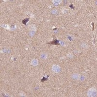


Comprehensive Human Protein Atlas IHC scoring per tissue (reliability: Approved — Low consistency between antibody staining and RNA expression data. At least one protein variant secreted, tissue location of RNA and protein might differ and correlation is complex.). Rows are taken directly from the HPA tissue chart — click any row's HPA link to view the source.
| Tissue | Cell type | Level | Evidence | Source |
|---|---|---|---|---|
| Testis | Leydig cells | High | Protein (IHC) | HPA → |
| Adrenal gland | Glandular cells | Medium | Protein (IHC) | HPA → |
| Appendix | Glandular cells | Medium | Protein (IHC) | HPA → |
| Cerebellum | Cells in granular layer | Medium | Protein (IHC) | HPA → |
| Cerebral cortex | Endothelial cells | Medium | Protein (IHC) | HPA → |
| Tissue | Cell type | Level | Evidence | Source |
|---|---|---|---|---|
| Adipose tissue | Adipocytes | Not detected | Protein (IHC) | HPA → |
| Bone marrow | Hematopoietic cells | Not detected | Protein (IHC) | HPA → |
| Breast | Adipocytes | Not detected | Protein (IHC) | HPA → |
| Caudate | Glial cells | Not detected | Protein (IHC) | HPA → |
| Esophagus | Squamous epithelial cells | Not detected | Protein (IHC) | HPA → |
Troubleshoot paraffin section chromogenic IHC for NTN4 by checking retrieval, tissue pattern, controls, and scoring against its secreted basement membrane biology (UniProt Q9HB63).
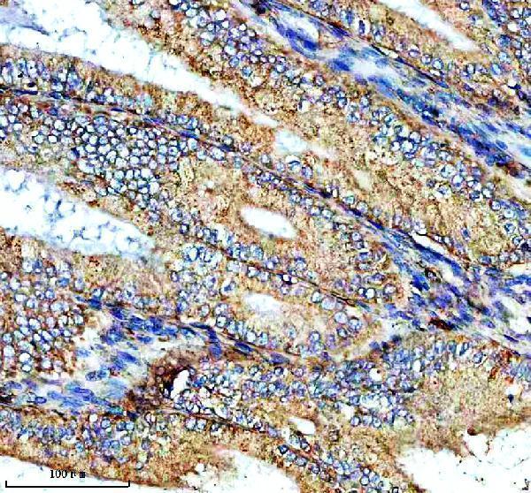
One human-reactive anti-NTN4 antibody has a real IHC image from a paraffin-embedded human endometrial cancer section (A07052-1 image caption); no IF image is supplied (catalog image list).
A07052-1 is listed for human IHC (catalog applications and reactivity). Its IHC image shows a paraffin-embedded human endometrial cancer section with chromogenic detection (A07052-1 image caption).
Which to pick: Choose A07052-1 for human paraffin-section IHC; its image caption documents EDTA retrieval at pH 8.0 and primary antibody at 2 μg/ml (A07052-1 image caption). The fixative is unreported (A07052-1 image caption). No SKU in this payload is listed for IF/ICC or for species beyond human (catalog applications and reactivity).