This website uses cookies to ensure you get the best experience on our website.


- Table of Contents
Plan NTRK3 IHC in paraffin sections using strongly stained cortical neurons as a reference (HPA tissue IHC). Assess the observed nuclear and cytoplasmic pattern alongside the receptor’s membrane topology, and check epitope coverage across its 5 isoforms (HPA tissue IHC; UniProt).

Expected localisation, validated protocols, controls and antibodies — the at-a-glance facts below, then the full design guide.
| Expected localisation | Nuclear and cytoplasmic in cerebral cortex (HPA tissue IHC) | |
| Staining pattern | Cortical neurons show strong nuclear and cytoplasmic staining (HPA tissue IHC) | |
| Antigen retrieval | Citrate pH 6.0 HIER, 95–98 °C, 20 min (rule: cytoplasmic / membrane antigen) | |
| Positive control | Cerebral cortex+4 more · see all | |
| Negative control | Adipose tissue+4 more · see all |
| Fixation | Keep fixation consistent across sections (standard IHC practice; not target-specific) | |
| Caveat | Nuclear staining differs from membrane receptor topology (HPA tissue IHC; UniProt) | |
| Regulation | Isoform 2 is higher in adult than fetal brain (UniProt) | |
| Isoform / epitope | 5 isoforms; check extracellular vs cytoplasmic epitope coverage (UniProt) |
Compare the catalog antibody’s IHC-P protocol with a published TrkC IHC protocol for medullary thyroid cancer sections (PMC5299608).
| Sample | Tissue sections; selected-image fixative not specified (standard IHC workflow) |
| Fixation | Image fixative and duration unreported (datasheet PA1992-1); verify before use. |
| Sectioning | 4–5 µm sections on charged slides (standard) |
| Deparaffinisation | Xylene, graded ethanol series to water (standard) |
| Antigen retrieval | Heat-induced epitope retrieval in citrate buffer, pH 6.0, 20 min at 95–98 °C (standard rule: cytoplasmic / membrane antigen) |
| Peroxidase block | 3% H2O2, 10 min, room temperature (standard) |
| Blocking | 10% normal serum of the secondary host, 30 min, room temperature (standard) |
| Primary antibody | Rabbit anti-NTRK3, 0.5-1μg/ml (datasheet PA1992-1) |
| Primary incubation | Overnight at 4 °C (standard) |
| Detection | HRP-polymer secondary, DAB chromogen 5–10 min (standard) |
| Counterstain | Hematoxylin, blue, dehydrate and mount (standard) |
| Expected result | NTRK3-positive staining in neuronal cells of cerebral cortex (HPA tissue IHC: High). HPA tissue profile: High nuclear and cytoplasmic expression in cerebral cortex. No signal in the no-primary control. |
NTRK3 is a transmembrane receptor with an extracellular region and a cytoplasmic kinase region (UniProt Q16288 topology). In paraffin section IHC, expect the strongest reported staining in cerebral cortex neuronal cells, where HPA describes high nuclear and cytoplasmic expression (HPA tissue IHC). Interpret that distribution alongside the membrane annotation; HPA rates the tissue staining Approved, with medium consistency between antibody staining and RNA expression (HPA tissue IHC).
| Strong nuclear and cytoplasmic staining in cerebral cortex neuronal cells. | This matches HPA's reported high IHC pattern (HPA tissue IHC). UniProt places NTRK3 at the membrane, so describe the observed compartments without assuming every nuclear signal is receptor at the cell surface (UniProt Q16288 topology; HPA tissue IHC). |
| Staining appears only in a compartment unsupported by the reference patterns, especially if it also appears in controls. | Suspect an artefact and check the no primary control, tissue structure and antibody specificity (general IHC practice). Nuclear staining alone is not grounds for rejection: HPA reports it in cortex IHC and reports nucleolar locations in ICC-IF (HPA tissue IHC; HPA subcellular ICC-IF). |
| Strong staining appears in a cell population reported as not detected, such as adipocytes or bladder urothelial cells. | Investigate cross-reactivity or endogenous detection activity before assigning NTRK3 positivity (general IHC practice). HPA reports NTRK3 as not detected in those specific cell populations; that finding does not make every cell in either tissue a negative control (HPA tissue IHC). |
| Uniform color covers tissue, stroma and spaces between cells, obscuring cell boundaries. | Treat this as diffuse background until controls show otherwise. Check no primary staining, blocking, washing and chromogen development (general IHC practice). HPA's reported pattern is cell specific, with high staining in cortex neuronal cells and variable levels elsewhere (HPA tissue IHC). |
| Cerebral cortex neuronal cells show no detectable stain while the slide controls behave as expected. | The result conflicts with HPA's high cortical neuronal signal (HPA tissue IHC). Review whether the chosen antibody is validated for IHC, then check the section and detection workflow (general IHC practice). A blank slide alone cannot establish biological absence. |
| Membrane topology | NTRK3 has an extracellular region at residues 32–429, a transmembrane segment at 430–453 and a cytoplasmic region at 454–839 (UniProt Q16288 topology). Interpret a compartment claim in light of the antibody's known epitope, if available; this payload provides no epitope location. |
| Tissue and cell context | HPA reports high staining in cerebral cortex neuronal cells; medium staining includes caudate neuronal cells, cerebellar granular layer cells and heart cardiomyocytes (HPA tissue IHC). These observations provide comparison patterns, not guaranteed results for every section or antibody. |
| Negative comparison cells | HPA reports no detection in adipocytes, skin fibroblasts, tonsil germinal center cells and bladder urothelial cells, among other listed populations (HPA tissue IHC). Score the specified cells rather than labeling an entire mixed tissue negative. |
| Evidence strength | HPA calls its tissue IHC result Approved and describes medium consistency with RNA expression (HPA tissue IHC). Its listed IHC antibody CAB009233 is Approved; the two listed ICC antibodies are Approved for ICC, with no IHC status supplied (HPA antibodies). These labels do not validate a different catalog antibody. |
| IF/ICC pattern? | On its separate ICC-IF evidence, HPA reports mainly nucleoli and nucleoli rim, with additional nuclear membrane localization; images are listed for A-549, U-251MG and U2OS (HPA subcellular ICC-IF). This answers the IF/ICC localization question but does not prescribe an IF protocol or override tissue IHC observations. |
| Isoforms and processing | UniProt lists 5 isoforms and a signal peptide at residues 1–31, followed by a receptor chain at 32–839 (UniProt Q16288). An antibody's binding across isoforms or to processed material depends on its epitope; neither epitope coverage nor shedding is established here. |
| Situation | Likely cause | Next action |
|---|---|---|
| Cortex neuronal cells are blank. | The observation disagrees with HPA's high cortical neuronal staining; the cause cannot be assigned from a blank result alone (HPA tissue IHC). | Confirm the correct cells and a working positive control, then check IHC validation, retrieval conditions and detection reagents for the chosen antibody (general IHC practice). |
| Only weak staining appears in a proposed positive comparison tissue. | HPA reports medium, rather than high, staining for several comparison cells, including caudate neuronal cells and cardiomyocytes (HPA tissue IHC). | Compare the same cell type and score its actual intensity before changing the assay. Use cerebral cortex neuronal cells as the high signal reference when available (HPA tissue IHC; general IHC practice). |
| Nuclear color is dismissed as off target because NTRK3 is a membrane receptor. | UniProt annotates membrane topology, while HPA reports nuclear and cytoplasmic cortex IHC staining (UniProt Q16288 topology; HPA tissue IHC). | Record the compartments separately and compare cell specificity and controls. Do not infer surface localization or reject nuclear staining solely from receptor topology (general IHC practice). |
| The no primary control develops color. | Signal can arise from endogenous detection activity or other assay background without primary antibody (general IHC practice). | Resolve the control signal by checking blocking and the detection system, then repeat interpretation on a clean control set (general IHC practice). Do not count the affected stain as NTRK3 specific. |
| Strong color appears in adipocytes or bladder urothelial cells. | HPA reports NTRK3 as not detected in those cell types; unexpected color may reflect cross-reactivity or detection background (HPA tissue IHC; general IHC practice). | Verify cell identity and inspect no primary and negative comparison controls. Reassess antibody specificity before calling those cells positive (general IHC practice). |
| An ICC-IF image appears nucleolar, unlike the paraffin section pattern. | HPA's ICC-IF summary reports nucleoli and nucleoli rim, while its tissue IHC profile reports nuclear and cytoplasmic cortex staining (HPA subcellular ICC-IF; HPA tissue IHC). | Compare each result with its own application and cell context. Keep the ICC-IF observation as localization evidence; evaluate the paraffin section against tissue IHC controls (general IHC practice). |
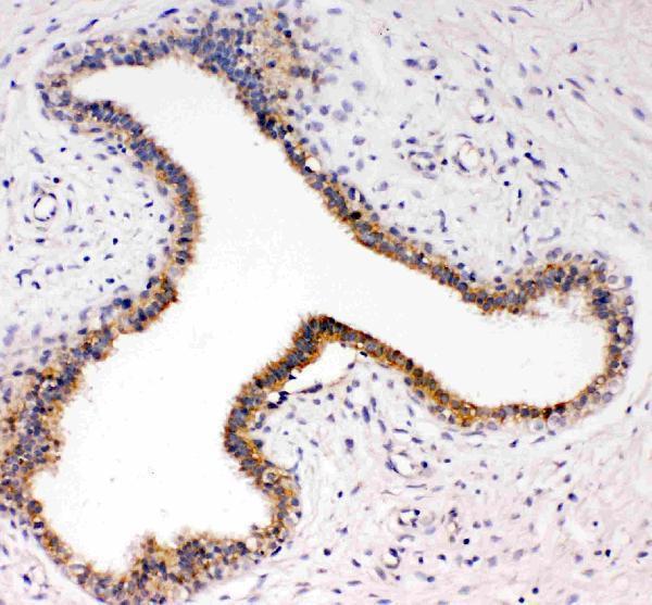
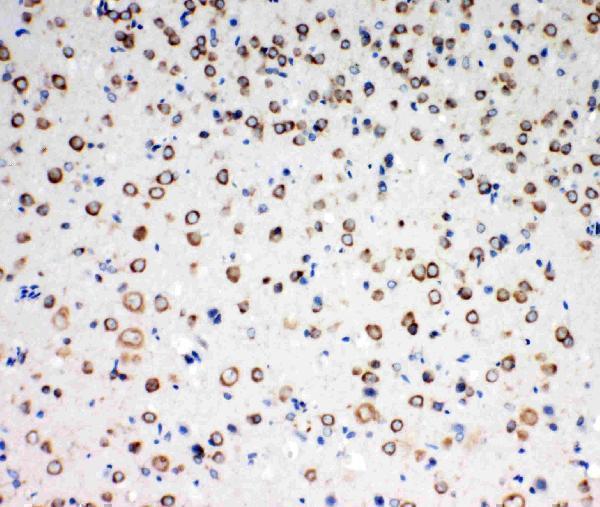
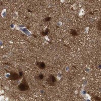


Comprehensive Human Protein Atlas IHC scoring per tissue (reliability: Approved — Medium consistency between antibody staining and RNA expression data.). Rows are taken directly from the HPA tissue chart — click any row's HPA link to view the source.
| Tissue | Cell type | Level | Evidence | Source |
|---|---|---|---|---|
| Cerebral cortex | Neuronal cells | High | Protein (IHC) | HPA → |
| Adrenal gland | Glandular cells | Medium | Protein (IHC) | HPA → |
| Bone marrow | Hematopoietic cells | Medium | Protein (IHC) | HPA → |
| Bronchus | Respiratory epithelial cells | Medium | Protein (IHC) | HPA → |
| Caudate | Neuronal cells | Medium | Protein (IHC) | HPA → |
| Tissue | Cell type | Level | Evidence | Source |
|---|---|---|---|---|
| Adipose tissue | Adipocytes | Not detected | Protein (IHC) | HPA → |
| Appendix | Glandular cells | Not detected | Protein (IHC) | HPA → |
| Fallopian tube | Glandular cells | Not detected | Protein (IHC) | HPA → |
| Parathyroid gland | Glandular cells | Not detected | Protein (IHC) | HPA → |
| Seminal vesicle | Glandular cells | Not detected | Protein (IHC) | HPA → |
Troubleshooting NTRK3 staining in paraffin sections requires attention to retrieval, compartment, antibody epitope and cell type.
PA1992-1 has IHC images of human mammary cancer and rat brain tissue (catalog image captions); A02502Y516 lists IF for human, mouse and rat (catalog applications and reactivity).
PA1992-1 is the card that will render, with a human mammary cancer paraffin-section IHC figure (card image caption). Its catalog also shows rat brain IHC and lists human, mouse and rat reactivity (catalog image caption and reactivity).
Which to pick: For tissue IHC, choose PA1992-1: its figure documents a human mammary cancer paraffin section, with a listed dilution of 0.5–1 μg/ml; the fixative is unreported (card image caption; catalog dilution). For IF, consider A02502Y516, a rabbit polyclonal antibody with IF listed at 1:50; ICC validation and an IF image are unreported (catalog applications, dilution and image fields). Both SKUs list human, mouse and rat reactivity for cross-species work; PA1992-1 also has a rat brain IHC image (catalog reactivity and image caption).