This website uses cookies to ensure you get the best experience on our website.


- Table of Contents
Plan NUCKS1 paraffin-section IHC around its ubiquitous nuclear pattern (HPA tissue IHC). Start with the catalog antibody at 2–5 μg/ml (datasheet A04997-3), and compare high-staining esophageal squamous cells with low-staining small-intestine glands (HPA tissue IHC).

Expected localisation, validated protocols, controls and antibodies — the at-a-glance facts below, then the full design guide.
| Expected localisation | Nuclear staining across tissues (HPA tissue IHC) | |
| Staining pattern | Nuclear signal in glandular, epithelial and other cells (HPA tissue IHC) | |
| Antigen retrieval | EDTA pH 8.0 HIER, heat-mediated (datasheet A04997-3) | |
| Positive control | Adrenal gland+4 more · see all | |
| Negative control | None in HPA (detected in all 45 tissues); use no-primary + isotype controls |
| Fixation | Keep fixation consistent across sections (standard IHC practice; not target-specific). Selected-image fixative and duration unreported (datasheet A04997-3); verify before use. | |
| Caveat | Small-intestine glands and marrow cells show low staining (HPA tissue IHC) | |
| Regulation | No specific expression regulator annotated (UniProt) | |
| Isoform / epitope | 2 isoforms; epitope differences unannotated (UniProt) |
Compare the catalog antibody’s IHC-P protocol with four published NUCKS1 tissue IHC methods (PMC10426139; PMC10393983; PMC4151634; PMC12174342).
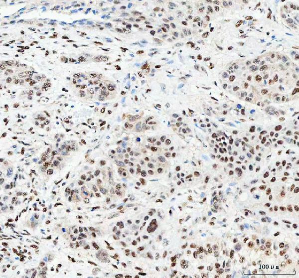
| Sample | Paraffin-embedded human esophageal squamous carcinoma tissue; fixative not specified (datasheet A04997-3) |
| Fixation | Image fixative and duration unreported (datasheet A04997-3); verify before use. |
| Sectioning | 4–5 µm sections on charged slides (standard) |
| Deparaffinisation | Xylene, graded ethanol series to water (standard) |
| Antigen retrieval | Heat retrieval: EDTA pH 8.0 (datasheet A04997-3); 20 min, 95–100 °C (standard) |
| Peroxidase block | 3% H2O2, 10 min, room temperature (standard) |
| Blocking | 10% goat serum (datasheet A04997-3) |
| Primary antibody | Rabbit anti-NUCKS1, 2-5 μg/ml (datasheet A04997-3) |
| Primary incubation | Overnight at 4 °C (datasheet A04997-3) |
| Detection | HRP-conjugated secondary, DAB chromogen (datasheet A04997-3) |
| Counterstain | Hematoxylin, blue, dehydrate and mount (standard) |
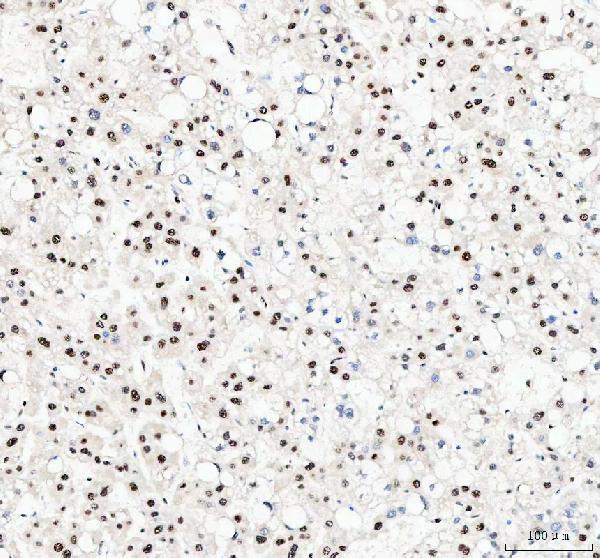
| Expected result | NUCKS1-positive staining in glandular cells of adrenal gland (HPA tissue IHC: High). HPA tissue profile: Ubiquitous nuclear expression. No signal in the no-primary control. |
NUCKS1 should stain nuclei across many cell types in paraffin sections: HPA describes ubiquitous nuclear expression with Supported tissue IHC reliability (HPA: tissue IHC). A nuclear or chromosome-associated pattern fits its annotated location, and the protein has no transmembrane segment (UniProt Q9H1E3: location and topology). Compare intensity with the specific cell population shown by HPA; ubiquitous expression does not mean every cell must stain equally (HPA: tissue IHC).
| Distinct nuclear chromogen in glandular or epithelial cells, with limited cytoplasmic staining. | This fits the reported nuclear pattern (HPA: tissue IHC). HPA records High staining in adrenal glandular cells and bronchial respiratory epithelial cells; those are useful tissue and cell comparisons (HPA: High in adrenal gland and bronchus). |
| Predominantly cytoplasmic or membrane-like staining, with nuclei largely clear. | That compartment disagrees with nuclear and chromosome localization (UniProt Q9H1E3: location) and HPA's ubiquitous nuclear IHC profile (HPA: tissue IHC). Review staining and counterstain before interpreting the cytoplasmic signal as NUCKS1. |
| Strong staining in a cell population expected to be Low in the selected tissue. | High signal in small-intestinal glandular cells or bone-marrow hematopoietic cells conflicts with their Low HPA entries (HPA: Low in small intestine and bone marrow). Consider cross-reactivity or endogenous detection activity; a discrepancy alone cannot identify the cause. |
| Diffuse chromogen across tissue, without discernible nuclear boundaries. | The pattern cannot be scored confidently as nuclear NUCKS1 (HPA: tissue IHC). General IHC practice is to inspect background, blocking, detection chemistry and counterstain alongside a control section before changing the interpretation. |
| No nuclear signal in a section expected to show High staining. | An unstained adrenal glandular or bronchial respiratory epithelial population conflicts with the listed High patterns (HPA: High in adrenal gland and bronchus). Check tissue preservation and the staining run; HPA does not establish NUCKS1-specific fixation sensitivity. |
| Tissue and cell context | HPA reports ubiquitous nuclear expression but different staining levels: selected glandular and epithelial populations are High, while small-intestinal glandular and bone-marrow hematopoietic cells are Low (HPA: tissue IHC). Compare the named cell population, not the entire section. |
| IHC antibody evidence | HPA062351 has Supported IHC status; HPA063843 has no IHC status listed (HPA: antibodies). The tissue profile has Supported reliability, described as high consistency between staining and RNA data (HPA: tissue IHC). These labels do not validate every staining condition. |
| Protein location and structure | NUCKS1 is annotated in the nucleus and on chromosomes, has no transmembrane segment, and is a single annotated chain from residues 1–243 (UniProt Q9H1E3). These facts support a nuclear readout; they do not establish antigen-retrieval needs. |
| Isoforms and modifications | UniProt lists 2 isoforms and 23 modified residues (UniProt Q9H1E3). Their presence alone does not predict which forms an IHC antibody detects or whether retrieval changes staining; no epitope-specific evidence is supplied. |
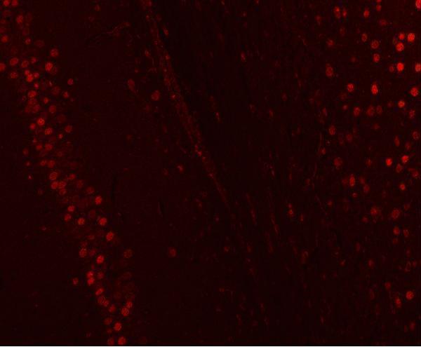
| IF/ICC Q: Where should fluorescence appear? | A: Mainly in the nucleoplasm, with additional nucleolar signal (HPA: supported ICC-IF locations). This is an interpretation cue only; IF/ICC workflow belongs to its separate guide page. |
| Situation | Likely cause | Next action |
|---|---|---|
| Nuclei are clear, but cytoplasm or cell borders are strongly colored. | The compartment conflicts with the reported nuclear location (HPA: tissue IHC; UniProt Q9H1E3: location). | Check the nuclear counterstain and compare a known HPA High cell population in the same run (HPA: High in adrenal gland or bronchus). Review detection background before calling the signal specific. |
| Small-intestinal glandular or bone-marrow hematopoietic cells stain as strongly as a High comparator. | Those populations are listed as Low; cross-reactivity or endogenous detection activity is possible, but the pattern does not distinguish them (HPA: tissue IHC). | Compare the same cell types with a detection control and assess whether staining remains nuclear. Treat the unexpected intensity as unresolved until controls explain it. |
| Chromogen is widespread and nuclear edges are difficult to distinguish. | Diffuse background can obscure the nuclear pattern (HPA: tissue IHC). The source record does not identify a NUCKS1-specific background mechanism. | Use general IHC controls to assess nonspecific detection, blocking and chromogen development. Adjust the staining workflow only after checking that the counterstain and a positive tissue remain interpretable. |
| An expected High population has no visible nuclear staining. | This conflicts with HPA's High entry for that cell population (HPA: High in adrenal gland or bronchus); the record does not establish a fixation-related explanation. | Verify tissue identity, run controls, reagent performance and the chosen IHC antibody's status. HPA062351 is Supported for IHC, whereas HPA063843 has no IHC status listed (HPA: antibodies). |
| A weakly stained field is called negative because few cells are conspicuous. | NUCKS1 is broadly nuclear, yet HPA reports Low staining in some populations (HPA: tissue IHC). Weak staining in a Low population alone cannot validate a negative call. | Score nuclei within identified cell populations and compare a High reference population from the same run (HPA: tissue IHC). Record weak signal separately from absent signal. |
| Nucleolar emphasis in IF/ICC is treated as an IHC failure. | HPA supports nucleoplasmic localization with additional nucleolar localization in ICC-IF (HPA: subcellular). Fluorescence detail need not appear identically with chromogenic IHC. | For this IHC guide, judge whether the chromogenic result is predominantly nuclear (HPA: tissue IHC). Use the separate IF/ICC guide for fluorescence workflow and its finer localization readout. |


Comprehensive Human Protein Atlas IHC scoring per tissue (reliability: Supported — High consistency between antibody staining and RNA expression data.). Rows are taken directly from the HPA tissue chart — click any row's HPA link to view the source.
| Tissue | Cell type | Level | Evidence | Source |
|---|---|---|---|---|
| Adrenal gland | Glandular cells | High | Protein (IHC) | HPA → |
| Breast | Adipocytes | High | Protein (IHC) | HPA → |
| Bronchus | Respiratory epithelial cells | High | Protein (IHC) | HPA → |
| Cerebellum | Cells in molecular layer | High | Protein (IHC) | HPA → |
| Cerebral cortex | Glial cells | High | Protein (IHC) | HPA → |
| Tissue | Cell type | Level | Evidence | Source |
|---|---|---|---|---|
| None in HPA: NUCKS1 is detected in all 45 scored tissues. Use a no-primary (secondary-only) and an isotype control instead. | ||||
Troubleshoot chromogenic NUCKS1 staining in paraffin sections by checking nuclear localisation, retrieval conditions, controls and scoring consistency (UniProt Q9H1E3; HPA tissue IHC).
A04997-3 has human paraffin-section IHC images and mouse and rat brain-section IF images (catalog image captions); its listed reactivity covers human, mouse and rat (catalog reactivity).
A04997-3 is listed for IHC and IF, with human paraffin-section IHC images from esophageal squamous carcinoma, liver cancer, lung cancer and rectum adenocarcinoma tissue (catalog applications and IHC image captions). Its IF images show paraffin-embedded mouse and rat brain sections (catalog IF image captions).
Which to pick: For tissue IHC, choose A04997-3: its human paraffin-section images use EDTA retrieval at pH 8.0 and 2 μg/ml primary antibody (catalog IHC image captions); the fixative is unreported (catalog IHC image captions). For IF, A04997-3 has mouse and rat brain-section images at 5 μg/ml (catalog IF image captions); ICC validation is unreported (catalog applications and image captions). For cross-species planning, A04997-3 lists human, mouse and rat reactivity, while its IHC dilution listing specifies human and mouse and its IF images show mouse and rat (catalog reactivity, dilution listing and IF image captions).