This website uses cookies to ensure you get the best experience on our website.


- Table of Contents
Plan paraffin-section NUDT5 IHC using the IHC-validated antibody and liver hepatocytes as a positive tissue reference (datasheet A07530; HPA tissue IHC). Score cytoplasmic staining while recording any nuclear signal separately, since the tissue profile and molecular annotation differ (HPA tissue IHC; UniProt).

Expected localisation, validated protocols, controls and antibodies — the at-a-glance facts below, then the full design guide.
| Expected localisation | Cytoplasmic in tissue; nucleus annotated molecularly (HPA tissue IHC; UniProt) | |
| Staining pattern | Cytoplasmic staining in several cell types, most abundant in liver (HPA tissue IHC) | |
| Antigen retrieval | EDTA pH 8.0 HIER, heat-mediated (datasheet A07530) | |
| Positive control | Liver+1 more · see all | |
| Negative control | Adipose tissue+4 more · see all |
| Fixation | Matched tissue-IHC evidence does not establish the fixation claim. Validate the specimen-specific method before use. (selected-SKU IHC image A07530) | |
| Caveat | Nuclear annotation differs from observed tissue cytoplasm (UniProt; HPA tissue IHC) | |
| Regulation | Broadly expressed; most abundant in liver (UniProt) | |
| Isoform / epitope | 0 annotated isoforms; one chain spanning residues 1–219 (UniProt) |
The catalog antibody’s IHC-P protocol (datasheet: A07530) accompanies four published NUDT5 IHC protocols (PMC7258951; PMC12108576; PMC7877577; PMC10845800).
| Sample | Paraffin-embedded human liver cancer tissue; fixative not specified (datasheet A07530) |
| Fixation | Image fixative and duration unreported (datasheet A07530); verify before use. |
| Sectioning | 4–5 µm sections on charged slides (standard) |
| Deparaffinisation | Xylene, graded ethanol series to water (standard) |
| Antigen retrieval | Heat retrieval: EDTA pH 8.0 (datasheet A07530); 20 min, 95–100 °C (standard) |
| Peroxidase block | 3% H2O2, 10 min, room temperature (standard) |
| Blocking | 10% goat serum (datasheet A07530) |
| Primary antibody | Rabbit anti-NUDT5, 2-5 μg/ml (datasheet A07530) |
| Primary incubation | Overnight at 4 °C (datasheet A07530) |
| Detection | HRP-conjugated secondary, DAB chromogen (datasheet A07530) |
| Counterstain | Hematoxylin, blue, dehydrate and mount (standard) |
| Expected result | NUDT5-positive staining in hepatocytes of liver (HPA tissue IHC: Medium). HPA tissue profile: Cytoplasmic expression in several cell types, most abundant in liver. No signal in the no-primary control. |
For paraffin-section IHC, expect cytoplasmic staining in hepatocytes and elongated or late spermatids, with medium staining reported in each (HPA tissue IHC). NUDT5 is also assigned to the nucleus and has no transmembrane segment (UniProt Q9UKK9). Interpret compartment differences cautiously: HPA tissue IHC is Approved, but reports low consistency between antibody staining and RNA expression; external characterization supports the antibody staining (HPA tissue IHC).
| Distinct cytoplasmic staining in hepatocytes, with a similar pattern in elongated or late spermatids. | This matches the reported medium-level positive cells (HPA tissue IHC). Compare signal with adjacent cells and the counterstain before scoring. A medium HPA category describes its reference observation; it does not prescribe a universal chromogen intensity or exposure setting (HPA tissue IHC; general IHC practice). |
| Strong staining confined to cell surfaces, extracellular material, or other unexpected structures. | Treat this as a localization warning because NUDT5 has no transmembrane segment and UniProt assigns it to the nucleus (UniProt Q9UKK9). Nuclear staining alone needs more careful review: it is biologically plausible, although the supplied tissue IHC profile emphasizes cytoplasm (UniProt Q9UKK9; HPA tissue IHC). |
| Strong, widespread staining in cells that HPA records as not detected, such as adipocytes. | Investigate cross-reactivity or endogenous detection activity before calling these cells positive (HPA tissue IHC; general IHC practice). HPA's 'not detected' is an assay observation, not proof that every cell lacks NUDT5; UniProt describes the protein as widely expressed (HPA tissue IHC; UniProt Q9UKK9). |
| Uniform chromogen haze across tissue compartments, including areas without clear cellular boundaries. | This does not establish NUDT5 localization. Review primary-antibody omission and detection controls, blocking, wash steps, and chromogen development as general IHC checks. Resolve the haze before comparing hepatocyte or spermatid staining with the HPA reference pattern (general IHC practice; HPA tissue IHC). |
| No discernible signal in hepatocytes of an otherwise interpretable liver section. | The result conflicts with the reported medium hepatocyte staining (HPA tissue IHC). Check that the IHC-validated antibody and detection system work on an appropriate control section, then review retrieval and antibody conditions. These are general assay checks; the supplied sources report no NUDT5-specific fixation sensitivity. |
| Tissue and cell selection | Liver hepatocytes and testicular elongated or late spermatids provide reported medium IHC examples (HPA tissue IHC). Kidney tubule cells, bronchial respiratory epithelial cells, skin fibroblasts, and tonsil squamous epithelial cells are reported low, so a faint result there is less decisive (HPA tissue IHC). |
| Compartment evidence | The tissue IHC summary describes cytoplasmic expression, whereas UniProt assigns NUDT5 to the nucleus (HPA tissue IHC; UniProt Q9UKK9). Score the compartment actually seen and disclose this difference; neither source makes every nuclear or cytoplasmic signal independently specific. |
| Antibody evidence | HPA019827 is Approved for IHC and ICC, while tissue IHC reliability is Approved with low antibody-staining/RNA consistency and supporting external characterization (HPA antibodies; HPA tissue IHC). Agreement with a reported pattern supports interpretation, but the supplied record does not establish IHC Enhanced validation. |
| Topology and processing | NUDT5 has no transmembrane segment, signal peptide, propeptide, or reported processed chain beyond residues 1–219 (UniProt Q9UKK9). These annotations give no basis to expect a membrane-bound or secreted IHC pattern; they also do not establish epitope accessibility after fixation. |
| IF/ICC Q&A: What localization should an IF image show? | HPA reports approved main localization in the nucleoplasm and vesicles, with additional centrosome and mid-piece localization (HPA subcellular ICC-IF). This is context for reading an IF result; the supplied evidence does not provide an IF protocol option for this IHC-P guide. |
| Situation | Likely cause | Next action |
|---|---|---|
| Liver control lacks hepatocyte staining. | Possible assay failure; HPA reports medium hepatocyte staining, but no NUDT5-specific fixation effect is supplied (HPA tissue IHC). | Confirm section integrity and detection controls, then review the IHC-validated antibody conditions and antigen retrieval as general IHC practice. Record the conditions used before comparing sections. |
| Chromogen appears broadly in negative-control tissue or after primary-antibody omission. | Endogenous detection activity or reagent background can produce signal without specific primary binding (general IHC practice). | Check the omission control and the detection chemistry; apply the appropriate endogenous-activity block for that chemistry, then compare with the same tissue stained using primary antibody (general IHC practice). |
| Strong signal appears in adipocytes recorded as not detected. | Cross-reactivity or assay background is possible; HPA's category reflects its observed assay result, not guaranteed molecular absence (HPA tissue IHC). | Compare cell boundaries and matched controls, then seek independent confirmation before scoring these cells as NUDT5-positive (general IHC practice). |
| Only surface or extracellular deposits dominate the section. | That distribution is poorly supported by the no-transmembrane topology and nuclear UniProt annotation (UniProt Q9UKK9). | Inspect the counterstain and omission control for deposits; compare with the HPA cytoplasmic IHC pattern and repeat under checked detection conditions if needed (HPA tissue IHC; general IHC practice). |
| Nuclear staining appears alongside, or instead of, cytoplasmic staining. | The sources differ by assay: UniProt assigns a nuclear location, HPA tissue IHC describes cytoplasm, and HPA ICC-IF identifies nucleoplasm (UniProt Q9UKK9; HPA tissue IHC; HPA subcellular ICC-IF). | Report nuclear and cytoplasmic scores separately, examine controls, and avoid treating the compartment alone as proof of specificity (general IHC practice). |
| Low-signal tissue seems negative while liver is positive. | Some cell types are reported low, including kidney tubule cells and bronchial respiratory epithelial cells, while hepatocytes are medium (HPA tissue IHC). | Use the liver result to verify the staining run, then document the detection threshold and score the low-signal tissue conservatively; absence of visible chromogen does not establish biological absence (HPA tissue IHC; general IHC practice). |

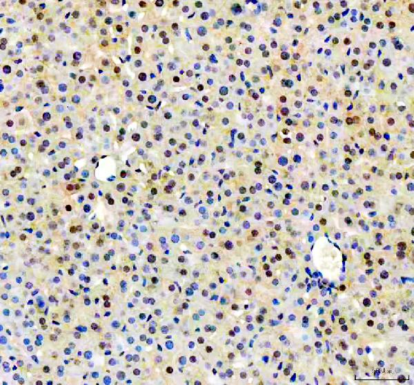
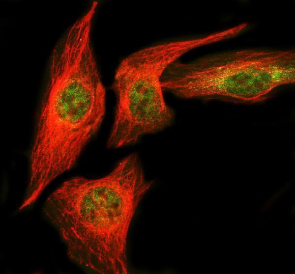


Comprehensive Human Protein Atlas IHC scoring per tissue (reliability: Approved — Low consistency between antibody staining and RNA expression data. External characterization data supports antibody staining.). Rows are taken directly from the HPA tissue chart — click any row's HPA link to view the source.
| Tissue | Cell type | Level | Evidence | Source |
|---|---|---|---|---|
| Liver | Hepatocytes | Medium | Protein (IHC) | HPA → |
| Testis | Elongated or late spermatids | Medium | Protein (IHC) | HPA → |
| Tissue | Cell type | Level | Evidence | Source |
|---|---|---|---|---|
| Adipose tissue | Adipocytes | Not detected | Protein (IHC) | HPA → |
| Adrenal gland | Glandular cells | Not detected | Protein (IHC) | HPA → |
| Appendix | Glandular cells | Not detected | Protein (IHC) | HPA → |
| Bone marrow | Hematopoietic cells | Not detected | Protein (IHC) | HPA → |
| Breast | Adipocytes | Not detected | Protein (IHC) | HPA → |
Troubleshoot NUDT5 chromogenic IHC by checking retrieval, cellular compartment, controls, and scoring before interpreting staining intensity.
A07530 has paraffin-section IHC images from human liver cancer and mouse and rat liver, plus IF/ICC data from U2OS cells (A07530 image captions). M07530 lists human, mouse and rat IHC reactivity (catalog).
A07530 has IHC images from paraffin sections of human liver cancer and mouse and rat liver, plus an IF/ICC image from U2OS cells (A07530 image captions). M07530 is listed for IHC in human, mouse and rat, with no IHC image supplied (catalog).
Which to pick: For tissue IHC, choose A07530 when documented paraffin-section staining matters: its captions specify EDTA retrieval at pH 8.0, but do not report the fixative (A07530 IHC captions). M07530 is a rabbit monoclonal, clone 26N82, listed for IHC without a supplied tissue image or processing details (catalog). For IF/ICC, choose A07530 based on its U2OS image at 5 μg/ml; for cross-species IHC, its images cover human, mouse and rat, while M07530 lists those species as reactive without supplied IHC images (A07530 image captions; catalog).