This website uses cookies to ensure you get the best experience on our website.


- Table of Contents
Plan NUMA1 staining in paraffin sections around the general nuclear pattern reported in tissue IHC (HPA tissue IHC). The guide covers fixation, controls, and scoring, including how mitotic relocalization may affect interpretation (UniProt).

Expected localisation, validated protocols, controls and antibodies — the at-a-glance facts below, then the full design guide.
| Expected localisation | Nuclear staining in tissue sections (HPA tissue IHC) | |
| Staining pattern | General nuclear staining across tissue cell types (HPA tissue IHC) | |
| Antigen retrieval | EDTA pH 8.0 HIER, heat-mediated (datasheet A02018-1) | |
| Positive control | Adipose tissue+4 more · see all | |
| Negative control | None in HPA (detected in all 45 tissues); use no-primary + isotype controls |
| Fixation | Matched tissue-IHC evidence does not establish the fixation claim. Validate the specimen-specific method before use. (selected-SKU IHC image A02018-1) | |
| Caveat | Mitotic relocalization can affect nuclear scoring (UniProt) | |
| Regulation | Cell-cycle-dependent redistribution (UniProt) | |
| Isoform / epitope | 5 isoforms; confirm epitope coverage (UniProt) |
The catalog antibody uses heat-mediated EDTA retrieval at pH 8.0 (datasheet A02018-1). The published IHC protocols below cover Wilms tumors, mouse tissue, and ovarian cancer specimens (PMC6925259; PMC8686194; PMC12537492).
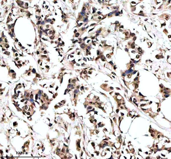
| Sample | Paraffin-embedded human acinar adenocarcinoma of prostate tissue; fixative not specified (datasheet A02018-1) |
| Fixation | Image fixative and duration unreported (datasheet A02018-1); verify before use. |
| Sectioning | 4–5 µm sections on charged slides (standard) |
| Deparaffinisation | Xylene, graded ethanol series to water (standard) |
| Antigen retrieval | Heat retrieval: EDTA pH 8.0 (datasheet A02018-1); 20 min, 95–100 °C (standard) |
| Peroxidase block | 3% H2O2, 10 min, room temperature (standard) |
| Blocking | 10% goat serum (datasheet A02018-1) |
| Primary antibody | Rabbit anti-NUMA1, 1-2 μg/ml (datasheet A02018-1) |
| Primary incubation | Overnight at 4 °C (datasheet A02018-1) |
| Detection | HRP-conjugated secondary, DAB chromogen (datasheet A02018-1) |
| Counterstain | Hematoxylin, blue, dehydrate and mount (standard) |
| Expected result | NUMA1-positive staining in adipocytes of adipose tissue (HPA tissue IHC: High). HPA tissue profile: General nuclear expression. No signal in the no-primary control. |
NUMA1 is predominantly nuclear in interphase and relocates to spindle poles and the polar cell cortex during mitosis (UniProt Q14980 localization). In paraffin section IHC, expect nuclear staining across many cell types, including adipocytes, glandular cells, hematopoietic cells and respiratory epithelial cells reported as strongly positive (HPA: general nuclear expression; High in these cell types). HPA rates its tissue pattern Enhanced, with high consistency between antibody staining and RNA expression (HPA: reliability Enhanced).
| Clear nuclear staining in adipocytes, glandular cells, hematopoietic cells or respiratory epithelium. | This matches the reported general nuclear pattern and High staining in those cell types (HPA: tissue IHC). Judge the cell nucleus against its counterstain; staining need not be identical in every cell (standard IHC practice). |
| Predominantly cytoplasmic or widespread membrane staining in cells with intact nuclei. | Treat this as a compartment mismatch for interphase cells and investigate artefact or antibody specificity (HPA: general nuclear expression; UniProt Q14980: interphase nuclear matrix). A limited cortical pattern in a mitotic cell has a different interpretation (UniProt Q14980: mitotic relocalization). |
| Strong staining confined to an unexpected cell population, with little staining in an HPA positive population. | Cross-reactivity or endogenous detection activity is possible (standard IHC practice). HPA reports low tissue specificity and supplies no negative tissue in this payload, so an unlisted cell type alone cannot be called NUMA1 negative (HPA: RNA specificity; tissue IHC). |
| Haze across nuclei, cytoplasm and surrounding tissue obscures cell boundaries. | Diffuse background prevents a reliable nuclear call (standard IHC practice). Compare with an appropriate detection control, then reassess blocking, antibody concentration and wash conditions; the supplied sources do not identify a NUMA1 specific cause (standard IHC practice). |
| No nuclear signal in a section containing an HPA positive cell population. | A negative result conflicts with the reported High staining in that population (HPA: tissue IHC). First establish that the run's positive control worked before interpreting the specimen as biologically negative (standard IHC practice). |
| Cell cycle stage (UniProt Q14980: localization). | Interphase NUMA1 occupies the nuclear matrix; mitotic NUMA1 can appear at spindle poles and the polar cortex (UniProt Q14980: localization). Interpret an isolated extranuclear mitotic figure in its cell cycle context rather than applying the interphase pattern to it. |
| Antibody validation (HPA: antibody records). | HPA019841, HPA019859 and HPA029912 each have Enhanced IHC status (HPA: antibody records). This supports the reported pattern, but the status does not validate an unrelated catalog antibody or establish its dilution or retrieval conditions. |
| Isoforms and epitope coverage (UniProt Q14980: five isoforms). | UniProt lists 5 NUMA1 isoforms (UniProt Q14980: isoforms). The supplied record gives no antibody epitope, so it cannot predict which isoforms a particular stain detects or whether isoform coverage explains a weak result. |
| Antigen retrieval and detection controls (standard IHC practice). | Retrieval and chromogenic detection settings can affect whether an IHC run is interpretable (standard IHC practice). Neither HPA tissue staining nor UniProt topology establishes NUMA1 specific fixation sensitivity or an optimal retrieval setting (HPA: tissue IHC; UniProt Q14980: topology). |
| Situation | Likely cause | Next action |
|---|---|---|
| No staining in a known positive control tissue. | An unsuccessful IHC run is possible; HPA reports High staining in several listed cell populations (HPA: tissue IHC). The supplied sources do not identify which workflow step failed. | Check section integrity, reagent activity, antibody use conditions, retrieval and chromogen development against the run controls (standard IHC practice). Reassess specimen negativity only after the positive control works. |
| Uniform brown background obscures nuclear detail. | Nonspecific antibody binding or detection background may obscure the expected nuclear pattern (standard IHC practice; HPA: general nuclear expression). | Inspect a control omitting primary antibody, then adjust blocking, antibody concentration or washes as indicated by that control (standard IHC practice). Record whether the background is nuclear, cytoplasmic or extracellular. |
| Brown signal appears in unexpected cells or structures. | Cross-reactivity or endogenous peroxidase activity may contribute to chromogenic signal (standard IHC practice). HPA provides no negative cell list here, so unexpected cell identity alone does not prove artefact (HPA: tissue IHC). | Use a primary omission control to assess detection activity and an independent antibody or other specificity control if available (standard IHC practice; HPA: Enhanced IHC antibody records). |
| Interphase cells show chiefly cytoplasmic staining. | This conflicts with the predominant nuclear pattern (HPA: tissue IHC; UniProt Q14980: interphase nuclear matrix). A mitotic figure can instead show extranuclear NUMA1 (UniProt Q14980: localization). | Check nuclear morphology and mitotic state, then compare the compartment pattern with a working positive control and detection control before assigning the signal to NUMA1 (standard IHC practice). |
| A few dividing cells show spindle pole or cortical signal. | NUMA1 moves from the interphase nucleus to spindle poles and polar cortex during mitosis (UniProt Q14980: localization). | Interpret these cells separately from the surrounding interphase nuclei; use recognizable mitotic morphology to support the assignment (UniProt Q14980: localization; standard IHC interpretation). |
| IF/ICC Q&A: What localization should be expected? | HPA reports NUMA1 mainly in the nucleoplasm, with an enhanced subcellular location assignment (HPA: ICC-IF subcellular). UniProt also describes cell cycle dependent relocalization (UniProt Q14980: localization). | Expect predominantly nucleoplasmic signal in interphase cells; assess apparent spindle pole or cortical signal in the context of mitosis (HPA: ICC-IF subcellular; UniProt Q14980: localization). |
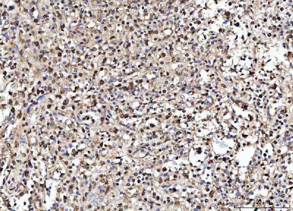
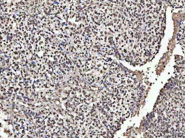
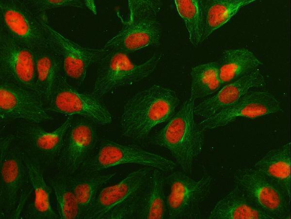
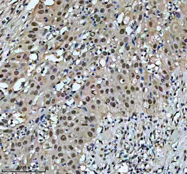


Comprehensive Human Protein Atlas IHC scoring per tissue (reliability: Enhanced — High consistency between antibody staining and RNA expression data.). Rows are taken directly from the HPA tissue chart — click any row's HPA link to view the source.
| Tissue | Cell type | Level | Evidence | Source |
|---|---|---|---|---|
| Adipose tissue | Adipocytes | High | Protein (IHC) | HPA → |
| Adrenal gland | Glandular cells | High | Protein (IHC) | HPA → |
| Appendix | Glandular cells | High | Protein (IHC) | HPA → |
| Bone marrow | Hematopoietic cells | High | Protein (IHC) | HPA → |
| Breast | Glandular cells | High | Protein (IHC) | HPA → |
| Tissue | Cell type | Level | Evidence | Source |
|---|---|---|---|---|
| None in HPA: NUMA1 is detected in all 45 scored tissues. Use a no-primary (secondary-only) and an isotype control instead. | ||||
Use the catalog antibody’s paraffin-section workflow as the starting point, then judge NUMA1 staining against its cell-cycle-dependent localisation (datasheet A02018-1; UniProt Q14980).
Both anti-NUMA1 antibodies have IHC images from human paraffin sections (catalog: A02018-1 and M02018-1 IHC captions); A02018-1 also has IF/ICC data from U2OS cells (catalog: A02018-1 IF caption).
A02018-1 has IHC images from human prostate, breast, esophageal and lung cancer sections, plus an IF/ICC image from U2OS cells (catalog: A02018-1 image captions). M02018-1 has IHC images from human bladder and breast cancer sections and rat testis; IF/ICC is listed as an application, with no IF image supplied (catalog: M02018-1 image captions and applications).
Which to pick: For tissue IHC, choose M02018-1 if a rabbit monoclonal with human and rat paraffin-section examples suits the study; its pictured IHC used EDTA retrieval at pH 8.0 and 1:50 primary dilution (catalog: M02018-1 clone and IHC captions). Choose A02018-1 when a pictured IF/ICC result is useful: its U2OS image used 5 μg/ml, while its human paraffin-section IHC images used EDTA retrieval at pH 8.0 and 2 μg/ml (catalog: A02018-1 IF and IHC captions). For broader listed species reactivity, A02018-1 includes monkey alongside human, mouse and rat, while M02018-1 lists human, mouse and rat; fixative is unreported for the pictured paraffin sections of both antibodies (catalog: reactivity and IHC captions).