This website uses cookies to ensure you get the best experience on our website.


- Table of Contents
Plan paraffin-section NUMB IHC around its membranous and cytoplasmic tissue staining (HPA tissue IHC). High staining in glandular cells supports positive-reference selection (HPA tissue IHC), while 9 isoforms warrant epitope review (UniProt).

Expected localisation, validated protocols, controls and antibodies — the at-a-glance facts below, then the full design guide.
| Expected localisation | Membranous and cytoplasmic staining in most tissues (HPA tissue IHC) | |
| Staining pattern | Strong glandular-cell staining; membrane and cytoplasm (HPA tissue IHC) | |
| Antigen retrieval | EDTA pH 8.0 HIER, heat-mediated (datasheet PB9301) | |
| Positive control | Appendix+4 more · see all | |
| Negative control | Adipose tissue+1 more · see all |
| Fixation | Keep fixation conditions consistent across sections (standard IHC practice; not target-specific) | |
| Caveat | IHC staining has medium concordance with RNA data (HPA tissue IHC) | |
| Regulation | Staining-linked expression regulation is unreported (UniProt) | |
| Isoform / epitope | 9 isoforms; check epitope conservation across forms (UniProt) |
Compare the catalog antibody’s IHC-P protocol (datasheet: PB9301) with 3 published NUMB IHC protocols (PMC5354663; PMC12035580; PMC5308634).
| Sample | Paraffin-embedded human liver cancer tissue; fixative not specified (datasheet PB9301) |
| Fixation | Image fixative and duration unreported (datasheet PB9301); verify before use. |
| Sectioning | 4–5 µm sections on charged slides (standard) |
| Deparaffinisation | Xylene, graded ethanol series to water (standard) |
| Antigen retrieval | Heat retrieval: EDTA pH 8.0 (datasheet PB9301); 20 min, 95–100 °C (standard) |
| Peroxidase block | 3% H2O2, 10 min, room temperature (standard) |
| Blocking | 10% goat serum (datasheet PB9301) |
| Primary antibody | Rabbit anti-NUMB, 2-5μg/ml (datasheet PB9301) |
| Primary incubation | Overnight at 4 °C (datasheet PB9301) |
| Detection | HRP-conjugated secondary, DAB chromogen (datasheet PB9301) |
| Counterstain | Hematoxylin, blue, dehydrate and mount (standard) |
| Expected result | NUMB-positive staining in glandular cells of appendix (HPA tissue IHC: High). HPA tissue profile: Membranous and cytoplasmic expression in most tissues. No signal in the no-primary control. |
NUMB should show membranous and cytoplasmic staining in many tissues, including high staining in appendix and colon glandular cells (HPA tissue IHC). Its membrane association and perinuclear endosome localization are consistent with that distribution; NUMB has no transmembrane segment (UniProt P49757). HPA rates tissue IHC as Approved, with medium agreement between staining and RNA data and external verification pending (HPA tissue IHC).
| Membranous and cytoplasmic signal in appendix or colon glandular cells. | This fits the reported high staining in those cells and the broad tissue pattern (HPA tissue IHC). Assess cell boundaries and cytoplasm separately when scoring; a perinuclear component can also fit NUMB's endosome localization (UniProt P49757). |
| Predominantly nuclear staining, with little membranous or cytoplasmic signal. | A nuclear-only pattern conflicts with the reported tissue distribution and localization (HPA tissue IHC; UniProt P49757). Suspect an artefact or off-target staining, then compare a known-positive section and the negative detection control before interpreting it as NUMB. |
| Strong signal confined to adipocytes or lymph-node germinal center cells. | HPA reports NUMB as not detected in those specific cell populations (HPA tissue IHC). Check whether staining persists when the primary antibody is omitted; unexpected cell-restricted signal can reflect cross-reactivity or endogenous detection activity (general IHC practice). |
| Diffuse color across tissue, stroma and empty spaces. | A field-wide deposit lacks the cell and compartment pattern reported for NUMB (HPA tissue IHC). Check blocking, washes, detection reagent and chromogen development on a control section; diffuse background cannot establish cellular NUMB expression (general IHC practice). |
| No signal in appendix or colon glandular cells. | These are reported high-staining cell populations, so an absent signal makes the run difficult to interpret (HPA tissue IHC). Check section quality, retrieval and detection with appropriate controls before concluding that the specimen lacks NUMB (general IHC practice). |
| Cellular compartment | HPA describes membranous and cytoplasmic tissue staining (HPA tissue IHC). UniProt places NUMB at cell and endosome membranes, including perinuclear endosomes, despite no transmembrane segment (UniProt P49757). Score the visible compartment rather than treating all cellular color alike. |
| Tissue and cell selection | Appendix and colon glandular cells are reported high, whereas adipocytes and lymph-node germinal center cells are reported not detected (HPA tissue IHC). Use these as pattern references; HPA's medium staining–RNA agreement and pending external verification limit how definitive any single section can be (HPA tissue IHC). |
| Isoforms and epitope coverage | UniProt lists 9 NUMB isoforms and a PID domain at residues 33–193 (UniProt P49757). The supplied sources do not map this assay's epitope or establish which isoforms it detects; check antibody documentation before comparing isoform-dependent staining. |
| Antibody validation and detection | HPA lists CAB011478 tissue IHC as Approved, while HPA002874 has no tissue-IHC status in the supplied record (HPA antibodies). Validation belongs to the tested antibody; background from endogenous enzyme activity or detection reagents must be assessed with controls (general IHC practice). |
| Situation | Likely cause | Next action |
|---|---|---|
| Known-positive glandular cells are blank. | The run may have failed; appendix and colon glandular cells are reported high (HPA tissue IHC). | Review tissue integrity, the antibody's IHC-P instructions, retrieval, dilution and detection; repeat with a positive control (general IHC practice). The supplied sources specify no retrieval condition or dilution. |
| Only nuclei stain strongly. | Nuclear-only staining disagrees with the membranous and cytoplasmic tissue pattern (HPA tissue IHC). | Compare with a positive tissue section and a no-primary control; review primary-antibody specificity and detection background before scoring (general IHC practice). |
| Adipocytes or germinal center cells stain strongly. | Both populations are reported not detected, although HPA validation remains pending external verification (HPA tissue IHC). | Check neighboring cell identity and a no-primary control. If signal persists without primary antibody, investigate endogenous detection activity; otherwise review antibody specificity (general IHC practice). |
| Brown deposit spreads beyond cell outlines. | Diffuse staining does not match HPA's cellular membrane and cytoplasm pattern (HPA tissue IHC); excess detection background is possible (general IHC practice). | Inspect the no-primary control, blocking and wash steps, then adjust detection and chromogen development according to reagent instructions (general IHC practice). |
| Positive and comparison tissues both look weak. | HPA reports different cell-level intensities, including high glandular staining and undetected adipocytes (HPA tissue IHC); weak color everywhere may indicate a technical issue (general IHC practice). | Verify the positive tissue and control staining first. Review retrieval and antibody concentration using the catalog antibody's IHC-P instructions; no NUMB-specific fixation sensitivity is reported in the supplied sources. |
| Does an IF/ICC junctional pattern validate this IHC-P result? | HPA calls IF/ICC junction localization uncertain, plasma membrane supported and cytosol uncertain; CAB011478 ICC is Uncertain (HPA subcellular; HPA antibodies). | Use the IF/ICC result as localization context only. Judge paraffin-section staining against tissue IHC cell patterns and the IHC validation for the antibody used (HPA tissue IHC; HPA antibodies). |


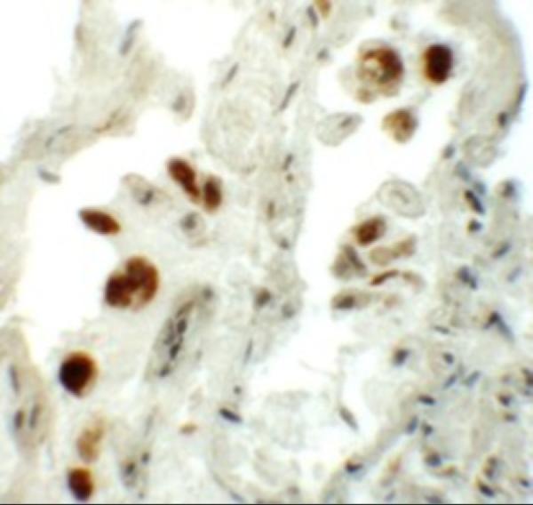
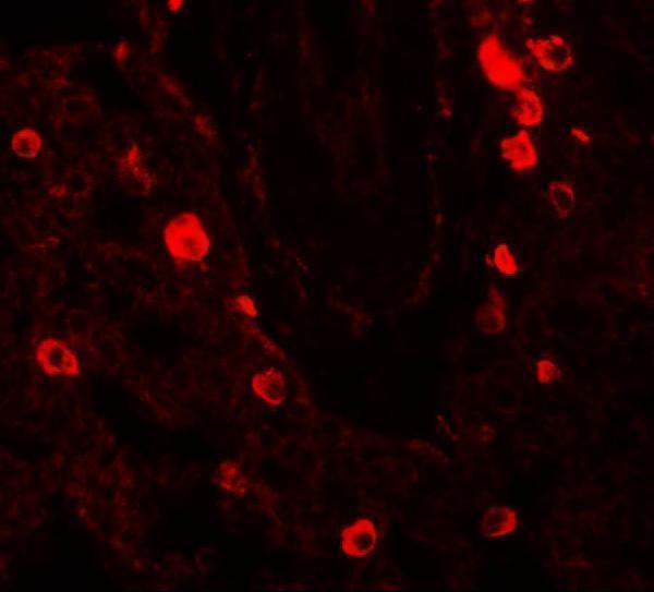


Comprehensive Human Protein Atlas IHC scoring per tissue (reliability: Approved — Medium consistency between antibody staining and RNA expression data. Pending external verification.). Rows are taken directly from the HPA tissue chart — click any row's HPA link to view the source.
| Tissue | Cell type | Level | Evidence | Source |
|---|---|---|---|---|
| Appendix | Glandular cells | High | Protein (IHC) | HPA → |
| Bone marrow | Hematopoietic cells | High | Protein (IHC) | HPA → |
| Breast | Glandular cells | High | Protein (IHC) | HPA → |
| Bronchus | Respiratory epithelial cells | High | Protein (IHC) | HPA → |
| Cerebellum | Cells in granular layer | High | Protein (IHC) | HPA → |
Troubleshoot NUMB staining by checking retrieval, compartment, cell type and controls before comparing signal across paraffin sections (UniProt P49757; HPA tissue IHC).
Two anti-NUMB antibodies have human tissue IHC images (catalog image captions); one also has a human lung IF image (A01206 image caption). Catalog reactivity spans human, monkey, mouse and rat (catalog applications/reactivity).
PB9301 shows IHC in paraffin sections of human liver cancer and lung cancer tissue (PB9301 IHC image captions). A01206 shows IHC and IF in human lung tissue (A01206 image captions).
Which to pick: For paraffin-section IHC, choose PB9301 if its documented retrieval and detection conditions are useful: EDTA pH 8.0 retrieval and DAB detection are shown in its own caption (PB9301 IHC image caption); the fixative is unreported (PB9301 IHC image caption). For tissue IF, choose A01206, which has a human lung IF image and lists IF as an application; ICC validation and the tissue fixative are unreported (A01206 IF image caption; A01206 applications). For cross-species planning, PB9301 lists human, monkey, mouse and rat reactivity, while A01206 lists human, mouse and rat; the cited IHC images show human tissue only, and clonality is unreported for both (catalog reactivity; catalog IHC image captions; catalog clone fields).