NUP210 / Nuclear pore membrane glycoprotein 210 · IHC design guide

Design Immunohistochemistry for NUP210

Plan NUP210 staining in paraffin sections using high-staining lung macrophages or prostate glandular cells as positive controls (HPA tissue IHC). Interpret chromogenic nuclear staining alongside the expected nuclear pore membrane location (HPA tissue IHC; UniProt).

Evidence assembled Oct 2026 · For research use; verify linked source records and product datasheet before use
Immunohistochemistry protocol sheet for NUP210 (IHC for NUP210): expected localisation General nuclear tissue staining (HPA tissue IHC), antibody A05308, validated IHC image, and IHC protocol steps
Printable NUP210 IHC protocol sheet — expected localisation General nuclear tissue staining (HPA tissue IHC), antibody A05308, controls and protocol steps. Open the full NUP210 IHC guide →

NUP210 Immunohistochemistry Experimental Design Guide

Expected localisation, validated protocols, controls and antibodies — the at-a-glance facts below, then the full design guide.

Must know before staining
Expected localisation General nuclear tissue staining (HPA tissue IHC)
Staining pattern Nuclear staining; high in lung macrophages and prostate glandular cells (HPA tissue IHC)
Antigen retrieval EDTA pH 8.0 HIER, heat-mediated (datasheet A05308)
Positive control ⓘ Lung+4 more · see all
Negative control ⓘ Adipose tissue+4 more · see all
Important caveats
Reasons your staining may differ from the expected pattern.
Fixation Keep fixation consistent across paraffin sections and controls. (selected-SKU IHC image A05308)
Caveat Staining and RNA show medium consistency (HPA tissue IHC)
Regulation Tissue-enhanced RNA in marrow/lymphoid tissue (HPA tissue RNA)
Isoform / epitope 2 isoforms; epitope unknown; check luminal vs cytoplasmic side (UniProt)
Section 1

Recommended NUP210 IHC & IF Protocols

Start with the catalog antibody’s IHC-P protocol, then compare the published tissue and organoid methods below (PMC13365274; PMC7214106; PMC8669001).

Recommended immunohistochemistry (IHC-P) protocol parameters
SampleParaffin-embedded human liver tissue; fixative not specified (datasheet A05308)
FixationImage fixative and duration unreported (datasheet A05308); verify before use.
Sectioning4–5 µm sections on charged slides (standard)
DeparaffinisationXylene, graded ethanol series to water (standard)
Antigen retrievalHeat retrieval: EDTA pH 8.0 (datasheet A05308); 20 min, 95–100 °C (standard)
Peroxidase block3% H2O2, 10 min, room temperature (standard)
Blocking10% goat serum (datasheet A05308)
Primary antibodyRabbit anti-NUP210, 2-5 μg/ml (datasheet A05308)
Primary incubationOvernight at 4 °C (datasheet A05308)
DetectionStreptavidin-biotin complex (SABC), DAB chromogen (datasheet A05308)
CounterstainHematoxylin, blue, dehydrate and mount (standard)
Expected resultNUP210-positive staining in macrophages of lung (HPA tissue IHC: High). HPA tissue profile: General nuclear expression. No signal in the no-primary control.
💡Decision noteTry heat-mediated EDTA pH 8.0 retrieval first (datasheet A05308); a published breast tissue protocol used pH 6 retrieval (PMC8669001).
Section 2

What Is the Expected NUP210 Staining Pattern?

NUP210 is a nuclear pore and nuclear envelope membrane protein with a single transmembrane segment at residues 1809–1829 (UniProt Q8TEM1 topology). In paraffin-section IHC, expect nuclear-associated staining in lung macrophages and prostate glandular cells, both reported as High by HPA (HPA: tissue IHC). HPA describes a general nuclear pattern, but rates agreement between antibody staining and RNA expression as medium (HPA: Approved; medium consistency).

What am I looking at on my slide?
Nuclear-associated staining in lung macrophages or prostate glandular cells, with stronger signal than nearby unstained structures.This fits HPA's High staining in those cell types and its general nuclear profile (HPA: tissue IHC). A rim-like or punctate edge can fit nuclear pore localization, but HPA tissue IHC does not establish that fine pattern (UniProt Q8TEM1 localization; HPA: tissue IHC).
Predominantly cytoplasmic or extracellular staining with little nuclear-associated signal.Treat this as a compartment mismatch: UniProt places NUP210 at nuclear pores, nuclear membrane and ER membrane, while HPA reports general nuclear IHC staining (UniProt Q8TEM1 localization; HPA: tissue IHC). Review morphology and controls before attributing broad cytoplasmic color to NUP210 (general IHC practice).
Strong staining in cells HPA lists as Not detected, such as adipocytes or cardiomyocytes.Consider cross-reactivity or endogenous detection activity, then check a no-primary control (HPA: adipocytes and cardiomyocytes Not detected; general IHC practice). “Not detected” describes HPA's assay result; it does not prove biological absence, especially given UniProt's reported ubiquitous expression (HPA: tissue IHC; UniProt Q8TEM1 tissue specificity).
Diffuse color across cells and surrounding tissue, obscuring nuclear boundaries.Interpret the slide as background-limited rather than assigning every stained area to NUP210 (general IHC practice). Review primary-antibody concentration, blocking, washing and chromogen development with a no-primary control; HPA's general nuclear profile cannot validate diffuse staining (general IHC practice; HPA: tissue IHC).
No visible signal in a lung section containing macrophages or in prostate glandular cells.A blank known-positive cell population makes the run inconclusive: HPA reports High staining in both populations, although its overall IHC reliability has medium RNA agreement (HPA: tissue IHC). Check tissue identity, preservation, detection reagents and a working positive control before calling the sample NUP210-negative (general IHC practice).
💡Expected NUP210 appearanceCall an IHC result positive when identifiable lung macrophages or prostate glandular cells show clear nuclear-associated chromogen above local background (HPA: High; general nuclear expression); isolated diffuse, extracellular or compartment-discordant color is suspect (UniProt Q8TEM1 localization; general IHC practice).
How each factor affects the staining
Tissue and cell selectionHPA reports High staining in lung macrophages and prostate glandular cells, Medium in bone marrow hematopoietic cells and several neural or glandular populations, and Not detected in adipocytes and cardiomyocytes (HPA: tissue IHC). Score the named cell population rather than assigning one intensity to an entire organ (general IHC practice).
Subcellular interpretationNUP210 has one transmembrane segment at 1809–1829; residues 27–1808 face the perinuclear space and 1830–1887 face the cytoplasm (UniProt Q8TEM1 topology). The antibody epitope is unspecified, so topology cannot predict which retrieval or permeabilisation condition will work (UniProt Q8TEM1 topology; general IHC practice).
Isoforms and processingUniProt lists 2 isoforms and removal of the 1–26 signal peptide, leaving the 27–1887 chain (UniProt Q8TEM1 isoforms and processing). Without an epitope map, do not infer that this antibody detects both isoforms or a specific processed region (general IHC practice).
Antibody evidence and IF/ICC Q&AQ: Can this IHC pattern be checked against HPA IF images? A: HPA's ICC-IF summary says “Membrane,” but lists no main location or cell lines with ICC-IF images (HPA: subcellular). Its listed antibody, HPA066888, is IHC Approved with no ICC status supplied; that does not establish an IF/ICC staining pattern (HPA: antibodies).
Why is my staining missing, weak or wrong?
SituationLikely causeNext action
Positive-control cells stain weakly or remain blank.The assay may have low sensitivity, or the expected cell population may be missing from the section (general IHC practice). HPA reports High staining specifically in lung macrophages and prostate glandular cells (HPA: tissue IHC).Confirm the relevant cells on the counterstained section; check primary-antibody application, retrieval, detection and chromogen steps against the validated IHC workflow (general IHC practice). No NUP210-specific retrieval condition is supplied.
Nuclear color appears in unexpected cells.NUP210 is reported as ubiquitously expressed by UniProt, while HPA measures different staining levels by cell population; identity or intensity may be misread (UniProt Q8TEM1 tissue specificity; HPA: tissue IHC).Identify the stained cells morphologically and compare with the corresponding HPA cell entry. Use no-primary and appropriate assay controls before calling unexpected color cross-reactivity (general IHC practice).
Color is mainly cytoplasmic or spread through the section.Broad signal may reflect nonspecific binding, endogenous detection activity or excess chromogen (general IHC practice). It does not match HPA's general nuclear IHC profile (HPA: tissue IHC).Compare a no-primary section, review blocking and washing, and shorten development if background obscures cellular boundaries (general IHC practice). Interpret any residual signal in light of nuclear pore and ER localization (UniProt Q8TEM1 localization).
A supposed negative-control tissue stains.A tissue-level label can hide mixed cell populations; HPA's Not detected calls apply to specified cells, such as adipocytes, cardiomyocytes and smooth muscle cells (HPA: tissue IHC).Score the exact HPA-listed cell type, inspect morphology and run a no-primary control (general IHC practice). Treat “Not detected” as an assay observation rather than proof that every cell in that tissue lacks NUP210 (HPA: tissue IHC).
Rim-like staining is absent, but nuclei have distinct signal.HPA reports general nuclear staining in tissue IHC; its tissue summary does not require a resolved nuclear rim (HPA: tissue IHC). Nuclear pore localization supplies biological context, not a guaranteed chromogenic image pattern (UniProt Q8TEM1 localization).Judge whether signal is nuclear-associated and enriched in HPA-positive cell populations, then compare controls and background (HPA: tissue IHC; general IHC practice). Avoid rejecting a slide solely because individual pores cannot be resolved.
Results differ between IHC and a separate IF/ICC experiment.HPA supplies an IHC Approved antibody entry but no ICC validation status or ICC-IF image-bearing cell lines for NUP210 (HPA: antibodies; HPA: subcellular). The two assays therefore lack a supplied matched-image benchmark.Evaluate each assay with its own controls and localization readout; use the dedicated IF/ICC guide for that experiment (general IHC/IF practice). Do not infer an IF/ICC protocol or validated IF pattern from the HPA IHC designation (HPA: antibodies).

Sample controls for NUP210 IHC & IF

🧪Run lung first and score macrophages, which show High NUP210 staining (HPA: High in lung macrophages). Use adipose tissue as the negative comparator, where adipocytes are Not detected (HPA: Not detected in adipocytes); on the lung slide, treat cells without a distinct nuclear-envelope signal as internal background comparators, not as confirmed NUP210-negative cells (UniProt Q8TEM1: nuclear pore and nuclear membrane localisation).
Positive control tissue: Lung (Macrophages, HPA High)
Negative control tissue: Adipose tissue (HPA Not detected)
ICC-IF cell lines (HPA subcellular resource): HPA carries no ICC-IF cell line for NUP210; derive a cell-line control from the positive tissue's cell type (Macrophages) and confirm it by RNA or western blot first.
Technical controls: Include a no-primary (secondary-only) section, an isotype-matched rabbit IgG control for the catalog antibody, and a NUP210 knockout specimen or validated peptide-block control (hero caption: rabbit primary; standard IHC practice). Block endogenous peroxidase and check endogenous biotin when using the caption’s biotin–streptavidin/DAB detection, and distinguish macrophage pigment from DAB signal in lung (hero caption: biotinylated secondary and DAB; standard IHC practice).
⚠️Feasibility: No target-specific fixation window or fixation effect is reported, and the selected A05308 liver paraffin-section caption does not state a fixative (hero caption: fixative unreported). The caption uses heat-mediated retrieval in EDTA at pH 8.0, but does not establish that retrieval is required for every specimen (hero caption: EDTA retrieval). Frozen-section and IF ease cannot be compared from the supplied evidence; lung macrophage pigment can complicate chromogenic scoring (HPA: High in lung macrophages; standard IHC practice).

HPA tissue IHC evidence for NUP210

Comprehensive Human Protein Atlas IHC scoring per tissue (reliability: Approved — Medium consistency between antibody staining and RNA expression data.). Rows are taken directly from the HPA tissue chart — click any row's HPA link to view the source.

Positive expression · recommended positive controls

TissueCell typeLevelEvidenceSource
Lung Macrophages High Protein (IHC) HPA →
Prostate Glandular cells High Protein (IHC) HPA →
Appendix Lymphoid tissue Medium Protein (IHC) HPA →
Bone marrow Hematopoietic cells Medium Protein (IHC) HPA →
Caudate Glial cells Medium Protein (IHC) HPA →

Undetected expression · recommended negative controls

TissueCell typeLevelEvidenceSource
Adipose tissue Adipocytes Not detected Protein (IHC) HPA →
Heart muscle Cardiomyocytes Not detected Protein (IHC) HPA →
Seminal vesicle Glandular cells Not detected Protein (IHC) HPA →
Smooth muscle Smooth muscle cells Not detected Protein (IHC) HPA →
Soft tissue Fibroblasts Not detected Protein (IHC) HPA →
Section 3

Advanced NUP210 IHC Tips

Use compartment-aware controls and the catalog antibody’s tissue-IHC conditions to troubleshoot NUP210 staining in paraffin sections.

What retrieval should I try first when NUP210 staining is weak?
Start with heat-mediated retrieval in EDTA at pH 8.0 for the catalog antibody (datasheet A05308). The illustrated paraffin-section workflow used 2 μg/ml primary antibody overnight at 4°C, so check those conditions before changing retrieval (caption A05308). Compare retrieved and unretrieved sections in the same run, with matched detection and exposure to DAB (standard IHC practice). Excessive heating can damage tissue morphology or increase nonspecific staining, so inspect section integrity alongside signal (standard IHC practice). If staining remains weak, test an alternative retrieval buffer as a controlled fallback, documenting its pH and heating conditions (standard IHC practice).
Could fixation explain weak or patchy NUP210 staining?
NUP210-specific sensitivity to fixation is unknown from the supplied evidence; the selected paraffin-section caption does not state a fixative (caption A05308). Record each specimen’s fixative, fixation interval, processing history and section age before comparing staining across cases (standard IHC practice). Run sections processed together with the same EDTA pH 8.0 retrieval and detection conditions to isolate a possible processing effect (datasheet A05308; standard IHC practice). Check morphology and a no-primary control when signal is patchy, because tissue damage and detection background can complicate interpretation (standard IHC practice). Do not assign a NUP210-specific fixation effect without a controlled comparison (supplied fixation evidence).
Should NUP210 appear inside nuclei or around their edges?
Evaluate staining at the nuclear boundary first: NUP210 is assigned to the nuclear pore complex, nuclear membrane and endoplasmic reticulum membrane (UniProt Q8TEM1 localisation). Its transmembrane segment spans residues 1809–1829, while residues 27–1808 face the perinuclear space (UniProt Q8TEM1 topology). A nuclear-edge pattern is therefore compatible with its pore-membrane position, although chromogenic resolution may blur a thin rim (UniProt Q8TEM1 localisation; standard IHC practice). HPA describes a general nuclear staining profile, which can guide review but does not establish subnuclear resolution in your sections (HPA tissue IHC). Compare suspect diffuse cytoplasmic signal against a no-primary control and preserved nuclear morphology (standard IHC practice).
How might epitope placement affect staining of NUP210 isoforms?
Check the antibody’s documented immunogen or epitope before interpreting a negative section; the supplied caption does not map the A05308 epitope (caption A05308). NUP210 has 2 annotated isoforms, so detection of both cannot be assumed without sequence coverage or direct validation (UniProt Q8TEM1 isoforms; caption A05308). Most of the mature chain, residues 27–1808, lies on the perinuclear side of the membrane, whereas residues 1830–1887 are cytoplasmic (UniProt Q8TEM1 topology). The record lists 12 glycosylation sites and several C-terminal phosphorylation sites, but their effects on this antibody’s IHC binding are untested (UniProt Q8TEM1 modifications; caption A05308). Interpret discordant staining with an independently mapped antibody or orthogonal expression evidence (standard IHC practice).
How should I assess NUP210 localisation by multiplex IF?
For the separate IF/ICC guide, pair NUP210 with a marker that identifies the expected cell type in the specimen and use a nuclear counterstain to locate each nuclear boundary (standard IF practice). If examining lung, macrophages are a documented high-staining population to identify in the multiplex image (HPA tissue IHC). Choose fluorophores after inspecting unstained tissue autofluorescence and include single-label controls for spectral overlap (standard IF practice). Match permeabilisation to the mapped epitope: residues 27–1808 are perinuclear and residues 1830–1887 are cytoplasmic (UniProt Q8TEM1 topology). The supplied evidence does not establish A05308 IF performance or map its epitope, so validate localisation with appropriate IF controls (caption A05308; standard IF practice).
What should I check when DAB staining obscures nuclear edges?
Start with a no-primary section to separate detection-system staining from antibody-dependent signal (standard IHC practice). The illustrated workflow used 10% goat serum blocking, a biotinylated secondary, a streptavidin–biotin complex and DAB (caption A05308). Apply a peroxidase block and assess endogenous biotin or enzyme-related signal when that detection chemistry gives background (standard chromogenic IHC practice). If the no-primary control is clean, compare antibody concentration, wash stringency and DAB development time on matched sections (standard IHC practice). Judge improvements by preservation of interpretable nuclear-edge staining and tissue morphology, consistent with NUP210’s nuclear pore and membrane localisation (UniProt Q8TEM1 localisation; standard IHC practice).
How can I score NUP210 IHC across mixed cell populations? ⚠ ANSWER MARKED FOR VERIFICATION
Define the cell population and nuclear-boundary scoring rule before comparing sections, because NUP210 is assigned to the nuclear pore and nuclear membrane (UniProt Q8TEM1 localisation; standard IHC practice). Report the % of positive cells and, if intensity is reproducible, an H-score within each annotated population (standard IHC practice). For spatial analyses, report positive-cell density per mm² of viable tissue and the corresponding total cell count or assessed area (standard IHC practice). Normalise comparisons to the same cell type and viable section area, rather than pooling regions with different cellular composition (standard IHC practice). HPA reports high staining in lung macrophages and prostate glandular cells, so document which cells were actually scored (HPA tissue IHC).
How do I distinguish convincing NUP210 staining from artefact?
Give greatest weight to reproducible nuclear-boundary staining in intact cells, consistent with NUP210’s nuclear pore and nuclear membrane assignments (UniProt Q8TEM1 localisation; standard IHC practice). HPA reports high staining in lung macrophages and prostate glandular cells, while adipocytes and cardiomyocytes are listed as not detected; use those observations as context, not universal controls (HPA tissue IHC). Treat staining confined to section edges or necrotic areas cautiously, and inspect a no-primary section for detection background (standard IHC practice). With DAB detection, check whether endogenous enzyme activity contributes to apparent positivity (standard chromogenic IHC practice). HPA rates its tissue profile Approved with medium staining–RNA consistency, so resolve unexpected patterns with independent evidence (HPA tissue IHC).
Boster reagents

Best NUP210 / Nuclear pore membrane glycoprotein 210 IHC Antibodies

The catalog antibody has IHC data from human paraffin tissue sections and IF/ICC data from human cells; its listed reactivity includes human and rat (catalog applications and reactivity; image captions).

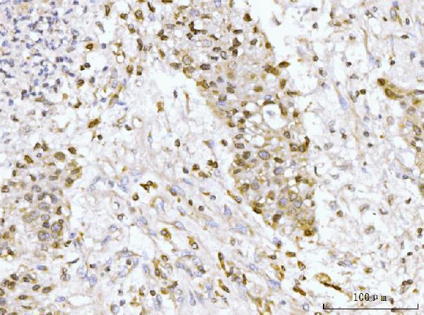
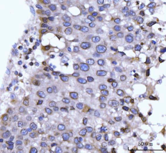
Real IHC data IHC analysis of GP210/NUP210 using anti-GP210/NUP210 antibody (A05308). GP210/NUP210 was detected in a paraffin-embedded section of human liver tissue. Heat mediated antigen retrieval was performed in EDTA buffer (pH 8.0, epitope retrieval solution). The tissue section was blocked with 10% goat serum. The tissue section was then incubated with 2 μg/ml rabbit anti-GP210/NUP210 Antibody (A05308) overnight at 4°C. Biotinylated goat anti-rabbit IgG was used as secondary antibody and incubated for 30 minutes at 37°C. The tissue section was developed using Strepavidin-Biotin-Complex (SABC) (Catalog # SA1022) with DAB as the chromogen.
Anti-GP210/NUP210 Antibody ®
Cat # A05308

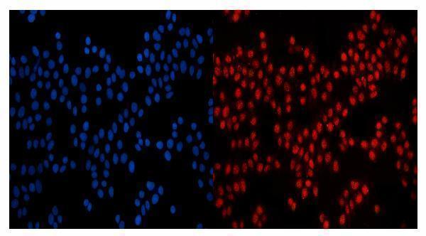
A05308 was shown by IHC in paraffin sections of human liver and appendiceal adenocarcinoma (A05308 IHC captions). A05308 was shown by IF/ICC in MCF-7 cells (A05308 IF caption).

Which to pick: Choose A05308 for human paraffin-section IHC: its caption documents EDTA retrieval at pH 8.0 and a primary concentration of 2 μg/ml; the fixative is unreported (A05308 IHC caption). For IF/ICC, A05308 has an MCF-7 image at 5 μg/ml (A05308 IF caption). For rat samples, A05308 lists rat reactivity, but the supplied IHC and IF images show human material; clonality is unreported (A05308 catalog reactivity, image captions and clone field).

Each figure is that product's own IHC / IF validation image from its datasheet.

References

  1. UniProt Consortium. UniProt entry Q8TEM1 (PO210_HUMAN, Nuclear pore membrane glycoprotein 210).
  2. Human Protein Atlas. NUP210 tissue IHC expression (reliability: Approved).
  3. Human Protein Atlas. NUP210 subcellular location (ICC-IF): Membrane.
  4. Human Protein Atlas. NUP210 antibody validation summary (1 antibodies).
  5. Super-enhancer-associated lncRNA HDAC11-AS1 aggravates hepatocellular carcinoma progression by modulating HDAC11 and NUP210 expression via promoting super-enhancer activity. Cellular oncology (Dordrecht, Netherlands) 2026 — PMC13365274.
  6. The Prognosis-Predictive and Immunoregulatory Role of SUMOylation Related Genes: Potential Novel Targets in Prostate Cancer Treatment. International journal of molecular sciences 2023 — PMC10488061.
  7. NUP210 and MicroRNA-22 Modulate Fas to Elicit HeLa Cell Cycle Arrest. Yonsei medical journal 2020 — PMC7214106.
  8. Nuclear pore protein NUP210 depletion suppresses metastasis through heterochromatin-mediated disruption of tumor cell mechanical response. Nature communications 2021 — PMC8669001.
  9. PubMed PMID:16641997 — UniProt-cited evidence.
  10. PubMed PMID:15489334 — UniProt-cited evidence.
  11. PubMed PMID:14702039 — UniProt-cited evidence.