NUP37 / Nucleoporin Nup37 · IHC design guide

Design Immunohistochemistry for NUP37

Plan NUP37 IHC in paraffin sections using the nuclear membrane staining seen in most cells as the expected tissue pattern (HPA tissue IHC). Compare staining with appropriate controls because agreement between antibody staining and RNA expression is medium (HPA tissue IHC).

Evidence assembled Oct 2026 · For research use; verify linked source records and product datasheet before use
Immunohistochemistry protocol sheet for NUP37 (IHC for NUP37): expected localisation Nuclear membranes in most cells (HPA tissue IHC), antibody A11877-1, validated IHC image, and IHC protocol steps
Printable NUP37 IHC protocol sheet — expected localisation Nuclear membranes in most cells (HPA tissue IHC), antibody A11877-1, controls and protocol steps. Open the full NUP37 IHC guide →

NUP37 Immunohistochemistry Experimental Design Guide

Expected localisation, validated protocols, controls and antibodies — the at-a-glance facts below, then the full design guide.

Must know before staining
Expected localisation Nuclear membranes in most cells (HPA tissue IHC)
Staining pattern Nuclear membrane staining across most cell types (HPA tissue IHC)
Antigen retrieval EDTA pH 8.0 HIER, heat-mediated (datasheet A11877-1)
Positive control ⓘ Caudate+4 more · see all
Negative control ⓘ Liver+4 more · see all
Important caveats
Reasons your staining may differ from the expected pattern.
Fixation Keep fixation consistent across sections. (standard IHC practice; not target-specific)
Caveat Staining and RNA data have medium consistency (HPA tissue IHC)
Regulation No expression regulator annotated (UniProt)
Isoform / epitope 0 isoforms; no processing annotated (UniProt)
Section 1

Recommended NUP37 IHC & IF Protocols

The catalog antibody protocol uses EDTA pH 8.0 heat retrieval (datasheet A11877-1). The published NUP37 IHC protocols below report additional tissue staining conditions (PMC8335818; PMC5716709; PMC8800332).

Recommended immunohistochemistry (IHC-P) protocol parameters
SampleParaffin-embedded human bladder urothelial carcinoma tissue; fixative not specified (datasheet A11877-1)
FixationImage fixative and duration unreported (datasheet A11877-1); verify before use.
Sectioning4–5 µm sections on charged slides (standard)
DeparaffinisationXylene, graded ethanol series to water (standard)
Antigen retrievalHeat retrieval: EDTA pH 8.0 (datasheet A11877-1); 20 min, 95–100 °C (standard)
Peroxidase block3% H2O2, 10 min, room temperature (standard)
Blocking10% goat serum (datasheet A11877-1)
Primary antibodyRabbit anti-NUP37, 2-5 μg/ml (datasheet A11877-1)
Primary incubationOvernight at 4 °C (datasheet A11877-1)
DetectionHRP-conjugated secondary, DAB chromogen (datasheet A11877-1)
CounterstainHematoxylin, blue, dehydrate and mount (standard)
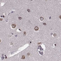
Expected resultNUP37-positive staining in neuronal cells of caudate (HPA tissue IHC: High). HPA tissue profile: Expression in the nuclear membranes in most cells. No signal in the no-primary control.
💡Decision noteStart with EDTA pH 8.0 heat retrieval (datasheet A11877-1); the glioma protocol also specifies microwave retrieval in EDTA pH 8.0 (PMC8335818).
Section 2

What Is the Expected NUP37 Staining Pattern?

NUP37 is a nuclear pore complex component with no transmembrane segment (UniProt Q8NFH4). In paraffin sections, expect staining at nuclear membranes in many cells, including strong staining in neuronal cells of the caudate and cerebral cortex (HPA tissue IHC). HPA rates the tissue pattern Supported, with medium agreement between antibody staining and RNA data and external verification pending (HPA tissue IHC).

What am I looking at on my slide?
Nuclear-rim staining in neuronal cells of the caudate or cerebral cortex; staining may vary among neighboring cells.This fits HPA's nuclear-membrane pattern and High staining in those neuronal populations (HPA tissue IHC). Judge the stained cells and compartment together: HPA reports low tissue specificity, so a positive result need not be confined to one tissue (HPA tissue IHC).
Predominantly cytoplasmic, extracellular, or tissue-edge color, with little convincing nuclear-rim staining.That distribution conflicts with the nuclear-membrane IHC profile and nuclear pore localisation (HPA tissue IHC; UniProt Q8NFH4). Treat it as a possible artefact and inspect morphology, background, and controls before calling NUP37 positive (standard IHC practice).
Strong color in alveolar cells, cholangiocytes, or rectal glandular cells while the expected nuclear pattern is unclear.HPA lists these specific cell populations as Not detected (HPA tissue IHC). An unexpected signal warrants a check for cross-reactivity or endogenous detection activity (standard IHC practice). It does not make the entire lung, liver, or rectum a validated negative tissue (HPA tissue IHC).
Diffuse color covers several compartments and obscures nuclear boundaries across the section.The compartment cannot be scored reliably against HPA's nuclear-membrane pattern (HPA tissue IHC). Broad chromogenic background can arise from detection or blocking problems (standard IHC practice); compare the no-primary control and review staining conditions before interpreting intensity.
No convincing signal in caudate neuronal cells or cerebellar Bergmann glia nuclei.Both are reported High by HPA, making absent staining a reason to investigate the run (HPA tissue IHC). Check tissue preservation, controls, retrieval settings, antibody conditions, and detection using general IHC workflow checks; no NUP37-specific fixation sensitivity is established here (standard IHC practice; supplied UniProt/HPA record).
💡Expected NUP37 appearanceCall a positive result when identifiable cells show nuclear-membrane chromogen, especially HPA High neuronal populations; isolated diffuse cytoplasmic or extracellular color without that pattern is suspect (HPA tissue IHC; standard IHC practice).
How each factor affects the staining
Where should signal sit?UniProt places NUP37 at the nuclear pore complex and also at centromeres/kinetochores; its sequence has no transmembrane segment (UniProt Q8NFH4). For routine tissue IHC, HPA's observed nuclear-membrane profile is the practical reference; a visible mitotic kinetochore pattern is not required by the supplied IHC evidence (HPA tissue IHC).
How strong is the tissue reference?HPA reports High staining in caudate and cerebral cortex neuronal cells and cerebellar Bergmann glia nuclei, Medium in breast glandular and bronchial respiratory epithelial cells, and Not detected in selected cell populations (HPA tissue IHC). These are cell-specific observations, not guarantees for every section or cell (HPA tissue IHC).
How firm is antibody validation?The tissue profile is Supported but has medium antibody/RNA consistency and awaits external verification (HPA tissue IHC). HPA073708 is Supported for IHC; HPA056300 is Supported for ICC, with no IHC status supplied for that antibody (HPA antibodies). Keep those application-specific assessments separate when judging an unexpected result.
What should IF/ICC show?HPA reports supported nucleoplasmic localisation in ICC-IF, whereas its tissue IHC description emphasizes nuclear membranes (HPA subcellular; HPA tissue IHC). This assay-specific difference calls for interpreting each image against its own reported pattern; the supplied sources do not establish that one pattern is caused by fixation or antigen retrieval.
Do processing or fixation explain a weak result?UniProt lists one NUP37 chain spanning residues 1–326, with no signal peptide, propeptide, annotated glycosylation sites, or isoforms (UniProt Q8NFH4). Those annotations do not identify an antibody epitope or predict staining after fixation; target-specific fixation sensitivity is unreported in the supplied UniProt/HPA evidence.
Why is my staining missing, weak or wrong?
SituationLikely causeNext action
A known-positive neuronal population is blank.The run may have a retrieval, antibody, detection, or tissue-quality problem; the source data do not identify which step failed (standard IHC practice; HPA tissue IHC).Confirm the expected cells are present, inspect a positive control, and review the validated IHC conditions for the antibody (standard IHC practice). HPA lists caudate and cerebral cortex neuronal cells as High references (HPA tissue IHC).
Color is widespread but nuclear rims are indistinct.Background or overly strong detection can obscure compartment scoring (standard IHC practice).Compare a no-primary control, then review blocking, antibody concentration, washes, and chromogen development as general IHC variables (standard IHC practice). Score NUP37 only when the nuclear distribution is interpretable against HPA's profile (HPA tissue IHC).
Unexpected color appears in an HPA Not detected cell type.Cross-reactivity or endogenous detection activity is possible; HPA's result is cell-specific and has pending external verification (standard IHC practice; HPA tissue IHC).Check a no-primary control and the staining compartment, then repeat with appropriate detection controls if needed (standard IHC practice). Compare the same named cell type with its HPA entry before treating the discrepancy as biological (HPA tissue IHC).
A section shows only cytoplasmic or extracellular deposits.That location conflicts with the reported nuclear-membrane IHC profile and nuclear pore association (HPA tissue IHC; UniProt Q8NFH4).Review section edges, tissue morphology, counterstain, and controls for non-specific deposition (standard IHC practice). Do not score deposits without a credible nuclear pattern as NUP37-positive cells (HPA tissue IHC; standard IHC practice).
An IF/ICC image looks nucleoplasmic rather than rim-like.HPA reports supported nucleoplasmic ICC-IF localisation and a nuclear-membrane tissue IHC profile (HPA subcellular; HPA tissue IHC).Interpret the IF/ICC image with its own HPA reference and antibody's ICC validation status (HPA subcellular; HPA antibodies). Do not infer a NUP37-specific fixation or retrieval mechanism from the difference; none is supplied by these sources.
A low-staining tissue gives a faint nuclear signal.HPA reports Low staining for glandular cells in thyroid, parathyroid, adrenal gland, stomach, and duodenum, and for esophageal squamous cells (HPA tissue IHC).Check the named cell population and nuclear distribution, then compare with a High reference and controls (HPA tissue IHC; standard IHC practice). Avoid assigning a tissue-wide positive or negative call from a faint signal in one cell group.

Sample controls for NUP37 IHC & IF

🧪Run caudate first and assess staining in neuronal cells (HPA: High in caudate neuronal cells); use liver cholangiocytes as the negative tissue comparison (HPA: Not detected in liver cholangiocytes). On the caudate slide, document adjacent cells with no specific nuclear staining above control background as internal negative comparators; HPA does not identify a particular negative cell type within caudate (HPA: caudate neuronal cells, High).
Positive control tissue: Caudate (Neuronal cells, HPA High)
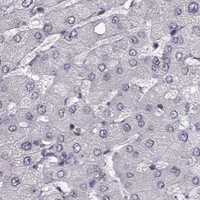
Negative control tissue: Liver (HPA Not detected)
ICC-IF cell lines (HPA subcellular resource): HPA ICC-IF images show NUP37 in A-431, U-251MG, U2OS, NIH 3T3, with annotated localisation: Nucleoplasm (supported) (HPA subcellular).
Technical controls: Include a no-primary, secondary-only control, a concentration-matched nonimmune rabbit IgG control for the rabbit primary antibody (selected-SKU tissue-IHC caption: rabbit anti-NUP37), and NUP37 knockout material as a biological specificity control. Quench endogenous peroxidase and inspect caudate sections for pigment that could be mistaken for DAB signal (standard chromogenic IHC practice).
⚠️Feasibility: A target-specific fixation window or fixation effect is unreported, and the selected-SKU paraffin-section caption does not report a fixative (selected-SKU tissue-IHC caption: fixative not stated). The demonstrated paraffin IHC procedure uses heat retrieval in EDTA at pH 8.0, followed by 2 μg/ml primary antibody overnight at 4°C; the evidence does not establish that retrieval is essential (selected-SKU tissue-IHC caption). Frozen sections have no supplied protocol, while ICC-IF images support nucleoplasmic localization in A-431, U-251MG, U2OS and NIH 3T3; neither route is established as easier than paraffin IHC, and pigment should be checked when scoring caudate DAB staining (HPA: Nucleoplasm, supported; HPA: ICC-IF image cell lines; standard chromogenic IHC practice).

HPA tissue IHC evidence for NUP37

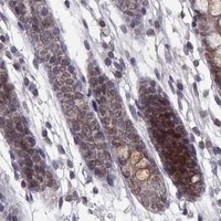
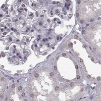
Comprehensive Human Protein Atlas IHC scoring per tissue (reliability: Supported — Medium consistency between antibody staining and RNA expression data. Pending external verification.). Rows are taken directly from the HPA tissue chart — click any row's HPA link to view the source.

Positive expression · recommended positive controls

TissueCell typeLevelEvidenceSource
Caudate Neuronal cells High Protein (IHC) HPA →
Cerebellum Bergmann glia - nucleus High Protein (IHC) HPA →
Cerebral cortex Neuronal cells High Protein (IHC) HPA →
Bone marrow Hematopoietic cells Medium Protein (IHC) HPA →
Breast Glandular cells Medium Protein (IHC) HPA →

Undetected expression · recommended negative controls

TissueCell typeLevelEvidenceSource
Liver Cholangiocytes Not detected Protein (IHC) HPA →
Lung Alveolar cells Not detected Protein (IHC) HPA →
Oral mucosa Squamous epithelial cells Not detected Protein (IHC) HPA →
Prostate Glandular cells Not detected Protein (IHC) HPA →
Rectum Glandular cells Not detected Protein (IHC) HPA →
Section 3

Advanced NUP37 IHC Tips

Troubleshoot NUP37 staining by checking retrieval, nuclear localisation, antibody controls and consistent scoring across paraffin sections.

What retrieval should I try when NUP37 staining is weak?
Start with heat-mediated antigen retrieval in EDTA at pH 8.0 for paraffin sections (datasheet A11877-1). This is the retrieval used for the catalog antibody’s illustrated NUP37 tissue staining; the caption does not report a heating time, so record the time and temperature chosen for each run (datasheet A11877-1; standard IHC practice). After retrieval, compare intact nuclear staining with a matched section processed in the same run, while keeping detection and counterstain conditions fixed (HPA: nuclear-membrane expression; standard IHC practice). If staining remains weak, adjust heating duration in small steps and check tissue preservation before testing another buffer as a fallback (standard IHC practice).
Could fixation explain faint or uneven NUP37 staining?
NUP37-specific fixation sensitivity is unknown because the selected paraffin-section caption does not state a fixative (datasheet A11877-1). Record fixation type and duration for each specimen, and compare sections prepared under matched conditions before assigning faint staining to biology (standard IHC practice). Excessive fixation can reduce antibody access, while uneven fixation can produce regional staining differences in paraffin sections (standard IHC practice). If staining varies across a block, examine tissue morphology and compare sections through the same pH 8.0 EDTA retrieval and detection run; interpret persistent differences only after processing differences have been assessed (datasheet A11877-1; standard IHC practice).
Where should convincing NUP37 signal appear in a paraffin section?
Expect predominantly nuclear-associated staining: NUP37 belongs to the nuclear pore complex and also localises to centromeres and kinetochores (UniProt Q8NFH4). Tissue IHC describes expression at nuclear membranes in most cells, while subcellular IF supports nucleoplasmic localisation (HPA: tissue IHC profile; HPA: subcellular summary). With a chromogenic stain, assess whether signal follows nuclei consistently across well-preserved cells rather than treating every faint perinuclear rim as definitive (HPA: tissue IHC profile; standard IHC practice). Diffuse cytoplasmic or extracellular DAB without corresponding nuclear-associated staining warrants review of background, tissue integrity and antibody controls (UniProt Q8NFH4; standard IHC practice).
Could an isoform or hidden epitope explain discordant NUP37 results?
The supplied NUP37 record lists a 326-residue chain and no annotated isoforms, domains, glycosylation sites or modified residues (UniProt Q8NFH4). Those annotations do not establish which epitope the catalog antibody recognises or whether paraffin processing exposes it equally in every specimen (UniProt Q8NFH4; standard IHC practice). Check the antibody’s stated immunogen or epitope information, if available, before attributing a staining difference to an isoform or modification (standard IHC practice). For the illustrated antibody, begin comparisons with pH 8.0 EDTA retrieval and 2 μg/ml primary antibody, then change one condition at a time while monitoring nuclear-associated staining (datasheet A11877-1; HPA: tissue IHC profile).
How can IF help investigate ambiguous chromogenic NUP37 staining?
Use IF as a separate localisation check: NUP37 is reported at nuclear pores and kinetochores, and supported subcellular IF localisation is nucleoplasmic (UniProt Q8NFH4; HPA: subcellular summary). For multiplexing, pair NUP37 with a validated neuronal-cell marker when examining neuronal cells, which are listed as high in the caudate and cerebral cortex (HPA: tissue IHC). Select a spectrally separated, preferably far-red NUP37 fluorophore after checking tissue autofluorescence and single-colour controls (standard IF practice). Because NUP37 has no transmembrane segment, choose permeabilisation according to the antibody epitope’s accessibility and use mild detergent when intracellular nuclear access is needed; the supplied evidence does not map that epitope (UniProt Q8NFH4; standard IF practice).
What should I check when DAB obscures nuclear NUP37 staining?
First inspect a no-primary control for staining caused by endogenous peroxidase, secondary reagent or chromogen, and include a peroxidase-blocking step in the chromogenic workflow (standard IHC practice). The illustrated paraffin-section assay used 10% goat-serum blocking, 2 μg/ml primary antibody overnight at 4°C, an anti-rabbit peroxidase secondary for 30 minutes at 37°C, and DAB (datasheet A11877-1). If diffuse stain persists, review blocking, washing, primary concentration and DAB development while changing one factor at a time (standard IHC practice). Score nuclear-associated signal only where cell boundaries and morphology remain interpretable, since background can mimic a broad nuclear rim (HPA: tissue IHC profile; standard IHC practice).
How should I score NUP37 across differently processed sections? ⚠ ANSWER MARKED FOR VERIFICATION
Define the nuclear-associated compartment and cell population before scoring, then use the same threshold and imaging settings for every comparison (HPA: tissue IHC profile; standard IHC practice). An H-score combines the percentage of cells at each intensity and spans 0–300; alternatively report percentage of positive cells or positive-cell density per mm² of evaluable tissue (standard IHC practice). Normalise counts to the number of intact target cells or measured viable tissue area, and exclude folds, necrosis and section edges using prespecified rules (standard IHC practice). Report retrieval, primary concentration and DAB development alongside scores so processing differences can be distinguished from apparent biological differences (standard IHC practice).
When is a NUP37-positive region credible rather than artefactual?
A credible result has reproducible nuclear-associated staining in morphologically intact cells, consistent with the nuclear pore and tissue IHC evidence for NUP37 (UniProt Q8NFH4; HPA: tissue IHC profile; standard IHC practice). Interpret the cell type as well as intensity: neuronal cells in caudate and cerebral cortex are listed as high, whereas liver cholangiocytes are listed as not detected in the supplied tissue profile (HPA: tissue IHC). Diffuse cytoplasmic stain, sharp section-edge enhancement, necrotic deposits or signal in a no-primary control call for review before scoring (UniProt Q8NFH4; standard IHC practice). Treat disagreement with the HPA profile as a prompt to check morphology, processing and controls; its tissue IHC reliability is Supported with external verification pending (HPA: tissue IHC reliability).
Boster reagents

Best NUP37 / Nucleoporin Nup37 IHC Antibodies

A11877-1 has IHC images from human and rat paraffin sections (catalog IHC captions) and IF/ICC data from U2OS cells (catalog IF caption).

Real IHC data IHC analysis of NUP37 using anti-NUP37 antibody (A11877-1). NUP37 was detected in a paraffin-embedded section of human bladder urothelial carcinoma tissue. Heat mediated antigen retrieval was performed in EDTA buffer (pH 8.0, epitope retrieval solution). The tissue section was blocked with 10% goat serum. The tissue section was then incubated with 2 μg/ml rabbit anti-NUP37 Antibody (A11877-1) overnight at 4°C. Peroxidase Conjugated Goat Anti-rabbit IgG was used as secondary antibody and incubated for 30 minutes at 37°C. The tissue section was developed using HRP Conjugated Rabbit IgG Super Vision Assay Kit (Catalog # SV0002) with DAB as the chromogen.
Anti-NUP37 Antibody ®
Cat # A11877-1

A11877-1 will render with an IHC image from a human bladder urothelial carcinoma paraffin section (card image caption). Its catalog also documents IHC in human large B-cell lymphoma, human ovarian cancer, and rat brain sections, plus IF/ICC in U2OS cells (catalog IHC and IF captions).

Which to pick: Choose A11877-1 for paraffin-section IHC: its caption documents EDTA retrieval at pH 8.0 and primary antibody at 2 μg/ml (card image caption). Choose the same SKU for IF/ICC in cells, supported by its U2OS image at 5 μg/ml (catalog IF caption), or for human and rat IHC, supported by the catalog reactivity list and IHC captions. Clonality and the IHC section fixative are unreported (catalog payload and IHC captions).

Each figure is that product's own IHC / IF validation image from its datasheet.

References

  1. UniProt Consortium. UniProt entry Q8NFH4 (NUP37_HUMAN, Nucleoporin Nup37).
  2. Human Protein Atlas. NUP37 tissue IHC expression (reliability: Supported).
  3. Human Protein Atlas. NUP37 subcellular location (ICC-IF): Localized to the nucleoplasm..
  4. Human Protein Atlas. NUP37 antibody validation summary (2 antibodies).
  5. Significantly high expression of NUP37 leads to poor prognosis of glioma patients by promoting the proliferation of glioma cells. Cancer medicine 2021 — PMC8335818.
  6. NUP37, a positive regulator of YAP/TEAD signaling, promotes the progression of hepatocellular carcinoma. Oncotarget 2017 — PMC5716709.
  7. DEPDC1B is involved in the proliferation, metastasis, cell cycle arrest and apoptosis of colon cancer cells by regulating NUP37. Molecular medicine reports 2023 — PMC10206622.
  8. Demethylation at enhancer upregulates MCM2 and NUP37 expression predicting poor survival in hepatocellular carcinoma patients. Journal of translational medicine 2022 — PMC8800332.
  9. PubMed PMID:12196509 — UniProt-cited evidence.
  10. PubMed PMID:15489334 — UniProt-cited evidence.
  11. PubMed PMID:14702039 — UniProt-cited evidence.