This website uses cookies to ensure you get the best experience on our website.


- Table of Contents
Plan NUP62 IHC in paraffin sections around the nuclear membrane staining seen in most cell types, with cytoplasmic staining in some (HPA tissue IHC). Use the catalog antibody’s IHC protocol and compare staining across sections with consistent fixation (datasheet A03950-2; standard IHC practice).

Expected localisation, validated protocols, controls and antibodies — the at-a-glance facts below, then the full design guide.
| Expected localisation | Nuclear membrane in most cells; cytoplasm in some (HPA tissue IHC) | |
| Staining pattern | Most cells: nuclear membrane; some: cytoplasm (HPA tissue IHC) | |
| Antigen retrieval | EDTA pH 8.0 HIER, heat-mediated (datasheet A03950-2) | |
| Positive control | Caudate+4 more · see all | |
| Negative control | None in HPA (detected in all 45 tissues); use no-primary + isotype controls |
| Fixation | Keep fixation consistent across sections (standard IHC practice; not target-specific) | |
| Caveat | Parathyroid glandular cells show low staining (HPA tissue IHC) | |
| Regulation | No specific regulator annotated (UniProt) | |
| Isoform / epitope | No isoforms annotated; no transmembrane segment (UniProt) |
The catalog antibody’s IHC-P protocol uses EDTA retrieval (datasheet: A03950-2). The published protocols below describe chromogenic NUP62 staining in rat and human tissue (PMC8169113; PMC11684083; PMC5595097).
| Sample | Paraffin-embedded human ovarian cancer tissue; fixative not specified (datasheet A03950-2) |
| Fixation | Image fixative and duration unreported (datasheet A03950-2); verify before use. |
| Sectioning | 4–5 µm sections on charged slides (standard) |
| Deparaffinisation | Xylene, graded ethanol series to water (standard) |
| Antigen retrieval | Heat retrieval: EDTA pH 8.0 (datasheet A03950-2); 20 min, 95–100 °C (standard) |
| Peroxidase block | 3% H2O2, 10 min, room temperature (standard) |
| Blocking | 10% goat serum (datasheet A03950-2) |
| Primary antibody | Rabbit anti-NUP62, 2-5 μg/ml (datasheet A03950-2) |
| Primary incubation | Overnight at 4 °C (datasheet A03950-2) |
| Detection | HRP-conjugated secondary, DAB chromogen (datasheet A03950-2) |
| Counterstain | Hematoxylin, blue, dehydrate and mount (standard) |
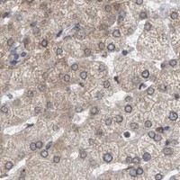
| Expected result | NUP62-positive staining in neuronal cells of caudate (HPA tissue IHC: High). HPA tissue profile: Expression in the nuclear membranes in most cell types. Also cytoplasmic expression in several cases. No signal in the no-primary control. |
NUP62 is a nuclear pore protein with no transmembrane segment (UniProt P37198: localization and topology). In paraffin section IHC, expect staining along the nuclear membrane in most cell types; cytoplasmic staining can also occur (HPA: tissue IHC profile). HPA rates the tissue pattern Supported, citing high consistency between antibody staining and RNA expression (HPA: tissue IHC reliability).
| A nuclear rim is visible in many cells, including strongly stained caudate neuronal cells, placental endothelial cells, or rectal glandular cells (HPA: tissue IHC, High). | This fits NUP62 at the nuclear pore complex (UniProt P37198: localization) and HPA's predominant nuclear membrane pattern (HPA: tissue IHC profile). Judge the rim against the counterstained nucleus; tissue labels alone do not establish the identity of every stained cell (general IHC practice). |
| Some cells show cytoplasmic staining in addition to a nuclear rim (HPA: tissue IHC profile). | Cytoplasmic signal is not automatically false: HPA reports it in several cases, and UniProt also places NUP62 at spindle poles and centrosomes (HPA: tissue IHC profile; UniProt P37198: localization). Assess whether the expected nuclear membrane pattern remains discernible before interpreting the extra signal. |
| Strong, uniform signal fills cells or extracellular areas while nuclear rims are indistinct. | Treat this as an uncertain pattern rather than evidence of NUP62 localization: the reported tissue pattern emphasizes nuclear membranes (HPA: tissue IHC profile). Diffuse chromogen, tissue pigment, or detection background can complicate compartment scoring (general IHC practice); inspect controls and morphology. |
| Staining appears mainly in an unexpected cell population, with little signal in the expected population. | Consider antibody cross-reactivity or endogenous detection activity (general IHC practice), but first check cell identification. HPA reports low staining in several sampled populations, not a list of negative tissues, and describes expression in most cell types (HPA: tissue IHC profile). A low-staining cell is therefore a weak exclusion control. |
| A known HPA High population has no discernible nuclear membrane staining. | The result needs a technical check before a biological absence call: HPA reports High staining in caudate neuronal, placental endothelial, and rectal glandular cells (HPA: tissue IHC). Check section quality, counterstain, antibody application, and detection with appropriate controls (general IHC practice). HPA staining levels do not establish fixation sensitivity. |
| Compartment and topology (UniProt P37198: localization and topology). | NUP62 occupies the nuclear pore complex and has no transmembrane segment (UniProt P37198). Score the nuclear boundary rather than assuming all nuclear or cytoplasmic color is equivalent; HPA's tissue pattern centers on the nuclear membrane (HPA: tissue IHC profile). |
| Cell and tissue context (HPA: tissue IHC). | Caudate neuronal cells, placental endothelial cells, and rectal glandular cells are High; appendix and breast glandular cells are Medium, among other listed Medium populations (HPA: tissue IHC). These are reported staining levels, not promised signal strengths for every section or run. |
| Cytoplasmic and mitotic context (HPA: tissue IHC; UniProt P37198: localization). | HPA reports cytoplasmic expression in several cases. UniProt describes association with mitotic spindle poles and centrosomes (HPA: tissue IHC profile; UniProt P37198: localization). Evaluate such staining with cell morphology and the nuclear rim pattern; neither source establishes that every cytoplasmic deposit is specific. |
| Evidence and antibody choice (HPA: antibody validation). | The listed antibodies HPA005435 and CAB020724 have Supported IHC status (HPA: antibody validation). Supported tissue reliability reflects agreement with RNA expression; it does not by itself prove every stained compartment in a new specimen is specific (HPA: tissue IHC reliability; general IHC practice). |
| Fixation evidence (HPA: tissue IHC; UniProt P37198). | No target-specific fixation sensitivity is supplied by these records. HPA staining levels and UniProt topology or modifications do not establish how fixation changes NUP62 detection (HPA: tissue IHC; UniProt P37198). Assess local processing with controls rather than assigning a target-specific fixation effect. |
| Situation | Likely cause | Next action |
|---|---|---|
| Positive control has no nuclear rim (HPA: tissue IHC pattern). | Antibody delivery, retrieval, detection, or section quality may have failed (general IHC practice); these sources do not identify a NUP62-specific fixation effect. | Repeat with a documented High population such as placental endothelial or rectal glandular cells (HPA: tissue IHC). Verify each IHC step and compare an appropriate run control (general IHC practice). |
| All cells or blank areas show diffuse chromogen. | Background from the detection workflow may obscure the nuclear boundary (general IHC practice). HPA's principal pattern is nuclear membrane staining (HPA: tissue IHC profile). | Inspect a no-primary control and tissue morphology; review blocking, washing, detection exposure, and counterstain using general IHC practice. Score only interpretable cellular staining. |
| Cytoplasm stains more strongly than the nuclear rim. | Some cytoplasmic NUP62 signal is plausible, while diffuse background is also possible (HPA: tissue IHC profile; general IHC practice). | Compare intact cells with the no-primary control and check whether a nuclear rim can still be resolved (HPA: tissue IHC profile; general IHC practice). Do not classify all cytoplasmic staining as artefact. |
| Only an HPA Low population is available as a comparison. | Low staining is not an absent-protein control; HPA lists low glandular staining in parathyroid and adrenal glands, among other Low populations (HPA: tissue IHC). | Add a documented High population to the same run where practical, and compare compartments within well-preserved cells (HPA: tissue IHC; general IHC practice). Avoid calling a Low population definitively negative. |
| A surprising cell population stains while the expected population does not. | Cell identification, cross-reactivity, or endogenous detection activity may account for the discrepancy (general IHC practice); HPA reports broad nuclear membrane expression (HPA: tissue IHC profile). | Recheck morphology and controls, then compare a listed High population and, if available, an independently validated antibody (HPA: tissue IHC and antibody validation; general IHC practice). |
| IF/ICC question: should fluorescence be read like the paraffin section IHC result? | HPA's ICC-IF summary places NUP62 mainly at the nuclear membrane and additionally in the nucleoplasm (HPA: subcellular ICC-IF); the tissue IHC profile also allows cytoplasmic staining in several cases (HPA: tissue IHC). | Use the nuclear membrane as the shared localization check, while interpreting nucleoplasmic fluorescence in the separate IF/ICC context (HPA: subcellular ICC-IF). Do not infer an IHC protocol choice from the ICC-IF images. |
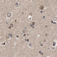



Comprehensive Human Protein Atlas IHC scoring per tissue (reliability: Supported — High consistency between antibody staining and RNA expression data.). Rows are taken directly from the HPA tissue chart — click any row's HPA link to view the source.
| Tissue | Cell type | Level | Evidence | Source |
|---|---|---|---|---|
| Caudate | Neuronal cells | High | Protein (IHC) | HPA → |
| Placenta | Endothelial cells | High | Protein (IHC) | HPA → |
| Rectum | Glandular cells | High | Protein (IHC) | HPA → |
| Appendix | Glandular cells | Medium | Protein (IHC) | HPA → |
| Bone marrow | Hematopoietic cells | Medium | Protein (IHC) | HPA → |
| Tissue | Cell type | Level | Evidence | Source |
|---|---|---|---|---|
| None in HPA: NUP62 is detected in all 45 scored tissues. Use a no-primary (secondary-only) and an isotype control instead. | ||||
Troubleshoot NUP62 staining in paraffin sections by checking retrieval, nuclear envelope localisation, controls and cell-specific scoring before interpreting signal.
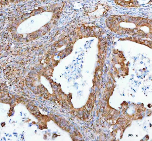
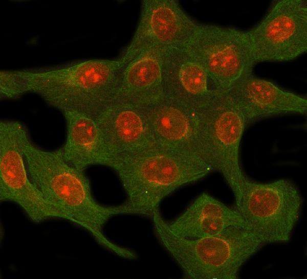
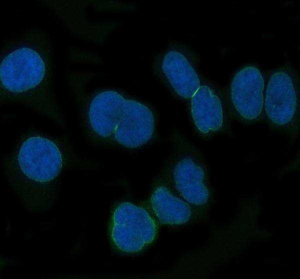
Anti-NUP62 antibodies have IHC data from a human paraffin-embedded ovarian cancer section and IF data from human U2OS and HeLa cells; both list Human, Mouse and Rat reactivity (catalog images; catalog reactivity).
A03950-2 will render with IHC data from a human paraffin-embedded ovarian cancer section; its catalog also lists IF/ICC and shows IF in U2OS cells (A03950-2 image captions; catalog applications). M03950 will render with IF data from HeLa cells and lists IF/ICC applications (M03950 image caption; catalog applications).
Which to pick: Choose A03950-2 for tissue IHC: its image shows staining in a paraffin-embedded section after EDTA pH 8.0 heat retrieval at 2 μg/ml; the fixative is unreported (A03950-2 IHC image caption). For IF/ICC, both SKUs are listed, with A03950-2 shown in U2OS cells at 5 μg/ml and rabbit monoclonal M03950 shown in HeLa cells at 1:50 (catalog applications; A03950-2 and M03950 IF image captions; M03950 catalog title). Both list Human, Mouse and Rat reactivity, but the supplied IHC and IF images show human samples only (catalog reactivity; image captions).