This website uses cookies to ensure you get the best experience on our website.


- Table of Contents
This guide uses high staining in bone marrow hematopoietic cells and undetected staining in adipocytes as cell-level references for paraffin-section NUSAP1 IHC (HPA tissue IHC). Interpret nuclear immune-cell staining alongside the reported off-target binding caveat (HPA tissue IHC).

Expected localisation, validated protocols, controls and antibodies — the at-a-glance facts below, then the full design guide.
| Expected localisation | Nuclear in immune cells; cytoplasmic/membranous elsewhere (HPA tissue IHC) | |
| Staining pattern | Immune-cell nuclei; cytoplasmic/membranous staining in other tissues (HPA tissue IHC) | |
| Antigen retrieval | EDTA pH 8.0 HIER, heat-mediated (datasheet A06066-2) | |
| Positive control | Appendix+4 more · see all | |
| Negative control | Adipose tissue+4 more · see all |
| Fixation | Keep paraffin-section fixation consistent across cases. (selected-SKU IHC image A06066-2) | |
| Caveat | Presumed off-target binding may confound staining (HPA tissue IHC) | |
| Regulation | Cell-cycle linked; intensity unreported (UniProt) | |
| Isoform / epitope | 7 isoforms; epitope coverage is unspecified (UniProt) |
The catalog antibody protocol uses EDTA pH 8.0 retrieval (datasheet A06066-2). The published IHC protocols below describe NUSAP1 staining in kidney and breast cancer samples (PMC11480612; PMC8667124; PMC4618922).
| Sample | Paraffin-embedded human prostate cancer tissue; fixative not specified (datasheet A06066-2) |
| Fixation | Image fixative and duration unreported (datasheet A06066-2); verify before use. |
| Sectioning | 4–5 µm sections on charged slides (standard) |
| Deparaffinisation | Xylene, graded ethanol series to water (standard) |
| Antigen retrieval | Heat retrieval: EDTA pH 8.0 (datasheet A06066-2); 20 min, 95–100 °C (standard) |
| Peroxidase block | 3% H2O2, 10 min, room temperature (standard) |
| Blocking | 10% goat serum (datasheet A06066-2) |
| Primary antibody | Rabbit anti-NUSAP1, 2-5 μg/ml recommended; image 1:100 (datasheet A06066-2) |
| Primary incubation | Overnight at 4 °C (datasheet A06066-2) |
| Detection | HRP-conjugated secondary, DAB chromogen (datasheet A06066-2) |
| Counterstain | Hematoxylin, blue, dehydrate and mount (standard) |
| Expected result | NUSAP1-positive staining in endocrine cells of appendix (HPA tissue IHC: High). HPA tissue profile: Cytoplasmic and membranous expression in several tissues, most abundant in the nuclear subcellular location of immune cells. No signal in the no-primary control. |
NUSAP1 can occupy the cytoplasm and nucleolus between divisions, then associate with chromosomes and the mitotic spindle during division (UniProt Q9BXS6). In tissue IHC, expect cell-specific staining, including strong staining in bone marrow hematopoietic cells (HPA: High). HPA rates its tissue pattern Supported, with medium agreement with RNA data and a warning about possible off-target staining (HPA: Supported). NUSAP1 has no transmembrane segment (UniProt Q9BXS6 topology).
| Strong staining in bone marrow hematopoietic cells, with staining in nuclei of immune cells. | This fits an HPA high-staining cell population and its description of nuclear staining as most abundant in immune cells (HPA: High in bone marrow hematopoietic cells; HPA: tissue IHC profile). Record the stained cell type and compartment: intensity alone is insufficient because HPA also reports cytoplasmic and membranous staining in several tissues (HPA: tissue IHC profile). |
| Signal follows a membrane rim alone, without convincing nuclear or division-associated staining. | Review the compartment call and controls before assigning this to NUSAP1. UniProt places NUSAP1 in the cytoplasm, nucleolus, spindle and chromosomes and reports no transmembrane segment (UniProt Q9BXS6). HPA nevertheless describes membranous staining in several tissues and warns of presumed off-target binding, so a rim is a concern rather than proof of artefact (HPA: tissue IHC profile). |
| A cell type reported as unstained shows strong chromogenic signal. | For example, HPA reports adipocytes as Not detected (HPA: adipose tissue). Check cell identification, primary-antibody controls and the detection system before calling the signal NUSAP1; cross-reactivity is plausible because HPA warns that antibodies target proteins from more than one gene and presumed off-target binding was observed (HPA: tissue IHC reliability). Endogenous detection activity is another general IHC possibility (standard IHC practice). |
| Brown deposit is widespread across cells or tissue spaces, obscuring cell boundaries. | Treat the pattern as background until it can be separated from cell-specific staining. Compare a no-primary control, washing and detection steps, and assess whether a recognizable nuclear or division-associated distribution remains (standard IHC practice; UniProt Q9BXS6 localization). HPA's Supported rating has only medium consistency with RNA data and includes an off-target warning (HPA: tissue IHC reliability). |
| No signal appears in a section containing bone marrow hematopoietic cells. | First confirm that the expected cells are present and that the detection run worked with appropriate controls (standard IHC practice). HPA rates these cells High, so absence merits review of the IHC-validated antibody, retrieval conditions and detection steps (HPA: bone marrow hematopoietic cells High; standard IHC practice). The supplied sources do not establish NUSAP1-specific fixation sensitivity. |
| Cell identity and tissue selection | HPA reports High staining in bone marrow hematopoietic cells, testis preleptotene spermatocytes and skin spinous-layer cells, among others (HPA: tissue IHC). It reports adipocytes and cardiomyocytes as Not detected (HPA: tissue IHC). Score the specified cells, since a tissue name alone does not define the expected result. |
| Cell-cycle stage | Interphase localization includes cytoplasm and nucleolus; during mitosis NUSAP1 redistributes to spindle and chromosome-associated regions (UniProt Q9BXS6, partly By similarity). A section with few dividing cells may offer few interpretable spindle profiles. Do not require every positive cell to display a visible spindle. |
| Antibody evidence | HPA calls the tissue IHC pattern Supported, describes medium agreement with RNA data, and warns that antibodies target proteins from more than one gene (HPA: tissue IHC reliability). HPA042904 is IHC Supported, whereas no IHC status is supplied for HPA074847 (HPA: antibody validation). Interpret unexpected staining with controls. |
| Epitope and molecular forms | UniProt lists seven isoforms and multiple modified residues, but the payload gives no antibody epitope or isoform coverage (UniProt Q9BXS6). Those annotations cannot establish which forms the IHC-validated antibody detects or predict a retrieval condition. Validate any change in staining against controls rather than attributing it to a specific modification. |
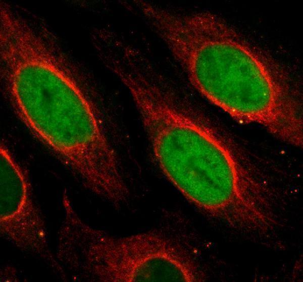
| IF/ICC: where should fluorescence appear? | HPA reports approved nucleoplasm, nucleoli and nucleoli fibrillar-center localization in ICC-IF, with a warning that the evidence uses antibodies targeting proteins from multiple genes (HPA: subcellular ICC-IF). This answers the localization question for IF/ICC; interpret paraffin-section chromogenic staining against the tissue IHC evidence (HPA: tissue IHC). |
| Situation | Likely cause | Next action |
|---|---|---|
| The expected positive cells are absent from the field. | The selected field cannot test the stated cell-level expectation; HPA's High call applies to specified cells (HPA: tissue IHC). | Locate and document the relevant cells before judging staining. For example, identify hematopoietic cells when using bone marrow (HPA: High in bone marrow hematopoietic cells). |
| Expected positive cells are present, but staining is absent. | A failed staining or detection step is possible (standard IHC practice); the sources give no NUSAP1-specific fixation-effect evidence. | Check run controls, antibody application, retrieval and chromogenic detection using the established IHC workflow (standard IHC practice). Reassess the known-positive cells (HPA: bone marrow hematopoietic cells High). |
| Signal appears mainly as a crisp membrane outline. | NUSAP1 lacks a transmembrane segment, although HPA describes membranous tissue staining and possible off-target binding (UniProt Q9BXS6 topology; HPA: tissue IHC profile and reliability). | Review the same field for nuclear or division-associated staining and compare controls before assigning the outline to NUSAP1 (UniProt Q9BXS6 localization; standard IHC practice). |
| Reported negative cells stain strongly. | Cross-reactivity is plausible under HPA's off-target warning; endogenous chromogenic activity is a separate general possibility (HPA: tissue IHC reliability; standard IHC practice). | Verify the cell type, compare a no-primary control and review detection blocking. Use HPA's Not detected calls for the specified cells as a reference, not a whole-tissue exclusion (HPA: tissue IHC). |
| Diffuse color obscures the expected compartments. | Nonspecific reagent retention or detection background can impair interpretation (standard IHC practice). | Inspect no-primary and run controls, washing and detection conditions; score NUSAP1 only where cells and compartments remain discernible (standard IHC practice; UniProt Q9BXS6 localization). |
| Staining intensity varies among adjacent positive cells. | NUSAP1 localization changes with cell-cycle stage, and HPA assigns staining levels to particular cell types (UniProt Q9BXS6; HPA: tissue IHC). | Record cell identity and compartment alongside intensity; examine visible dividing cells for spindle or chromosome-associated signal without requiring every cell to show it (UniProt Q9BXS6, partly By similarity). |
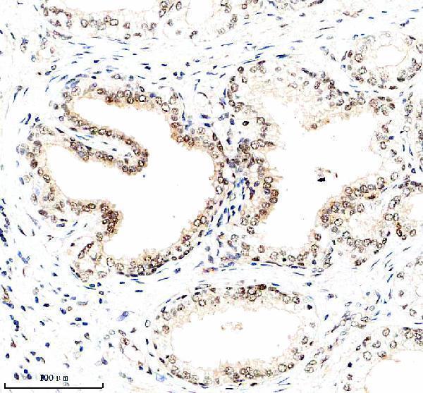
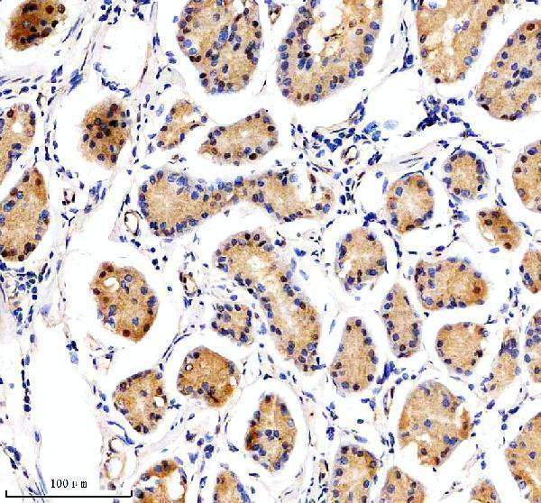


Comprehensive Human Protein Atlas IHC scoring per tissue (reliability: Supported — Medium consistency between antibody staining and RNA expression data. Caution, targets protein from more than one gene. Presumed off target binding observed and disregarded.). Rows are taken directly from the HPA tissue chart — click any row's HPA link to view the source.
| Tissue | Cell type | Level | Evidence | Source |
|---|---|---|---|---|
| Appendix | Endocrine cells | High | Protein (IHC) | HPA → |
| Bone marrow | Hematopoietic cells | High | Protein (IHC) | HPA → |
| Colon | Endocrine cells | High | Protein (IHC) | HPA → |
| Duodenum | Endocrine cells | High | Protein (IHC) | HPA → |
| Rectum | Endocrine cells | High | Protein (IHC) | HPA → |
| Tissue | Cell type | Level | Evidence | Source |
|---|---|---|---|---|
| Adipose tissue | Adipocytes | Not detected | Protein (IHC) | HPA → |
| Caudate | Glial cells | Not detected | Protein (IHC) | HPA → |
| Cerebellum | Cells in granular layer | Not detected | Protein (IHC) | HPA → |
| Cerebral cortex | Endothelial cells | Not detected | Protein (IHC) | HPA → |
| Heart muscle | Cardiomyocytes | Not detected | Protein (IHC) | HPA → |
Use the catalog antibody’s paraffin section evidence to set up NUSAP1 IHC, then assess compartment, cell type, and staining controls together.
A06066-2 has IHC images of human prostate and stomach cancer paraffin sections, plus IF images of U2OS cells and human stomach cancer paraffin sections (catalog image captions).
A06066-2 is listed for human IHC and has prostate and stomach cancer paraffin-section images (catalog applications/reactivity; IHC image captions). The same SKU is listed for IF/ICC and has IF images of U2OS cells and human stomach cancer paraffin sections (catalog applications; IF image captions).
Which to pick: Choose A06066-2 for human tissue IHC: its rabbit antibody is listed at 2–5 μg/ml, and its own IHC captions show paraffin sections; the fixative is unreported (catalog host/reactivity/dilution; IHC image captions). For IF/ICC, A06066-2 is listed at 5 μg/ml and has U2OS-cell and human tissue IF images (catalog applications/dilution; IF image captions). For cross-species work, its declared reactivity is Human, although separate IHC captions show mouse testis and epididymis (catalog reactivity; IHC image captions).