This website uses cookies to ensure you get the best experience on our website.


- Table of Contents
Plan paraffin IHC for OAS1 using the cytoplasmic tissue pattern (HPA tissue IHC) and its interferon-induced expression (UniProt). Interpret staining with the catalog antibody’s OAS1/3 designation (datasheet A01684-3) and the uncertain tissue staining reliability in mind (HPA tissue IHC).

Expected localisation, validated protocols, controls and antibodies — the at-a-glance facts below, then the full design guide.
| Expected localisation | Cytoplasmic staining across tissues (HPA tissue IHC) | |
| Staining pattern | Cytoplasmic staining in diverse tissue cell types (HPA tissue IHC) | |
| Antigen retrieval | EDTA pH 8.0 HIER, heat-mediated (datasheet A01684-3) | |
| Positive control | Adipose tissue+4 more · see all | |
| Negative control | None in HPA (detected in all 45 tissues); use no-primary + isotype controls |
| Fixation | Keep fixation consistent across sections (standard IHC practice; not target-specific) | |
| Caveat | OAS1/3 antibody limits OAS1-specific scoring (datasheet A01684-3) | |
| Regulation | Interferon induced (UniProt) | |
| Isoform / epitope | Four isoforms; antibody epitope coverage is unspecified (UniProt) |
The catalog antibody’s IHC-P protocol is supplemented by published OAS1 chromogenic IHC methods for lung, mouse spinal cord, and pancreatic tissue (PMC11437775; PMC4690264; PMC9309611).
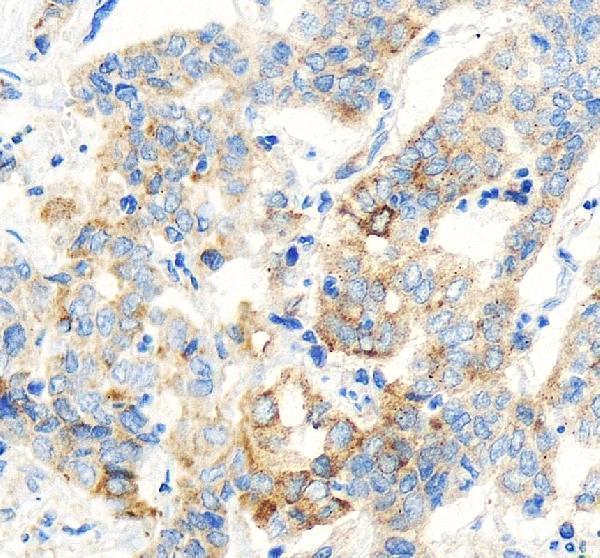
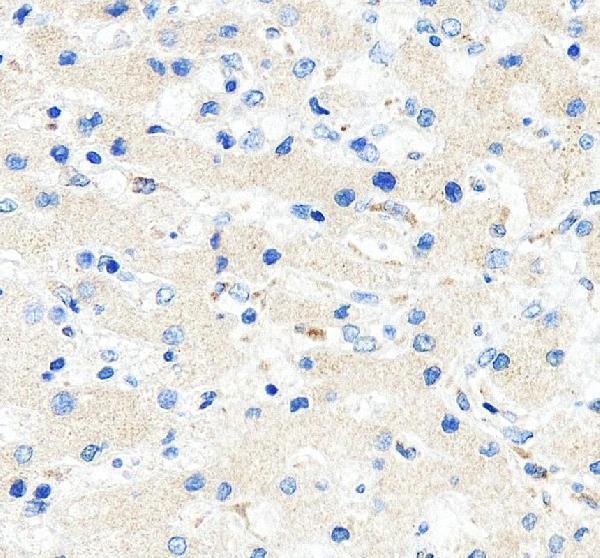
| Sample | Paraffin-embedded human liver cancer tissue; fixative not specified (datasheet A01684-3) |
| Fixation | Image fixative and duration unreported (datasheet A01684-3); verify before use. |
| Sectioning | 4–5 µm sections on charged slides (standard) |
| Deparaffinisation | Xylene, graded ethanol series to water (standard) |
| Antigen retrieval | Heat retrieval: EDTA pH 8.0 (datasheet A01684-3); 20 min, 95–100 °C (standard) |
| Peroxidase block | 3% H2O2, 10 min, room temperature (standard) |
| Blocking | 10% goat serum (datasheet A01684-3) |
| Primary antibody | Rabbit anti-OAS1, 1:50 recommended; image 2 μg/ml (datasheet A01684-3) |
| Primary incubation | Overnight at 4 °C (datasheet A01684-3) |
| Detection | HRP-conjugated secondary, DAB chromogen (datasheet A01684-3) |
| Counterstain | Hematoxylin, blue, dehydrate and mount (standard) |
| Expected result | OAS1-positive staining in adipocytes of adipose tissue (HPA tissue IHC: High). HPA tissue profile: Ubiquitous cytoplasmic expression. No signal in the no-primary control. |
OAS1 is observed mainly in the cytoplasm across many tissues (HPA tissue IHC: ubiquitous cytoplasmic expression). High staining is reported in bronchial respiratory epithelium and bone marrow hematopoietic cells, among others (HPA tissue IHC). Cytosol and nucleoplasm are supported IF locations (HPA subcellular). OAS1 has no transmembrane segment (UniProt P00973 topology). Interpret tissue IHC cautiously: its reliability is Uncertain because antibody staining and RNA data have low consistency (HPA tissue IHC).
| Cytoplasmic stain in bronchial respiratory epithelial cells or bone marrow hematopoietic cells, with recognizable cell outlines. | This fits reported High staining in those cells and the broad cytoplasmic tissue profile (HPA tissue IHC). Compare cells within the same section before calling intensity abnormal. A matching pattern supports interpretation, but the tissue IHC evidence remains Uncertain and awaits external verification (HPA tissue IHC). |
| Stain is confined to cell borders or luminal surfaces, with little intracellular signal. | Recheck this as a possible staining artefact: OAS1 has no transmembrane segment, while the reported tissue profile is cytoplasmic (UniProt P00973 topology; HPA tissue IHC). Do not reject nuclear staining solely by location; nucleoplasm is supported by IF, and UniProt also lists nuclear association (HPA subcellular; UniProt P00973 subcellular). |
| Smooth muscle cells stain as strongly as adjacent cells reported High, or an implausibly uniform stain spans unrelated structures. | Smooth muscle cells are reported Low, whereas many other cell types are reported High (HPA tissue IHC). Check for cross-reactivity or endogenous chromogenic activity before assigning the stain to OAS1 (general IHC practice). Broad staining alone is insufficient to reject a result because the reported tissue profile is ubiquitous (HPA tissue IHC). |
| A diffuse haze covers tissue, stroma, and empty spaces, obscuring cell boundaries. | Treat the haze as background until a no-primary control and detection controls are reviewed (general IHC practice). OAS1 can be secreted, so extracellular signal cannot be ruled out by location alone (UniProt P00973 subcellular); nevertheless, haze without identifiable positive cells does not reproduce the reported cytoplasmic tissue pattern (HPA tissue IHC). |
| No cellular signal appears in bronchial respiratory epithelium or bone marrow hematopoietic cells. | Both are reported High by tissue IHC (HPA tissue IHC). First check section quality, retrieval, primary antibody, and detection with controls (general IHC practice). An absent result is not proof of absent OAS1: the HPA tissue IHC assessment is Uncertain, and OAS1 is interferon induced (HPA tissue IHC; UniProt P00973 function). |
| Tissue and antibody evidence | The tissue profile is broadly cytoplasmic, yet its reliability is Uncertain; both listed antibodies have Uncertain IHC status (HPA tissue IHC; HPA antibodies). Use reported High and Low cells as comparisons, not guaranteed controls. |
| Cellular compartment | Cytosol and nucleoplasm are supported IF locations (HPA subcellular). UniProt additionally lists mitochondrial, microsomal, endoplasmic reticulum, and secreted associations (UniProt P00973 subcellular). Avoid treating a single compartment as mandatory in every preparation. |
| Isoforms and epitope | Four isoforms are listed: p46, p42, p48, and p44 (UniProt P00973 isoforms). The supplied evidence gives no antibody epitope, so it cannot establish which isoforms either IHC antibody detects; do not infer isoform identity from stain location. |
| Processing and topology | The listed chain spans residues 1–400, with no signal peptide, propeptide, or transmembrane segment (UniProt P00973 processing and topology). UniProt also lists a secreted form (UniProt P00973 subcellular); the supplied record does not establish a tissue IHC pattern for that form. |
| IF/ICC? | Supported nucleoplasmic and cytosolic localization is reported, with images from A-431, SK-MEL-30, and U-251MG (HPA subcellular). HPA003657 has Supported ICC status but Uncertain IHC status (HPA antibodies). Interpret IF observations through the separate IF/ICC guide. |
| Antigen retrieval and fixation | Target-specific fixation effects are not established by the supplied assay evidence. Verify with a matched IHC source before attributing a result to fixation. |
| Situation | Likely cause | Next action |
|---|---|---|
| Reported High cells are blank while the counterstain and tissue morphology remain readable. | Primary incubation, retrieval, or detection may have failed (general IHC practice); a High designation is not a guaranteed result because HPA tissue IHC reliability is Uncertain (HPA tissue IHC). | Run a documented positive tissue alongside the specimen and verify antibody dilution, retrieval, detection reagents, and reagent order against the antibody's IHC-P instructions (general IHC practice). |
| Brown signal persists in the no-primary control. | Endogenous enzyme activity or nonspecific detection-reagent binding can create chromogenic signal (general IHC practice). | Review the detection chemistry, apply the appropriate endogenous-activity block, and compare with a no-primary section before scoring OAS1 (general IHC practice). |
| The section has widespread brown haze that hides cell boundaries. | Excess primary or detection reagent, insufficient blocking, or inadequate washing can raise background (general IHC practice). | Adjust blocking, antibody concentration, and washes one variable at a time; retain a no-primary section for comparison (general IHC practice). |
| Only crisp membrane outlines stain, without convincing cytoplasm. | A membrane-only pattern conflicts with the reported cytoplasmic tissue profile and lack of a transmembrane segment (HPA tissue IHC; UniProt P00973 topology). | Inspect no-primary and reagent controls, review morphology, and repeat with a separately validated antibody if available; keep the HPA Uncertain IHC status in view (general IHC practice; HPA antibodies). |
| Smooth muscle stains more strongly than nearby reported High cell types. | This reverses the reported Low smooth-muscle pattern, though tissue IHC reliability is Uncertain (HPA tissue IHC). Cross-reactivity or detection background are possible (general IHC practice). | Compare matched regions and no-primary controls, then seek independent antibody or orthogonal evidence before calling the result OAS1-specific (general IHC practice). |
| Nuclear stain accompanies cytoplasmic signal. | Nucleoplasm and cytosol are both supported IF locations, while tissue IHC is described as cytoplasmic overall (HPA subcellular; HPA tissue IHC). | Record the two compartments separately and check controls; avoid labeling every nuclear signal artefactual or treating IF localization as IHC validation (general IHC practice; HPA antibodies). |


Comprehensive Human Protein Atlas IHC scoring per tissue (reliability: Uncertain — Low consistency between antibody staining and RNA expression data. Pending external verification.). Rows are taken directly from the HPA tissue chart — click any row's HPA link to view the source.
| Tissue | Cell type | Level | Evidence | Source |
|---|---|---|---|---|
| Adipose tissue | Adipocytes | High | Protein (IHC) | HPA → |
| Adrenal gland | Glandular cells | High | Protein (IHC) | HPA → |
| Appendix | Glandular cells | High | Protein (IHC) | HPA → |
| Bone marrow | Hematopoietic cells | High | Protein (IHC) | HPA → |
| Breast | Glandular cells | High | Protein (IHC) | HPA → |
| Tissue | Cell type | Level | Evidence | Source |
|---|---|---|---|---|
| None in HPA: OAS1 is detected in all 45 scored tissues. Use a no-primary (secondary-only) and an isotype control instead. | ||||
Troubleshoot OAS1 staining in paraffin sections using the catalog antibody’s IHC evidence, while accounting for its OAS1/3 label and uncertain tissue staining reliability.
A01684-3 has real IHC images from paraffin-embedded human liver and liver cancer tissue (A01684-3 IHC captions); its catalog lists human/mouse reactivity and IF/ICC applications (A01684-3 catalog).
A01684-3 is the only rendered card; its IHC images show paraffin-embedded human liver and liver cancer sections (A01684-3 IHC captions). The catalog lists human/mouse reactivity and IHC, IF, and ICC applications, but supplies no IF image (A01684-3 catalog).
Which to pick: For tissue IHC, choose polyclonal A01684-3: its images document paraffin-embedded human sections, with the fixative unreported (A01684-3 catalog; A01684-3 IHC captions). For IF/ICC, A01684-3 lists both applications, while A01684 lists IF only; neither has a supplied IF image (A01684-3 catalog; A01684 catalog). Both list mouse reactivity, but the supplied IHC examples for A01684-3 are human tissue only (A01684 catalog; A01684-3 catalog; A01684-3 IHC captions).