This website uses cookies to ensure you get the best experience on our website.


- Table of Contents
Plan paraffin section IHC for OAZ1 using its reported cytoplasmic and membranous tissue pattern (HPA tissue IHC). The guide covers controls and interpretation in light of the uncertain tissue staining reliability (HPA tissue IHC).

Expected localisation, validated protocols, controls and antibodies — the at-a-glance facts below, then the full design guide.
| Expected localisation | General cytoplasmic and membranous staining (HPA tissue IHC) | |
| Staining pattern | Cells show general cytoplasmic and membranous staining (HPA tissue IHC) | |
| Antigen retrieval | EDTA pH 8.0 HIER, heat-mediated (datasheet A06486-4) | |
| Positive control | Bronchus+4 more · see all | |
| Negative control | Bone marrow |
| Fixation | Keep fixation consistent across sections. (standard IHC practice; not target-specific) | |
| Caveat | Tissue staining reliability is uncertain; verify controls (HPA tissue IHC) | |
| Regulation | Responds to increased intracellular polyamines (UniProt) | |
| Isoform / epitope | 1 isoform; no annotated processing (UniProt) |
The catalog antibody protocol is paired with published OAZ1 IHC methods using a tissue microarray and paraffin-embedded tumor sections (PMC6534535; PMC12350266).
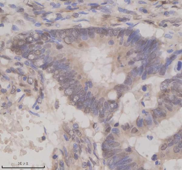
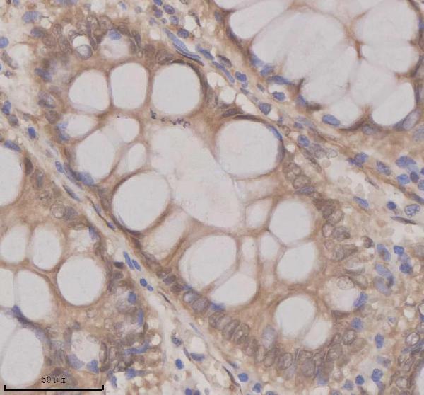
| Sample | Paraffin-embedded human colon cancer tissue; fixative not specified (datasheet A06486-4) |
| Fixation | Image fixative and duration unreported (datasheet A06486-4); verify before use. |
| Sectioning | 4–5 µm sections on charged slides (standard) |
| Deparaffinisation | Xylene, graded ethanol series to water (standard) |
| Antigen retrieval | Heat retrieval: EDTA pH 8.0 (datasheet A06486-4); 20 min, 95–100 °C (standard) |
| Peroxidase block | 3% H2O2, 10 min, room temperature (standard) |
| Blocking | 10% goat serum (datasheet A06486-4) |
| Primary antibody | Rabbit anti-OAZ1, 2-5 μg/ml (datasheet A06486-4) |
| Primary incubation | Overnight at 4 °C (datasheet A06486-4) |
| Detection | HRP-conjugated secondary, DAB chromogen (datasheet A06486-4) |
| Counterstain | Hematoxylin, blue, dehydrate and mount (standard) |
| Expected result | OAZ1-positive staining in respiratory epithelial cells of bronchus (HPA tissue IHC: High). HPA tissue profile: General cytoplasmic and membranous expression. No signal in the no-primary control. |
OAZ1 should show mainly cytoplasmic staining, sometimes with a membranous component, in cells where HPA reports expression (HPA tissue IHC: general cytoplasmic and membranous expression). High staining is reported in respiratory epithelial cells, neurons, glomerular cells, myocytes, peripheral nerve and stomach glandular cells (HPA tissue IHC: High). Interpret the pattern cautiously: tissue IHC reliability is Uncertain, and OAZ1 has no annotated transmembrane segment (HPA tissue IHC: Uncertain; UniProt P54368 topology).
| Cytoplasmic staining in bronchial respiratory epithelial cells or stomach glandular cells. | This fits the reported compartment and high-staining cell types (HPA tissue IHC: general cytoplasmic and membranous expression; High in both cell types). Compare cell-level staining with tissue structure; intensity alone cannot establish antibody specificity when tissue IHC reliability is Uncertain (HPA tissue IHC: Uncertain; general IHC practice). |
| A cytoplasmic signal with some membrane-associated staining in cells expected to be positive. | A membranous component is compatible with HPA's tissue profile, but it does not establish that OAZ1 spans the plasma membrane (HPA tissue IHC: cytoplasmic and membranous; UniProt P54368 topology: no transmembrane segment). Do not require the fine vesicular puncta seen by ICC-IF to be resolved in chromogenic sections (HPA ICC-IF: vesicles; general IHC practice). |
| Predominantly nuclear staining with little cytoplasmic signal in an expected positive cell population. | A nucleus-dominant pattern conflicts with the supplied tissue profile and approved ICC-IF locations (HPA tissue IHC: cytoplasmic and membranous; HPA ICC-IF: vesicles and centriolar satellites). Treat it as suspect, then compare controls and section morphology before calling it artefact (general IHC practice). |
| Strong staining in hematopoietic cells of bone marrow while expected positive cells do not stain. | That distribution conflicts with HPA's reported bone-marrow result and positive examples (HPA tissue IHC: Not detected in bone-marrow hematopoietic cells; High in respiratory epithelial cells). Cross-reactivity or endogenous detection activity is possible; compare reagent controls before assigning a cause (general IHC practice). HPA's Uncertain reliability makes the bone-marrow finding a comparison, not an absolute exclusion (HPA tissue IHC: Uncertain). |
| Uniform haze across cells, stroma and empty areas, or no signal in a reported high-staining tissue. | Neither provides convincing cell-specific OAZ1 staining (HPA tissue IHC: cell-level distribution; general IHC practice). Haze suggests background; absent signal may reflect the assay or specimen. Check controls and tissue preservation before interpreting either as OAZ1 biology (general IHC practice). |
| Tissue and cell selection | HPA reports high staining in several distinct cell populations, but low tissue RNA specificity (HPA tissue IHC: High examples; low tissue specificity). Score the named cells within the section instead of treating an entire organ as uniformly positive (general IHC practice). |
| Strength of the tissue-IHC evidence | The tissue profile is rated Uncertain because antibody staining and RNA expression have medium consistency, with external verification pending (HPA tissue IHC: reliability description). Use reported high and not-detected cells as provisional references, and document discordant patterns (general IHC practice). |
| Compartment interpretation | HPA describes cytoplasmic and membranous tissue staining, while UniProt lists no transmembrane segment (HPA tissue IHC: profile; UniProt P54368 topology). Membrane-associated staining can be recorded as observed; topology alone cannot identify its precise structure or prove membrane insertion. |
| Relationship to IF/ICC | HPA approves vesicles as the main ICC-IF location and centriolar satellites as an additional location (HPA ICC-IF: approved locations). Those finer structures provide context for an IF/ICC question, but chromogenic tissue IHC has its own, broader cytoplasmic and membranous profile (HPA tissue IHC: profile; general IHC practice). |
| Protein form | UniProt lists one isoform and a chain spanning residues 1–228, with no signal peptide, propeptide or annotated glycosylation sites (UniProt P54368: isoforms, processing, glycosylation). The supplied record therefore gives no basis for predicting a secreted or shed staining pattern; epitope-specific behavior remains unspecified. |
| Situation | Likely cause | Next action |
|---|---|---|
| No staining in bronchial respiratory epithelium or another reported high-staining population. | The result conflicts with an HPA positive reference, but the tissue-IHC evidence is Uncertain (HPA tissue IHC: High examples; Uncertain). Assay performance or specimen quality may account for loss of signal (general IHC practice). | Confirm the expected cells are present and assess morphology, counterstain, detection controls and the antibody's documented IHC-P conditions before interpreting a negative result (general IHC practice). |
| Diffuse chromogen covers stroma, empty areas and expected negative cells. | The distribution lacks the cell-specific pattern described by HPA and is compatible with nonspecific background or endogenous detection activity (HPA tissue IHC: profile; general IHC practice). | Compare reagent and detection controls, then review blocking, wash steps and chromogen development as general IHC variables (general IHC practice). Rescore only distinguishable cellular staining. |
| Staining is mainly nuclear. | That compartment is unsupported by the supplied HPA tissue and ICC-IF locations (HPA tissue IHC: cytoplasmic and membranous; HPA ICC-IF: vesicles and centriolar satellites). | Verify cell identity and compare controls; document the mismatch instead of scoring nuclei as expected OAZ1 signal (general IHC practice). |
| Bone-marrow hematopoietic cells stain strongly. | HPA reports them as not detected, although its tissue-IHC reliability is Uncertain (HPA tissue IHC: Not detected in hematopoietic cells; Uncertain). Cross-reactivity or endogenous activity is possible (general IHC practice). | Check a reagent control and compare staining in a reported positive cell population on the same run before judging specificity (general IHC practice). |
| A sharp membrane outline appears without convincing cytoplasmic staining. | HPA permits a membranous component, but its general profile also includes cytoplasm; UniProt reports no transmembrane segment (HPA tissue IHC: profile; UniProt P54368 topology). A membrane-only outline needs corroboration. | Inspect tissue edges and control sections for the same outline, then report the observed compartment precisely rather than inferring membrane insertion (general IHC practice). |
| IF/ICC shows puncta that are hard to recognize in paraffin-section IHC. | HPA approves vesicular and centriolar-satellite ICC-IF localisation, while its tissue-IHC description is broader (HPA ICC-IF: approved locations; HPA tissue IHC: profile). The readouts resolve different visual detail (general IHC practice). | Interpret each application against its own HPA pattern; score chromogenic tissue IHC by cellular compartment and cell type, without requiring ICC-IF puncta (HPA tissue IHC: profile; general IHC practice). |
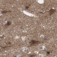


Comprehensive Human Protein Atlas IHC scoring per tissue (reliability: Uncertain — Medium consistency between antibody staining and RNA expression data. Pending external verification.). Rows are taken directly from the HPA tissue chart — click any row's HPA link to view the source.
| Tissue | Cell type | Level | Evidence | Source |
|---|---|---|---|---|
| Bronchus | Respiratory epithelial cells | High | Protein (IHC) | HPA → |
| Cerebellum | Purkinje cells | High | Protein (IHC) | HPA → |
| Cerebral cortex | Neuronal cells | High | Protein (IHC) | HPA → |
| Hippocampus | Neuronal cells | High | Protein (IHC) | HPA → |
| Kidney | Cells in glomeruli | High | Protein (IHC) | HPA → |
| Tissue | Cell type | Level | Evidence | Source |
|---|---|---|---|---|
| Bone marrow | Hematopoietic cells | Not detected | Protein (IHC) | HPA → |
Troubleshoot OAZ1 staining in paraffin sections using the catalog antibody’s documented conditions, tissue context, and controls appropriate to chromogenic IHC.
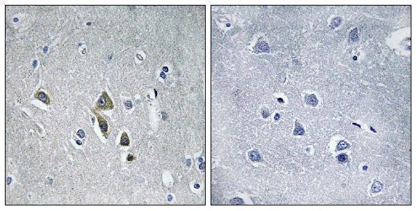
Two anti-OAZ1 antibodies have real IHC images from paraffin sections of human colon cancer and brain tissue (catalog image captions); IF is listed for A06486-2, with no IF image supplied (catalog applications; catalog IF images).
A06486-4 has IHC images from paraffin sections of human colon cancer, colon and lung cancer tissue, and lists human reactivity (A06486-4 image captions; catalog reactivity). A06486-2 has a paraffin-section IHC image from human brain with a peptide-blocked comparison; it lists human, mouse and rat reactivity and IF/ICC applications (A06486-2 image caption; catalog applications and reactivity).
Which to pick: For human tissue IHC, choose A06486-4 when its documented paraffin-section examples match your needs; its caption reports EDTA retrieval and 2 μg/ml primary antibody (A06486-4 image captions). For IF/ICC or mouse and rat samples, choose A06486-2 because those applications and species are listed; its IHC image shows a human brain paraffin section, and no IF image is supplied (catalog applications, reactivity and image captions). The fixative is unreported in both IHC captions (A06486-4 and A06486-2 image captions).