This website uses cookies to ensure you get the best experience on our website.


- Table of Contents
Plan chromogenic OCRL IHC on paraffin sections with 2–5 μg/ml of the IHC-validated antibody (datasheet A02042-2). Assess cytoplasmic staining in glandular and neuronal cells, where HPA reports high signal in several tissues (HPA tissue IHC).

Expected localisation, validated protocols, controls and antibodies — the at-a-glance facts below, then the full design guide.
| Expected localisation | General cytoplasmic staining (HPA tissue IHC) | |
| Staining pattern | Cytoplasmic signal in glandular and neuronal cells (HPA tissue IHC) | |
| Antigen retrieval | EDTA pH 8.0 HIER, heat-mediated (datasheet A02042-2) | |
| Positive control | Adrenal gland+4 more · see all | |
| Negative control | None in HPA (detected in all 45 tissues); use no-primary + isotype controls |
| Fixation | Keep fixation consistent (standard IHC practice; not target-specific); Matched tissue-IHC evidence does not establish the fixation claim. Validate the specimen-specific method before use. (selected-SKU IHC image A02042-2) | |
| Caveat | Staining and RNA levels show medium consistency (HPA tissue IHC) | |
| Regulation | Low tissue specificity (HPA tissue RNA) | |
| Isoform / epitope | 2 isoforms (A/B); epitope coverage unknown (UniProt) |
The catalog antibody protocol uses paraffin sections; one published article provides additional brain-section preparation and DAB staining details for IHC (PMC12192959).
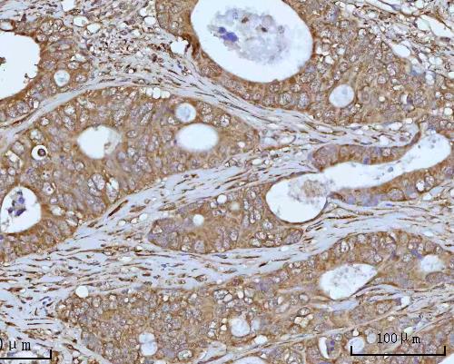
| Sample | Paraffin-embedded human colon adenocarcinoma tissue; fixative not specified (datasheet A02042-2) |
| Fixation | Image fixative and duration unreported (datasheet A02042-2); verify before use. |
| Sectioning | 4–5 µm sections on charged slides (standard) |
| Deparaffinisation | Xylene, graded ethanol series to water (standard) |
| Antigen retrieval | Heat retrieval: EDTA pH 8.0 (datasheet A02042-2); 20 min, 95–100 °C (standard) |
| Peroxidase block | 3% H2O2, 10 min, room temperature (standard) |
| Blocking | 10% goat serum (datasheet A02042-2) |
| Primary antibody | Rabbit anti-OCRL, 2-5 μg/ml (datasheet A02042-2) |
| Primary incubation | Overnight at 4 °C (datasheet A02042-2) |
| Detection | HRP-conjugated secondary, DAB chromogen (datasheet A02042-2) |
| Counterstain | Hematoxylin, blue, dehydrate and mount (standard) |
| Expected result | OCRL-positive staining in glandular cells of adrenal gland (HPA tissue IHC: High). HPA tissue profile: General cytoplasmic expression. No signal in the no-primary control. |
OCRL is a cytoplasmic, membrane-associated protein with no transmembrane segment; UniProt places it at endosomes, the trans-Golgi network, vesicles and cilia (UniProt Q01968). In paraffin-section IHC, expect mainly cytoplasmic staining in HPA-listed cells, including glandular, respiratory epithelial and neuronal cells (HPA tissue IHC). HPA calls the tissue pattern Approved, with medium consistency between staining and RNA expression (HPA tissue IHC).
| Cytoplasmic staining in adrenal gland or breast glandular cells. | This fits the general cytoplasmic profile and the High staining reported for those cells (HPA tissue IHC). Record the cell type and intensity rather than treating every positive cell as equally informative; HPA reports low tissue specificity (HPA tissue IHC). |
| Cytoplasmic staining in bronchial respiratory epithelium, or in caudate or cerebral-cortex neurons. | These are additional High-staining cell populations (HPA tissue IHC). Vesicular or perinuclear detail may be compatible with UniProt endosome and trans-Golgi locations, but a routine chromogenic section cannot identify an organelle from appearance alone (UniProt Q01968; general IHC practice). |
| Predominantly nuclear staining with little cytoplasmic signal. | This is discordant with the HPA general cytoplasmic IHC profile and UniProt subcellular locations (HPA tissue IHC; UniProt Q01968). Review controls and staining conditions before assigning that compartment to OCRL; morphology alone cannot establish antibody specificity (general IHC practice). |
| Strong staining in an unexpected cell population, especially when expected cells are unstained. | Possible explanations include cross-reactivity or endogenous chromogenic activity (general IHC practice). HPA lists High staining in selected populations but also low tissue specificity and no negative cell examples in this payload, so staining elsewhere is not automatically false (HPA tissue IHC). |
| Uniform haze across tissue, or no staining in an HPA High-staining population. | Haze obscuring cell boundaries suggests background rather than a readable OCRL pattern (general IHC practice). An absent signal in a listed High-staining population warrants a run check, but HPA's Approved rating and medium RNA–staining consistency do not guarantee every specimen will stain (HPA tissue IHC). |
| Tissue and cell selection | HPA reports High staining in adrenal, appendix and breast glandular cells; bronchial respiratory epithelial cells; bone-marrow hematopoietic cells; and selected brain cells (HPA tissue IHC). Match the assessment to the cell population actually present in the section (general IHC practice). |
| Subcellular resolution | UniProt lists endosomal, vesicular, trans-Golgi and ciliary locations (UniProt Q01968). HPA tissue IHC reports a general cytoplasmic profile, so do not require visible cilia or organelle-sized puncta to call a paraffin-section IHC result concordant (HPA tissue IHC; general IHC practice). |
| Antibody evidence | HPA012495 is Approved for IHC and Supported for ICC-IF; the overall tissue-IHC rating has medium staining–RNA consistency (HPA antibodies; HPA tissue IHC). These ratings support comparison with the reported pattern but do not establish specificity in every specimen (general IHC practice). |
| Isoforms and epitope | UniProt records two OCRL isoforms, A and B (UniProt Q01968). The supplied evidence gives no antibody epitope or isoform-specific staining claim, so an IHC difference cannot be assigned to one isoform from this record (UniProt Q01968; HPA antibodies). |
| Processing and fixation evidence | UniProt lists a single 1–901 chain, with no signal peptide, propeptide or glycosylation sites (UniProt Q01968). The supplied HPA and UniProt records do not report OCRL-specific fixation sensitivity; assess retrieval problems with ordinary run controls, without attributing them to OCRL (general IHC practice). |
| Situation | Likely cause | Next action |
|---|---|---|
| No signal in an HPA High-staining cell population. | The section, retrieval, primary-antibody step or detection run may have failed (general IHC practice); HPA does not establish an OCRL-specific fixation effect (HPA tissue IHC). | Confirm the target cells are present and inspect an appropriate positive-control section and run controls before changing one IHC condition at a time (general IHC practice). |
| Weak cytoplasmic staining is hard to score. | Low signal or excessive counterstain can reduce contrast (general IHC practice). HPA's High designation describes reported cells, not a guaranteed intensity in each specimen (HPA tissue IHC). | Compare equivalent cell populations with the positive control; check primary-antibody dilution, detection and counterstain against the validated workflow (general IHC practice). |
| Diffuse staining covers multiple compartments. | Nonspecific binding, excessive chromogen development or inadequate washing can obscure cellular boundaries (general IHC practice). | Review the no-primary control, blocking, washes and development time; score OCRL only where cell-associated staining remains distinguishable (general IHC practice; HPA tissue IHC). |
| Unexpected cells stain strongly. | Cross-reactivity or endogenous detection activity is possible (general IHC practice). The supplied HPA profile has no negative cell examples and reports low tissue specificity (HPA tissue IHC). | Check the no-primary and detection controls, then compare cell morphology and distribution with HPA's reported populations before labeling the finding false-positive (general IHC practice; HPA tissue IHC). |
| Nuclear signal dominates the section. | The pattern conflicts with reported general cytoplasmic IHC staining and UniProt's listed locations (HPA tissue IHC; UniProt Q01968). Background or nonspecific recognition is possible (general IHC practice). | Inspect controls and re-evaluate the cytoplasmic compartment in HPA-listed cells; do not infer a new OCRL nuclear location from chromogenic staining alone (general IHC practice; HPA tissue IHC). |
| Can IF/ICC resolve an uncertain IHC compartment? | HPA ICC-IF supports primary-cilium localization; its microtubule, cytosol, centriolar-satellite and basal-body assignments are uncertain (HPA subcellular ICC-IF). These cell-image findings do not make cilia a required feature of tissue IHC (HPA tissue IHC). | Use the separate IF/ICC guide for that application; compare any IF localization claim with HPA's supported primary-cilium assignment and preserve the uncertainty of its other assignments (HPA subcellular ICC-IF). |


Comprehensive Human Protein Atlas IHC scoring per tissue (reliability: Approved — Medium consistency between antibody staining and RNA expression data.). Rows are taken directly from the HPA tissue chart — click any row's HPA link to view the source.
| Tissue | Cell type | Level | Evidence | Source |
|---|---|---|---|---|
| Adrenal gland | Glandular cells | High | Protein (IHC) | HPA → |
| Appendix | Glandular cells | High | Protein (IHC) | HPA → |
| Bone marrow | Hematopoietic cells | High | Protein (IHC) | HPA → |
| Breast | Glandular cells | High | Protein (IHC) | HPA → |
| Bronchus | Respiratory epithelial cells | High | Protein (IHC) | HPA → |
| Tissue | Cell type | Level | Evidence | Source |
|---|---|---|---|---|
| None in HPA: OCRL is detected in all 45 scored tissues. Use a no-primary (secondary-only) and an isotype control instead. | ||||
Troubleshoot chromogenic OCRL staining in paraffin sections using the catalog antibody’s tissue result, reported localisation, and appropriate controls.
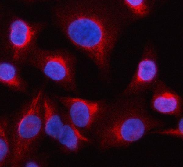
The catalog antibody A02042-2 has IHC data from human paraffin sections and IF/ICC data from A549 cells (catalog image captions).
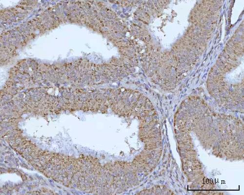
A02042-2 has IHC images from paraffin sections of human colon adenocarcinoma, endometrioid adenocarcinoma and liver cancer (A02042-2 IHC captions). It also has an IF/ICC image from A549 cells (A02042-2 IF caption).
Which to pick: Choose A02042-2 for human tissue IHC: its colon adenocarcinoma caption documents paraffin sections, EDTA retrieval at pH 8.0 and 2 μg/ml primary antibody; the fixative is unreported (A02042-2 IHC caption). For IF/ICC, A02042-2 has an A549 cell image, and its application list includes IF and ICC (A02042-2 IF caption; catalog applications). No cross-species or clonality-based choice is supported: the listed reactivity is Human and the clone field is unreported (A02042-2 catalog).