This website uses cookies to ensure you get the best experience on our website.


- Table of Contents
This guide maps expected spermatid cytoplasmic staining and tissue controls for ODF2 IHC (HPA tissue IHC). It covers the catalog antibody’s paraffin-section workflow and interpretation of presumed off-target staining (datasheet A05599-3; HPA tissue IHC).

Expected localisation, validated protocols, controls and antibodies — the at-a-glance facts below, then the full design guide.
| Expected localisation | Spermatid cytoplasm (HPA tissue IHC) | |
| Staining pattern | Cytoplasmic staining in spermatids (HPA tissue IHC) | |
| Antigen retrieval | EDTA pH 8.0 HIER, heat-mediated (datasheet A05599-3) | |
| Positive control | Bronchus+4 more · see all | |
| Negative control | Adipose tissue+4 more · see all |
| Fixation | Keep fixation consistent across sections (standard IHC practice; not target-specific) | |
| Caveat | Presumed off-target staining may confound interpretation (HPA tissue IHC) | |
| Regulation | High in late spermatids (HPA tissue IHC) | |
| Isoform / epitope | 10 isoforms; epitope coverage is unspecified (UniProt) |
The catalog antibody has an IHC-P protocol (datasheet A05599-3). One published mouse inner-ear protocol uses chromogenic DAB detection for ODF2 (PMC3368262).
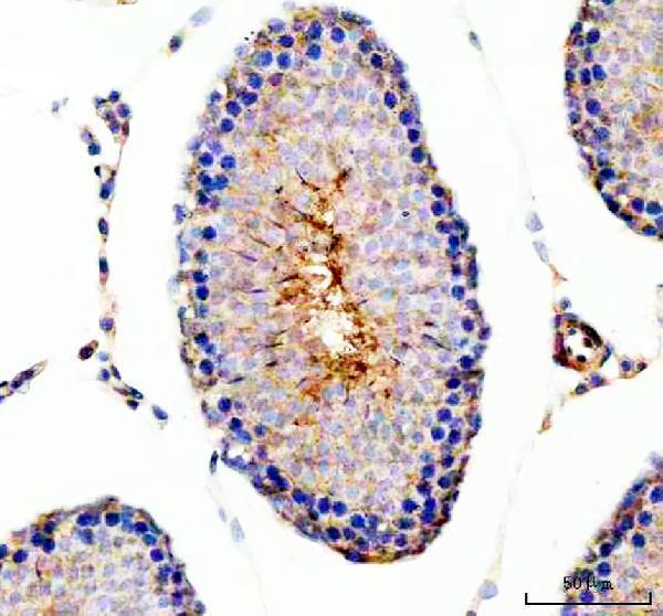
| Sample | Paraffin-embedded rat testis tissue; fixative not specified (datasheet A05599-3) |
| Fixation | Image fixative and duration unreported (datasheet A05599-3); verify before use. |
| Sectioning | 4–5 µm sections on charged slides (standard) |
| Deparaffinisation | Xylene, graded ethanol series to water (standard) |
| Antigen retrieval | Heat retrieval: EDTA pH 8.0 (datasheet A05599-3); 20 min, 95–100 °C (standard) |
| Peroxidase block | 3% H2O2, 10 min, room temperature (standard) |
| Blocking | 10% goat serum (datasheet A05599-3) |
| Primary antibody | Rabbit anti-ODF2, 2-5 μg/ml (datasheet A05599-3) |
| Primary incubation | Overnight at 4 °C (datasheet A05599-3) |
| Detection | HRP-conjugated secondary, DAB chromogen (datasheet A05599-3) |
| Counterstain | Hematoxylin, blue, dehydrate and mount (standard) |
| Expected result | ODF2-positive staining in ciliated cells (cilia axoneme) of bronchus (HPA tissue IHC: High). HPA tissue profile: Cytoplasmic expression in spermatids. No signal in the no-primary control. |
ODF2 is a cytoskeletal protein associated with centrosomes, centrioles, cilia and sperm tails; it has no transmembrane segment (UniProt Q5BJF6). In tissue IHC, expect cytoplasmic staining in spermatids and staining of cilia axonemes in ciliated cells (HPA: tissue IHC). HPA rates the tissue profile Enhanced, while noting medium consistency with RNA data and presumed off-target staining that was disregarded (HPA: reliability description).
| Strong cytoplasmic staining in elongated or late spermatids. | This matches a high-staining testis cell population (HPA: High in elongated or late spermatids). UniProt also reports abundant ODF2 in step 2 round spermatid cytoplasm, so spermatid stage and position should be recorded when comparing sections (UniProt Q5BJF6 tissue specificity). |
| Strong, narrow staining along the cilia of ciliated epithelial cells. | Cilia axonemes stain strongly in bronchus, fallopian tube and nasopharynx (HPA: High in ciliated cells, cilia axoneme). Score the ciliary border separately from cell bodies; ODF2 also has cilium and centrosome localizations (UniProt Q5BJF6 subcellular location). |
| Predominantly nuclear staining or broad staining across cells without a ciliary or spermatid pattern. | This does not match the supported tissue pattern or cytoskeletal localization (HPA: tissue IHC; UniProt Q5BJF6 subcellular location). Assess antibody specificity and detection artefacts before treating it as ODF2. HPA itself notes presumed off-target staining in its tissue assessment (HPA: reliability description). |
| Staining in cell types listed as undetected, such as adipocytes in adipose tissue. | HPA reports no detectable staining in those adipocytes (HPA: Not detected in adipose adipocytes). Check whether the signal follows tissue structures or the detection chemistry; unexpected staining could reflect cross-reactivity or endogenous detection activity (general IHC practice). |
| Weak haze across tissue, or no staining in an otherwise suitable testis control. | Diffuse haze is difficult to score as a compartment-specific ODF2 result (HPA: spermatid cytoplasmic profile). An unstained testis control is inconclusive without checking for spermatids and a working detection run; late spermatids are a high-staining population (HPA: High in elongated or late spermatids). |
| Which tissue and cell population is scored | HPA records high staining in late spermatids and ciliated-cell axonemes, medium staining in lung macrophages, and no detection in several specified cell populations (HPA: tissue IHC). Treat these as cell-specific observations, not whole-organ positive or negative labels. |
| Subcellular resolution in paraffin sections | ODF2 associates with centrosomes, mother-centriole appendages, cilia and sperm-tail structures (UniProt Q5BJF6 subcellular location). Fine structures may be hard to resolve in chromogenic sections (general IHC practice); judge the visible ciliary or spermatid pattern with tissue morphology. |
| Antibody validation and scope | HPA001874 has Enhanced IHC status, while HPA048841 has no listed IHC status (HPA: antibodies). The Enhanced tissue profile includes a caution about medium RNA concordance and disregarded presumed off-target staining (HPA: reliability description); validation does not make every stained cell specific. |
| Isoform and modification context | UniProt lists 10 ODF2 isoforms and phosphorylation sites (UniProt Q5BJF6 isoforms and modified residues). The supplied sources do not identify the IHC epitope or show that an isoform or modification changes staining; avoid explaining discordant slides through either mechanism without further evidence. |
| Topology and processing | ODF2 has no transmembrane segment, signal peptide or propeptide in this record (UniProt Q5BJF6 topology and processing). A crisp plasma-membrane outline or secreted-looking deposit therefore lacks support as the expected ODF2 pattern; investigate it before scoring. |
| IF/ICC Q: what localization should be expected? | A: HPA places ODF2 mainly at the primary cilium, centrosome, basal body and connecting piece; vesicle and microtubule assignments are uncertain (HPA: subcellular ICC-IF). These imaging observations aid interpretation but do not supply an IF/ICC protocol (HPA: subcellular ICC-IF). |
| Situation | Likely cause | Next action |
|---|---|---|
| No signal in a testis section expected to be positive. | The sampled area may lack the relevant spermatid stages, or the IHC run may have failed (HPA: High in elongated or late spermatids; general IHC practice). | Confirm spermatid stages on the counterstain, then review the run's positive control, antigen retrieval, primary-antibody step and chromogenic detection (general IHC practice). Do not infer ODF2 absence from this slide alone. |
| Cilia appear negative while adjacent cells show broad cytoplasmic color. | The distribution conflicts with the high axonemal staining reported for bronchus, fallopian tube and nasopharynx (HPA: ciliated cells, cilia axoneme). Background or cross-reactivity is possible (general IHC practice). | Identify intact cilia on the counterstain, compare the staining with a no-primary control, and review blocking and detection conditions (general IHC practice). Score axonemes separately from adjacent cytoplasm. |
| Many nuclei or unrelated tissue compartments stain strongly. | Predominant nuclear or widespread unrelated staining is inconsistent with ODF2's reported cytoskeletal locations and HPA tissue pattern (UniProt Q5BJF6 subcellular location; HPA: tissue IHC). | Check the no-primary control and antibody dilution, then compare with a suitable positive tissue section (general IHC practice). Flag the result as unresolved if the compartment remains discordant. |
| A pale brown haze obscures cell boundaries. | Nonspecific antibody binding or detection background can obscure compartment-specific staining (general IHC practice). Haze alone does not reproduce the reported spermatid or axonemal pattern (HPA: tissue IHC). | Review blocking, washing, primary-antibody concentration and detection time; compare a no-primary control processed in the same run (general IHC practice). |
| Lung macrophages stain, but the expected ciliary signal is unclear. | Macrophages have medium staining in lung, while ciliated-cell axonemes have high staining in the listed airway tissues (HPA: tissue IHC). Macrophage color alone is a limited specificity check given HPA's off-target caution (HPA: reliability description). | Score macrophages and cilia as distinct cell populations, inspect a cilia-rich positive section, and compare the no-primary control (general IHC practice; HPA: High in bronchial ciliated-cell cilia). |
| Two antibodies give different tissue patterns. | HPA lists Enhanced IHC status for HPA001874 but no IHC status for HPA048841; it also reports presumed off-target staining in the tissue assessment (HPA: antibodies; HPA: reliability description). | Record each antibody's identity and its staining by cell type and compartment. Recheck controls and compare the result with the validated spermatid and ciliary patterns before interpreting the difference (HPA: tissue IHC; general IHC practice). |


Comprehensive Human Protein Atlas IHC scoring per tissue (reliability: Enhanced — Medium consistency between antibody staining and RNA expression data. Presumed off target binding observed and disregarded.). Rows are taken directly from the HPA tissue chart — click any row's HPA link to view the source.
| Tissue | Cell type | Level | Evidence | Source |
|---|---|---|---|---|
| Bronchus | Ciliated cells (cilia axoneme) | High | Protein (IHC) | HPA → |
| Fallopian tube | Ciliated cells (cilia axoneme) | High | Protein (IHC) | HPA → |
| Nasopharynx | Ciliated cells (cilia axoneme) | High | Protein (IHC) | HPA → |
| Testis | Elongated or late spermatids | High | Protein (IHC) | HPA → |
| Lung | Macrophages | Medium | Protein (IHC) | HPA → |
| Tissue | Cell type | Level | Evidence | Source |
|---|---|---|---|---|
| Adipose tissue | Adipocytes | Not detected | Protein (IHC) | HPA → |
| Adrenal gland | Glandular cells | Not detected | Protein (IHC) | HPA → |
| Appendix | Endocrine cells | Not detected | Protein (IHC) | HPA → |
| Bone marrow | Hematopoietic cells | Not detected | Protein (IHC) | HPA → |
| Breast | Adipocytes | Not detected | Protein (IHC) | HPA → |
Troubleshoot chromogenic ODF2 staining in paraffin sections using the catalog antibody’s rat testis image, reported localisation, and matched controls.
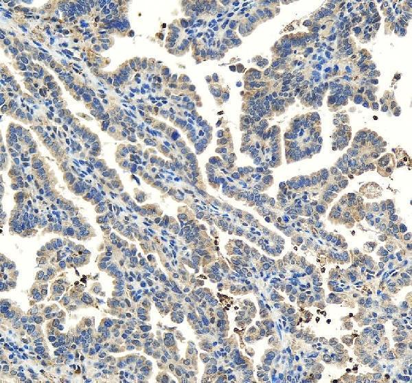
The catalog antibody A05599-3 has real IHC images from paraffin sections of rat testis and human ovarian cancer (catalog IHC image captions); no IF image is supplied (catalog IF image list).
A05599-3 is listed for IHC and reacts with human, mouse, and rat samples (catalog applications and reactivity). Its IHC captions show rat testis and human ovarian cancer paraffin sections (catalog IHC image captions).
Which to pick: Choose A05599-3 for paraffin-section IHC; its rat testis figure uses EDTA retrieval at pH 8.0 and 2 μg/ml primary antibody (A05599-3 IHC figure caption). The catalog lists human, mouse, and rat reactivity, but its IHC captions show rat and human tissue only; the fixative is unreported (catalog reactivity and IHC image captions). No SKU in this payload lists IF/ICC validation, so there is no supported IF/ICC pick here (catalog applications and IF image list).