This website uses cookies to ensure you get the best experience on our website.


- Table of Contents
Plan chromogenic IHC for OGA in paraffin sections using the catalog antibody’s tissue protocol (datasheet A32463). Compare nuclear and cytoplasmic staining with the HPA tissue profile, and account for its low consistency with RNA expression (HPA tissue IHC).

Expected localisation, validated protocols, controls and antibodies — the at-a-glance facts below, then the full design guide.
| Expected localisation | Cytoplasmic and nuclear staining in most tissues (HPA tissue IHC) | |
| Staining pattern | Most tissue cells: cytoplasmic and nuclear staining (HPA tissue IHC) | |
| Antigen retrieval | EDTA pH 8.0 HIER, heat-mediated (datasheet A32463) | |
| Positive control | Oral mucosa+4 more · see all | |
| Negative control | Adipose tissue+4 more · see all |
| Fixation | Matched tissue-IHC evidence does not establish the fixation claim. Validate the specimen-specific method before use. (selected-SKU IHC image A32463) | |
| Caveat | Staining shows low consistency with RNA expression (HPA tissue IHC) | |
| Regulation | No staining-linked regulator reported (UniProt) | |
| Isoform / epitope | 4 isoforms; check epitope coverage across variants (UniProt) |
The catalog antibody’s IHC-P protocol is paired with one published OGA IHC protocol (PMC11871760).
| Sample | Paraffin-embedded rat spleen tissue; fixative not specified (datasheet A32463) |
| Fixation | Image fixative and duration unreported (datasheet A32463); verify before use. |
| Sectioning | 4–5 µm sections on charged slides (standard) |
| Deparaffinisation | Xylene, graded ethanol series to water (standard) |
| Antigen retrieval | Heat retrieval: EDTA pH 8.0 (datasheet A32463); 20 min, 95–100 °C (standard) |
| Peroxidase block | 3% H2O2, 10 min, room temperature (standard) |
| Blocking | 10% goat serum (datasheet A32463) |
| Primary antibody | Rabbit anti-OGA, 2 μg/ml (datasheet A32463) |
| Primary incubation | Overnight at 4 °C (datasheet A32463) |
| Detection | Streptavidin-biotin complex (SABC), DAB chromogen (datasheet A32463) |
| Counterstain | Hematoxylin, blue, dehydrate and mount (standard) |
| Expected result | OGA-positive staining in squamous epithelial cells of oral mucosa (HPA tissue IHC: High). HPA tissue profile: Cytoplasmic and nuclear expression in most tissues. No signal in the no-primary control. |
OGA is a soluble protein without a transmembrane segment (UniProt O60502 topology). In paraffin tissue IHC, expect cytoplasmic and nuclear staining in many tissues (HPA tissue IHC: Approved; cytoplasmic and nuclear expression in most tissues). Judge signal by cell type: oral squamous epithelial cells and spermatogonia stain strongly, while some other sampled cells are undetected (HPA tissue IHC). HPA reports low consistency between antibody staining and RNA expression data (HPA tissue IHC reliability).
| Oral squamous epithelial cells or spermatogonia show strong cytoplasmic and/or nuclear chromogen, with recognizable cell boundaries (HPA tissue IHC: High in both cell types; cytoplasmic and nuclear profile). | This fits the reported positive pattern. Score the named cells and their compartments separately; neighboring cells need not share the same intensity (HPA tissue IHC: cell-specific levels). Treat intensity as an observed IHC category, not a quantitative OGA concentration (standard IHC practice). |
| Signal is mainly confined to cell membranes, extracellular material, or luminal deposits, without a credible intracellular pattern (UniProt O60502: nucleus, cytoplasm; no transmembrane segment). | That distribution conflicts with the recorded locations and warrants an artefact check (UniProt O60502 subcellular location and topology). Compare with a no-primary control and inspect pigment or precipitate before assigning OGA positivity (standard IHC practice). |
| Strong staining appears in adipocytes, cardiomyocytes, or placental trophoblastic cells (HPA tissue IHC: Not detected in these sampled cell types). | This is discordant with the cited HPA observations; cross-reactivity or endogenous detection activity is possible, not proven (HPA tissue IHC; standard IHC practice). Confirm cell identity and compare no-primary and detection-only controls before interpreting the signal. |
| Pale chromogen covers most cells and stroma uniformly, obscuring nuclear and cytoplasmic boundaries (HPA tissue IHC: cellular cytoplasmic and nuclear profile). | A diffuse veil cannot establish cell-specific OGA localisation (HPA tissue IHC: cell-specific staining levels). Check a no-primary control, blocking, wash quality, and detection exposure for nonspecific background (standard IHC practice). |
| Neither oral squamous epithelial cells nor spermatogonia stain in an otherwise interpretable section (HPA tissue IHC: High in these cells). | This weakens confidence in a negative call elsewhere, although HPA's Approved IHC assessment has low antibody–RNA consistency (HPA tissue IHC reliability). Review tissue preservation, retrieval, antibody dilution, and detection controls as general IHC checks (standard IHC practice). |
| Cell-specific tissue pattern (HPA tissue IHC) | High staining is recorded in oral squamous epithelial cells and spermatogonia; appendix glandular, bronchial respiratory epithelial, and cortical neuronal cells are Medium (HPA tissue IHC). Select a comparator by the cell population being scored, since a tissue label alone does not specify the positive cells (HPA tissue IHC). |
| Compartment evidence differs by assay (UniProt O60502; HPA tissue IHC and subcellular ICC-IF) | UniProt lists nucleus and cytoplasm, and tissue IHC reports both compartments (UniProt O60502; HPA tissue IHC). HPA ICC-IF supports cytosol as its main location; that assay-specific observation does not erase the tissue IHC nuclear pattern (HPA subcellular ICC-IF; HPA tissue IHC). |
| Antibody evidence and interpretation limit (HPA antibodies; HPA tissue IHC reliability) | HPA036141 is Approved for IHC, whereas HPA076501 is Supported for ICC-IF, with no IHC status supplied for the latter (HPA antibodies). The tissue assessment also reports low staining–RNA consistency, so a plausible pattern still needs appropriate controls (HPA tissue IHC reliability; standard IHC practice). |
| Q: What should IF/ICC show? (HPA subcellular ICC-IF) | A: HPA supports cytosolic localisation and lists images in A-431 and U2OS (HPA subcellular ICC-IF). This is an IF/ICC interpretation cue, not an IHC-P protocol or a basis for applying IF exposure settings to chromogenic tissue scoring (HPA subcellular ICC-IF; standard IHC practice). |
| Processing and fixation evidence (UniProt O60502; supplied HPA records) | UniProt lists a single 1–916 chain, with no signal peptide or propeptide and no transmembrane segment (UniProt O60502 processing and topology). Target-specific fixation sensitivity and an OGA retrieval condition are unreported in the supplied records; choose and document retrieval using general IHC practice. |
| Situation | Likely cause | Next action |
|---|---|---|
| A known positive cell population is blank (HPA tissue IHC: High oral squamous epithelial cells or spermatogonia). | An ineffective staining run or poorly preserved section is possible; the HPA pattern alone cannot identify the failed step (HPA tissue IHC; standard IHC practice). | Check a same-run positive control, section quality, retrieval, antibody dilution, and detection reagents; document each condition tested (standard IHC practice). |
| Everything is brown, including stroma (HPA tissue IHC: expected cellular profile). | Nonspecific background, excess detection signal, or endogenous enzyme activity can obscure cell boundaries (standard chromogenic IHC practice). | Compare no-primary and detection-only controls; adjust blocking, washes, or detection exposure according to the control result (standard IHC practice). |
| Only nuclei stain, with no visible cytoplasm (HPA tissue IHC: cytoplasmic and nuclear profile). | A nuclear component is compatible with UniProt, but exclusive nuclear staining is narrower than HPA's tissue profile (UniProt O60502 location; HPA tissue IHC). | Review cell boundaries and counterstain, then compare with a positive control and no-primary control before scoring nuclear-only signal (standard IHC practice). |
| Strong signal appears mainly in adipocytes or cardiomyocytes (HPA tissue IHC: Not detected in those cells). | Cell misidentification, cross-reactivity, or endogenous detection signal could explain the mismatch; HPA observations do not establish which cause applies (HPA tissue IHC; standard IHC practice). | Recheck morphology and controls, and seek corroboration with another validated assay before calling those cells OGA-positive (standard IHC practice). |
| Placenta appears negative despite UniProt listing high expression there (UniProt O60502 tissue specificity; HPA tissue IHC). | The statements concern different measures and cell resolution: HPA reports trophoblastic cells as Not detected in its IHC sample (HPA tissue IHC). | Record the cell type and assay with the result; use an HPA High IHC cell population as the slide-level positive comparator (HPA tissue IHC; standard IHC practice). |
| ICC-IF looks cytosolic, while tissue IHC includes nuclear staining (HPA subcellular ICC-IF; HPA tissue IHC). | The supplied HPA summaries differ by assay: supported cytosol in ICC-IF and cytoplasmic plus nuclear expression in tissue IHC (HPA subcellular ICC-IF; HPA tissue IHC). | Interpret each image against its own assay and controls; do not reject tissue nuclear staining solely because the ICC-IF summary names cytosol (HPA subcellular ICC-IF; HPA tissue IHC). |

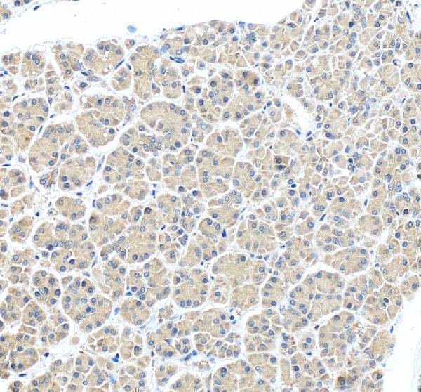

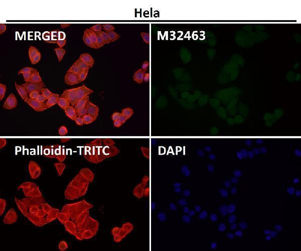


Comprehensive Human Protein Atlas IHC scoring per tissue (reliability: Approved — Low consistency between antibody staining and RNA expression data.). Rows are taken directly from the HPA tissue chart — click any row's HPA link to view the source.
| Tissue | Cell type | Level | Evidence | Source |
|---|---|---|---|---|
| Oral mucosa | Squamous epithelial cells | High | Protein (IHC) | HPA → |
| Testis | Spermatogonia cells | High | Protein (IHC) | HPA → |
| Appendix | Glandular cells | Medium | Protein (IHC) | HPA → |
| Bronchus | Respiratory epithelial cells | Medium | Protein (IHC) | HPA → |
| Cerebral cortex | Neuronal cells | Medium | Protein (IHC) | HPA → |
| Tissue | Cell type | Level | Evidence | Source |
|---|---|---|---|---|
| Adipose tissue | Adipocytes | Not detected | Protein (IHC) | HPA → |
| Bone marrow | Hematopoietic cells | Not detected | Protein (IHC) | HPA → |
| Cervix | Glandular cells | Not detected | Protein (IHC) | HPA → |
| Epididymis | Glandular cells | Not detected | Protein (IHC) | HPA → |
| Heart muscle | Cardiomyocytes | Not detected | Protein (IHC) | HPA → |
Troubleshoot OGA staining in paraffin sections by checking retrieval, compartment, cellular context, controls, and scoring before interpreting signal intensity.
Anti-OGA A32463 has IHC images from rat spleen and human pancreas paraffin sections (A32463 IHC captions) and IF in T-47D cells (A32463 IF caption); M32463 has an IF image without sample details (M32463 IF caption).
A32463 shows IHC in rat spleen and human pancreas paraffin sections and IF in T-47D cells (A32463 image captions). M32463 lists IHC and human/mouse reactivity, but its provided IF caption identifies neither a sample nor a species (M32463 catalog applications, reactivity and IF caption).
Which to pick: Choose A32463 for paraffin-section tissue IHC because its own captions document rat spleen and human pancreas staining; the fixative is unreported (A32463 IHC captions). Choose A32463 for IF/ICC because both applications are listed and its IF caption documents T-47D cells; M32463 lists IHC but has no provided IHC image caption, while its IF caption gives no sample details (A32463 catalog applications and IF caption; M32463 catalog applications and image captions). Choose A32463 when rat reactivity is needed alongside human and mouse; M32463 is a rabbit monoclonal listing human/mouse reactivity (A32463 catalog reactivity; M32463 catalog clone and reactivity).