This website uses cookies to ensure you get the best experience on our website.


- Table of Contents
Plan OGG1 staining in paraffin sections around its mainly nuclear tissue pattern (HPA tissue IHC). This guide covers control tissue selection (HPA tissue IHC), consistent fixation (standard IHC practice), and the catalog antibody’s 2–5 μg/ml IHC range (datasheet: A00768-1).

Expected localisation, validated protocols, controls and antibodies — the at-a-glance facts below, then the full design guide.
| Expected localisation | Mainly nuclear in most tissues (HPA tissue IHC) | |
| Staining pattern | Nuclear staining in most tissues (HPA tissue IHC) | |
| Antigen retrieval | EDTA pH 8.0 HIER, heat-mediated (datasheet A00768-1) | |
| Positive control | Bronchus+4 more · see all | |
| Negative control | Adipose tissue+2 more · see all |
| Fixation | Matched tissue-IHC evidence does not establish the fixation claim. Validate the specimen-specific method before use. (selected-SKU IHC image A00768-1) | |
| Caveat | Staining and RNA expression show medium consistency (HPA tissue IHC) | |
| Regulation | Ubiquitous expression (UniProt) | |
| Isoform / epitope | 8 isoforms; variant epitope coverage is unknown (UniProt) |
The catalog antibody protocol is paired with 4 published OGG1 IHC protocols from rat lens, rat spleen, mouse retina, and mouse liver (PMC2929939; PMC2614128; PMC2690988; PMC2565677).
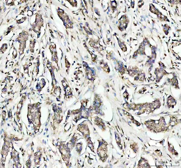
| Sample | Paraffin-embedded human breast cancer tissue; fixative not specified (datasheet A00768-1) |
| Fixation | Image fixative and duration unreported (datasheet A00768-1); verify before use. |
| Sectioning | 4–5 µm sections on charged slides (standard) |
| Deparaffinisation | Xylene, graded ethanol series to water (standard) |
| Antigen retrieval | Heat retrieval: EDTA pH 8.0 (datasheet A00768-1); 20 min, 95–100 °C (standard) |
| Peroxidase block | 3% H2O2, 10 min, room temperature (standard) |
| Blocking | 10% goat serum (datasheet A00768-1) |
| Primary antibody | Rabbit anti-OGG1, 2-5μg/ml (datasheet A00768-1) |
| Primary incubation | Overnight at 4 °C (datasheet A00768-1) |
| Detection | HRP-conjugated secondary, DAB chromogen (datasheet A00768-1) |
| Counterstain | Hematoxylin, blue, dehydrate and mount (standard) |
| Expected result | OGG1-positive staining in ciliated cells (cell body) of bronchus (HPA tissue IHC: High). HPA tissue profile: Mainly nuclear expression in most tissues. No signal in the no-primary control. |
OGG1 should stain mainly nuclei across many tissues (HPA tissue IHC: mainly nuclear; UniProt O15527: nucleus). Expect stronger staining in bronchial ciliated cells and placental trophoblastic cells, among other reported high-staining populations (HPA tissue IHC: High). Mitochondrial localisation is also annotated (UniProt O15527: mitochondrion). HPA rates tissue IHC as Approved but reports medium consistency between antibody staining and RNA expression (HPA tissue IHC: reliability).
| Distinct nuclear chromogen in bronchial ciliated cells or placental trophoblastic cells, with cell-to-cell variation. | This fits the mainly nuclear tissue pattern and reported High staining in those populations (HPA tissue IHC: profile; Bronchus and Placenta). Assess the named cells rather than treating every cell in the section as an equally strong positive. |
| Predominantly broad cytoplasmic staining, with little nuclear signal in an expected-positive population. | That distribution conflicts with the main tissue IHC pattern and nucleoplasmic ICC-IF localisation (HPA tissue IHC: profile; HPA subcellular: nucleoplasm). Investigate background or localisation before interpreting it as OGG1; mitochondrial localisation is annotated, so cytoplasmic signal alone is not conclusive (UniProt O15527: mitochondrion). |
| Strong chromogen in cardiomyocytes or adipocytes while the expected-positive cells stain weakly or not at all. | HPA reports OGG1 as Not detected in cardiomyocytes and adipocytes, but High in bronchial ciliated cells and placental trophoblastic cells (HPA tissue IHC: Heart muscle, Adipose tissue, Bronchus, Placenta). This mismatch raises concern for cross-reactivity or endogenous detection activity; it does not prove either cause. |
| Haze or granular colour covers nuclei, cytoplasm and extracellular areas without clear cell boundaries. | Diffuse colour cannot establish the reported mainly nuclear pattern (HPA tissue IHC: profile). Review controls, blocking, antibody concentration and detection conditions as general IHC background checks (general IHC practice); do not score the haze as nuclear OGG1. |
| No signal in bronchial ciliated cells or placental trophoblastic cells, including when section morphology is intact. | Those populations are reported High by HPA, so an absent result warrants a technical check (HPA tissue IHC: Bronchus; Placenta). Verify the positive control, retrieval and detection workflow before calling the specimen negative (general IHC practice). HPA's Approved rating has medium RNA-to-staining consistency (HPA tissue IHC: reliability). |
| Cell population and tissue | HPA reports High staining in bronchial and nasopharyngeal ciliated cells, placental trophoblasts, skin melanocytes and tonsillar squamous epithelium; it reports Not detected staining in adipocytes, cardiomyocytes and smooth muscle cells (HPA tissue IHC: listed populations). Score the relevant cell type. |
| Compartment | Mainly nuclear tissue staining and enhanced nucleoplasmic ICC-IF localisation support a nuclear reference pattern (HPA tissue IHC: profile; HPA subcellular: main location). UniProt also annotates mitochondria and nuclear speckles; speckle recruitment with APEX1 is described in UVA-irradiated cells (UniProt O15527: subcellular location). |
| Isoform coverage | UniProt lists 8 OGG1 isoforms (UniProt O15527: isoforms). The supplied HPA antibody summaries do not map epitopes to those isoforms (HPA antibodies: validation summaries); avoid assigning a staining difference to a particular isoform without further evidence. |
| Antibody evidence | HPA027514 and CAB047301 are both IHC Approved, while HPA's tissue-level reliability description says staining and RNA have medium consistency (HPA antibodies: IHC status; HPA tissue IHC: reliability). Treat a discordant result as a finding to check, not as automatic proof of absence or cross-reactivity. |
| Detection chemistry | If chromogenic detection uses HRP, endogenous peroxidase can add colour unrelated to primary-antibody binding; blocking and omission controls help identify it (general IHC practice). This is a workflow consideration, not an OGG1-specific effect reported by HPA or UniProt. |
| Situation | Likely cause | Next action |
|---|---|---|
| Expected-positive nuclei are blank in a paraffin section. | An unsuccessful retrieval, antibody-incubation or detection step is possible (general IHC practice); HPA reports High staining in bronchial ciliated cells and placental trophoblasts (HPA tissue IHC: Bronchus; Placenta). | Run a known-positive section alongside the specimen; check the catalog antibody's IHC-P instructions and verify retrieval, reagent activity and counterstain visibility (general IHC practice). |
| Most of the section has brown haze that obscures nuclear boundaries. | Excess background from antibody concentration, blocking or detection conditions is possible (general IHC practice); haze does not match HPA's mainly nuclear profile (HPA tissue IHC: profile). | Inspect a primary-antibody omission control, then adjust blocking, washes or antibody concentration using the IHC-P instructions (general IHC practice). Re-score only identifiable cells with discrete nuclear signal. |
| Colour is prominent in adipocytes or cardiomyocytes but absent in the expected-positive cells. | A nonspecific primary-antibody signal or endogenous chromogenic activity is possible (general IHC practice); HPA lists those cells as Not detected (HPA tissue IHC: Adipose tissue; Heart muscle). | Compare omission and positive controls, confirm the stained cell identities and review detection blocking (general IHC practice). Report the discrepancy rather than assigning OGG1 expression from colour alone. |
| The stain appears mainly cytoplasmic. | Background or a localisation mismatch is possible because HPA reports mainly nuclear tissue staining (HPA tissue IHC: profile). UniProt's mitochondrial annotation means cytoplasmic staining cannot be rejected solely by compartment (UniProt O15527: mitochondrion). | Check nuclear counterstain alignment and controls; assess whether discrete signal accompanies the expected nuclear pattern (general IHC practice; HPA tissue IHC: profile). Avoid calling diffuse cytoplasm a confirmed mitochondrial pattern. |
| A tissue expected to stain looks patchy or weak. | HPA reports expression by named cell population and gives its tissue IHC a medium RNA-to-staining consistency description (HPA tissue IHC: listed populations; reliability). Unequal representation of those cells can complicate a whole-section impression. | Identify and score the reported cell population separately, then compare it with a known-positive section processed in the same run (HPA tissue IHC: listed populations; general IHC practice). |
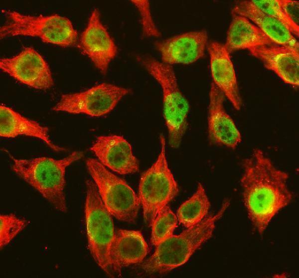
| IF/ICC Q&A: what localisation should a companion fluorescence image show? | HPA's ICC-IF summary places OGG1 in the nucleoplasm; its tissue IHC profile is mainly nuclear (HPA subcellular: nucleoplasm; HPA tissue IHC: profile). | Look for signal aligned with the nuclear counterstain and interpret fluorescence on the separate IF/ICC guide page (HPA subcellular: nucleoplasm; general IF practice). |


Comprehensive Human Protein Atlas IHC scoring per tissue (reliability: Approved — Medium consistency between antibody staining and RNA expression data.). Rows are taken directly from the HPA tissue chart — click any row's HPA link to view the source.
| Tissue | Cell type | Level | Evidence | Source |
|---|---|---|---|---|
| Bronchus | Ciliated cells (cell body) | High | Protein (IHC) | HPA → |
| Nasopharynx | Ciliated cells (cell body) | High | Protein (IHC) | HPA → |
| Placenta | Trophoblastic cells | High | Protein (IHC) | HPA → |
| Skin | Melanocytes | High | Protein (IHC) | HPA → |
| Tonsil | Squamous epithelial cells | High | Protein (IHC) | HPA → |
Troubleshoot OGG1 staining in paraffin sections by checking retrieval, nuclear localisation, controls, and cell-specific scoring before interpreting DAB signal.
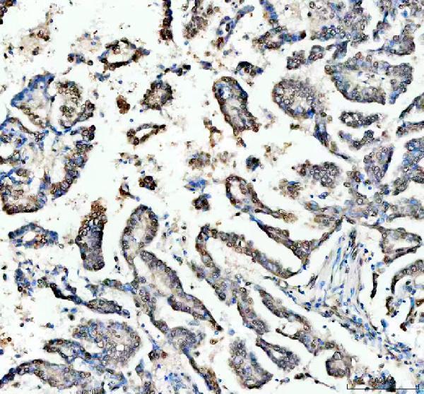
The catalog antibody has IHC data from human breast and lung cancer paraffin sections and IF/ICC data from HeLa cells (A00768-1 image captions).
A00768-1 will render with its IHC figure from a human breast cancer paraffin section (A00768-1 IHC image caption). The same SKU also has IHC data from a human lung cancer paraffin section and IF/ICC data from HeLa cells (A00768-1 image captions).
Which to pick: Choose A00768-1 for human tissue IHC on paraffin sections; its IHC captions document breast and lung cancer sections, but do not report the fixative (A00768-1 IHC image captions). Choose the same SKU for human IF/ICC based on its HeLa cell image; it is a rabbit antibody, and clonality is unreported (A00768-1 catalog and IF image caption). No cross-species choice is supported because the listed reactivity is human only (A00768-1 catalog).