This website uses cookies to ensure you get the best experience on our website.


- Table of Contents
Plan chromogenic IHC of paraffin sections with retinal photoreceptors as a positive reference (HPA tissue IHC). This guide covers fixation consistency, the catalog antibody’s 2–5 μg/ml IHC range (datasheet A08880-2), and interpretation of membrane staining (UniProt).

Expected localisation, validated protocols, controls and antibodies — the at-a-glance facts below, then the full design guide.
| Expected localisation | Membrane expected (UniProt); retinal photoreceptors stain (HPA tissue IHC) | |
| Staining pattern | Retinal photoreceptor cells and brain processes; compartment unspecified (HPA tissue IHC) | |
| Antigen retrieval | EDTA pH 8.0 HIER, heat-mediated (datasheet A08880-2) | |
| Positive control | Retina | |
| Negative control | Adipose tissue+4 more · see all |
| Fixation | Keep paraffin section fixation consistent (standard IHC practice; not target-specific); Matched tissue-IHC evidence does not establish the fixation claim. Validate the specimen-specific method before use. (selected-SKU IHC image A08880-2) | |
| Caveat | Testis-enriched RNA may mislead tissue choice; retinal staining is high (HPA tissue IHC) | |
| Regulation | Corneal injury induction, by similarity (UniProt) | |
| Isoform / epitope | No isoforms annotated; epitope side matters across 7 TM spans (UniProt) |
Start with the catalog antibody’s EDTA pH 8.0 heat retrieval (datasheet: A08880-2), then compare the published duck and gander hypothalamus IHC protocols (PMC10964073; PMC11758404).
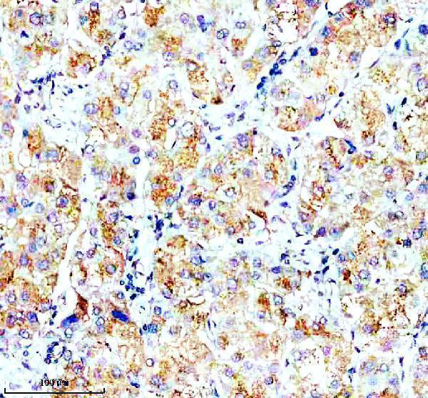
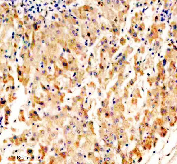
| Sample | Paraffin-embedded human liver cancer tissue; fixative not specified (datasheet A08880-2) |
| Fixation | Image fixative and duration unreported (datasheet A08880-2); verify before use. |
| Sectioning | 4–5 µm sections on charged slides (standard) |
| Deparaffinisation | Xylene, graded ethanol series to water (standard) |
| Antigen retrieval | Heat retrieval: EDTA pH 8.0 (datasheet A08880-2); 20 min, 95–100 °C (standard) |
| Peroxidase block | 3% H2O2, 10 min, room temperature (standard) |
| Blocking | 10% goat serum (datasheet A08880-2) |
| Primary antibody | Rabbit anti-OPN5, 2-5 μg/ml (datasheet A08880-2) |
| Primary incubation | Overnight at 4 °C (datasheet A08880-2) |
| Detection | HRP-conjugated secondary, DAB chromogen (datasheet A08880-2) |
| Counterstain | Hematoxylin, blue, dehydrate and mount (standard) |
| Expected result | OPN5-positive staining in photoreceptor cells of retina (HPA tissue IHC: High). HPA tissue profile: Expression in photoreceptor cells in retina and processes in brain. No signal in the no-primary control. |
OPN5 is a seven-pass cell-membrane protein (UniProt Q6U736 topology and subcellular location). In paraffin-section IHC, expect high staining in retinal photoreceptor cells and staining in brain processes (HPA tissue IHC). The HPA tissue pattern is rated Supported by external characterization; that rating does not establish the exact membrane domain stained within a photoreceptor (HPA tissue IHC; UniProt Q6U736 topology).
| Photoreceptor cells show strong, cell-associated staining in retina, with a membrane-like distribution. | This matches the reported high photoreceptor-cell signal and OPN5 membrane location (HPA tissue IHC; UniProt Q6U736). Record the retinal layer and cellular distribution; HPA does not specify a positive photoreceptor subcompartment (HPA tissue IHC). |
| Staining follows processes in brain sections. | Process staining fits the HPA tissue profile; UniProt also reports OPN5 detection in brain (HPA tissue IHC; UniProt Q6U736 tissue specificity). Interpret a process pattern in its anatomical context rather than treating every stained brain cell as a documented positive cell type (HPA tissue IHC). |
| A strong, isolated nuclear pattern dominates the retinal signal. | Nuclear-only staining conflicts with the reported cell-membrane location and seven-pass topology (UniProt Q6U736). Treat it as suspect, then compare its distribution with the positive retinal pattern and a no-primary control before assigning it to OPN5 (HPA tissue IHC; general IHC practice). |
| Cells reported as unstained show convincing chromogen, such as adipocytes in adipose tissue or glandular cells in adrenal gland. | Those cell types are listed as not detected in HPA tissue IHC. Widespread signal there raises concern for nonspecific antibody binding or endogenous detection activity; a negative HPA result is a reference pattern, not proof of absolute protein absence (HPA tissue IHC; general IHC practice). |
| The whole section has diffuse staining, or retinal photoreceptor cells have no discernible signal. | Diffuse staining obscures the cell-specific pattern, while no signal in a known-positive retina prevents interpretation of a negative test section (HPA tissue IHC; general IHC practice). First establish whether the positive control has selective photoreceptor staining before scoring other tissue (HPA tissue IHC; general IHC practice). |
| Topology and epitope location | OPN5 has 7 transmembrane segments and extracellular and cytoplasmic regions (UniProt Q6U736 topology). Interpret membrane-like signal accordingly; the supplied record gives no antibody epitope, so it cannot predict which face an antibody recognizes or a target-specific retrieval requirement (UniProt Q6U736; supplied antibody record). |
| Protein processing and variants | UniProt lists one chain spanning residues 1–354, no signal peptide or propeptide, and 0 annotated isoforms (UniProt Q6U736). These annotations give no basis for assigning a separate staining pattern to cleavage or an alternate isoform; they do not prove that every tissue shares identical protein processing (UniProt Q6U736). |
| Tissue evidence and antibody validation | HPA rates retinal photoreceptor staining High and the tissue IHC result Supported; the listed antibody, HPA026084, is also IHC Supported (HPA tissue IHC; HPA antibodies). HPA reports testis-enriched RNA, which alone does not establish a positive testis protein-staining pattern (HPA tissue IHC). |
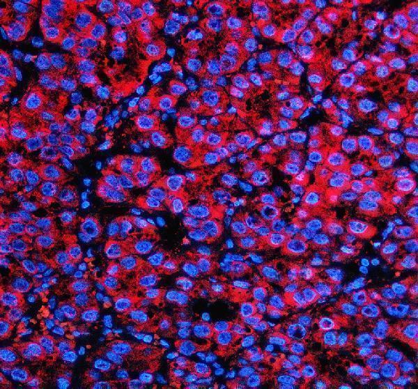
| Can ICC/IF confirm this IHC pattern? | HPA summarizes the subcellular location as membrane but provides no ICC/IF images, and ICC validation is unlisted for HPA026084 (HPA subcellular; HPA antibodies). Treat any IF result as a separate observation requiring its own controls, without inferring an established ICC staining pattern from the IHC evidence (HPA subcellular; HPA antibodies; general IF practice). |
| Situation | Likely cause | Next action |
|---|---|---|
| Retina positive control has no photoreceptor signal. | A missing expected signal may reflect a failed IHC run or unsuitable assay settings; the cause cannot be identified from the absence alone (HPA tissue IHC; general IHC practice). | Target-specific fixation effects are not established by the supplied assay evidence. Verify with a matched IHC source before attributing a result to fixation. |
| Most cells or extracellular spaces show similar brown color. | Section-wide color is inconsistent with the reported cell-selective photoreceptor pattern and may reflect background or endogenous detection activity (HPA tissue IHC; general IHC practice). | Compare with a no-primary control and assess detection-system blocking and washes; score OPN5 only where cellular localization remains distinguishable (general IHC practice). |
| Signal is strongest in HPA-negative reference cells. | Adipose adipocytes and adrenal glandular cells are reported not detected; strong signal there may indicate nonspecific binding or endogenous activity (HPA tissue IHC; general IHC practice). | Run a matched negative reference section and no-primary control, then compare their color distribution with retina before calling the test section positive (HPA tissue IHC; general IHC practice). |
| Predominantly nuclear staining appears in retina. | A nuclear-only pattern does not fit UniProt's cell-membrane assignment or seven-pass topology (UniProt Q6U736). | Review the counterstain and no-primary control, and compare nuclear color with cell-associated photoreceptor signal in the positive control (HPA tissue IHC; general IHC practice). |
| Brain staining is present but its cellular source is unclear. | HPA describes staining in brain processes without naming their cell of origin; UniProt reports detection in brain without assigning every stained process to a cell type (HPA tissue IHC; UniProt Q6U736 tissue specificity). | Report the anatomical site and process-like distribution seen on the slide; defer a specific cell-type claim until independently supported (HPA tissue IHC; general IHC practice). |
| A second section differs in intensity from the positive retina. | HPA's High rating applies to retinal photoreceptor cells, not to all tissues or preparations (HPA tissue IHC). | Compare sections within the same controlled IHC run and score location and cell type before intensity; do not transfer the retinal intensity threshold to an uncharacterized tissue (HPA tissue IHC; general IHC practice). |


Comprehensive Human Protein Atlas IHC scoring per tissue (reliability: Supported — External characterization data supports antibody staining.). Rows are taken directly from the HPA tissue chart — click any row's HPA link to view the source.
| Tissue | Cell type | Level | Evidence | Source |
|---|---|---|---|---|
| Retina | Photoreceptor cells | High | Protein (IHC) | HPA → |
| Tissue | Cell type | Level | Evidence | Source |
|---|---|---|---|---|
| Adipose tissue | Adipocytes | Not detected | Protein (IHC) | HPA → |
| Adrenal gland | Glandular cells | Not detected | Protein (IHC) | HPA → |
| Appendix | Glandular cells | Not detected | Protein (IHC) | HPA → |
| Bone marrow | Hematopoietic cells | Not detected | Protein (IHC) | HPA → |
| Breast | Adipocytes | Not detected | Protein (IHC) | HPA → |
Troubleshoot chromogenic OPN5 staining in paraffin sections using the catalog antibody workflow, tissue expression evidence, and controls matched to each specimen.
Anti-OPN5 antibody A08880-2 has IHC data from human paraffin liver and liver cancer sections (catalog IHC captions) and IF data from a human paraffin liver cancer section (catalog IF caption).
A08880-2 is listed for IHC and IF, with Human, Mouse and Rat reactivity (catalog applications and reactivity). Its IHC captions show human paraffin liver and liver cancer sections; its IF caption shows a human paraffin liver cancer section (catalog image captions).
Which to pick: For tissue IHC, choose A08880-2, a rabbit polyclonal antibody with a listed IHC concentration of 2–5 μg/ml; its IHC images use human paraffin sections, and the fixative is unreported (catalog antibody details; catalog IHC captions). For IF, A08880-2 has a listed concentration of 5 μg/ml and a paraffin-section IF image; ICC validation is unreported (catalog antibody details; catalog IF caption). For cross-species work, A08880-2 lists Human, Mouse and Rat reactivity, while the supplied IHC and IF images show human tissue only (catalog reactivity; catalog image captions).