This website uses cookies to ensure you get the best experience on our website.


- Table of Contents
Plan chromogenic IHC for OTC using liver hepatocytes and intestinal endocrine cells as positive tissue references (HPA tissue IHC). The guide covers fixation, staining interpretation, and a catalog antibody validated for IHC-P (datasheet: IHC-P).

Expected localisation, validated protocols, controls and antibodies — the at-a-glance facts below, then the full design guide.
| Expected localisation | Granular cytoplasm in hepatocytes (HPA tissue IHC) | |
| Staining pattern | Granular cytoplasm in hepatocytes and intestinal endocrine cells (HPA tissue IHC) | |
| Antigen retrieval | Citrate pH 6.0 HIER, 95–98 °C, 20 min (rule: cytoplasmic / membrane antigen) | |
| Positive control | Duodenum+4 more · see all | |
| Negative control | Adipose tissue+4 more · see all |
| Fixation | Keep formalin fixation consistent across FFPE sections (standard IHC practice; not target-specific) | |
| Caveat | Duodenal and small-intestinal positives are endocrine cells (HPA tissue IHC) | |
| Regulation | Expression regulation is unreported (UniProt) | |
| Isoform / epitope | No annotated isoforms; mature chain spans aa 33–354 (UniProt) |
The catalog antibody’s IHC-P protocol is accompanied by one published OTC staining protocol for hepatocellular carcinoma tissue (PMC8426309).
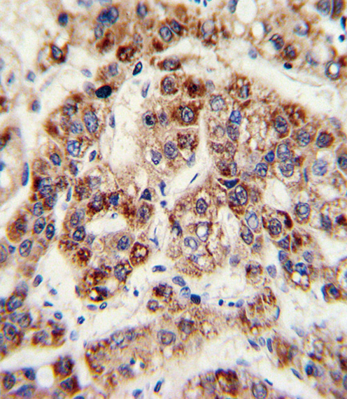
| Sample | FFPE human hepatoarcinoma tissue (datasheet A00721-2) |
| Fixation | Image formalin-fixed; duration unreported (datasheet A00721-2); verify before use. |
| Sectioning | 4–5 µm sections on charged slides (standard) |
| Deparaffinisation | Xylene, graded ethanol series to water (standard) |
| Antigen retrieval | Heat-induced epitope retrieval in citrate buffer, pH 6.0, 20 min at 95–98 °C (standard rule: cytoplasmic / membrane antigen) |
| Peroxidase block | 3% H2O2, 10 min, room temperature (standard) |
| Blocking | 10% normal serum of the secondary host, 30 min, room temperature (standard) |
| Primary antibody | Rabbit anti-OTC, 1:10-1:50 (datasheet A00721-2) |
| Primary incubation | Overnight at 4 °C (standard) |
| Detection | HRP-polymer secondary, DAB chromogen 5–10 min (standard) |
| Counterstain | Hematoxylin, blue, dehydrate and mount (standard) |
| Expected result | OTC-positive staining in endocrine cells of duodenum (HPA tissue IHC: High). HPA tissue profile: Granular cytoplasmic expression in hepatocytes, duodenum and small intestine. No signal in the no-primary control. |
OTC is a mitochondrial matrix protein without a transmembrane segment (UniProt P00480 topology). In paraffin-section IHC, expect granular cytoplasmic staining in hepatocytes and intestinal cells (HPA: tissue IHC profile). Liver hepatocytes and duodenal and small-intestinal endocrine cells are high-staining examples (HPA: High); the tissue IHC pattern has Enhanced reliability, reflecting high consistency with RNA expression (HPA: reliability).
| Granular cytoplasmic staining in hepatocytes, with appropriately stained intestinal cells. | This fits the mitochondrial matrix location (UniProt P00480) and HPA's tissue pattern. Hepatocytes and duodenal and small-intestinal endocrine cells are reported as High (HPA: tissue IHC). Judge the granules within identified cells; the expected result does not require uniform staining across every cell in the section (HPA: cell-specific levels). |
| Predominantly nuclear, membranous, or extracellular staining, especially without cytoplasmic granules. | These compartments conflict with the mitochondrial matrix assignment and lack of a transmembrane segment (UniProt P00480 topology). Treat an isolated wrong-compartment signal as suspect, then compare it with a known-positive hepatocyte region and a detection control before assigning it to OTC (HPA: High in hepatocytes; standard IHC practice). |
| Strong staining in cells reported as negative, such as adipocytes in adipose tissue. | HPA reports OTC as Not detected in those adipocytes (HPA: adipose tissue IHC). Reproducible signal there warrants a specificity check; antibody cross-reactivity or endogenous chromogenic activity are possible explanations, not conclusions established by HPA (standard IHC practice). Check cell identity and detection controls before scoring that signal as OTC. |
| Diffuse color across cells and tissue, with little distinction between granules and background. | A diffuse deposit obscures the granular cytoplasmic pattern used to interpret OTC staining (HPA: tissue IHC profile). It can reflect nonspecific reagent binding or the chromogenic detection system (standard IHC practice). Compare a primary-antibody omission control and tissue-free areas; neither background appearance nor a single control establishes OTC specificity by itself. |
| No staining in well-preserved hepatocytes on a putative positive-control section. | Hepatocytes are reported as High and are a useful positive reference (HPA: liver IHC). An absent result calls for a workflow check, including control-section identity, reagent performance, antigen retrieval, and detection steps (standard IHC practice). HPA's tissue pattern does not identify which step failed or establish OTC-specific fixation sensitivity. |
| Tissue and cell selection (HPA: tissue IHC). | Liver hepatocytes, duodenal endocrine cells, and small-intestinal endocrine cells are High; appendix endocrine cells and rectal enterocytes are Medium (HPA: tissue IHC). Use the stated cell type when comparing sections: an HPA level for one population does not describe every neighboring cell. |
| Compartment and molecular form (UniProt P00480). | OTC occupies the mitochondrial matrix, has no transmembrane segment, and has an annotated mature chain at residues 33–354 (UniProt P00480). These facts support intracellular granular interpretation; they do not locate the catalog antibody's epitope or predict its retrieval requirement. |
| Strength of the tissue-pattern evidence (HPA: reliability and antibodies). | HPA calls the tissue IHC pattern Enhanced and lists 2 antibodies with Enhanced IHC status, HPA000243 and HPA000570 (HPA: tissue IHC; HPA: antibodies). That supports the reported distribution, but it does not validate an unspecified catalog antibody or every staining run. |
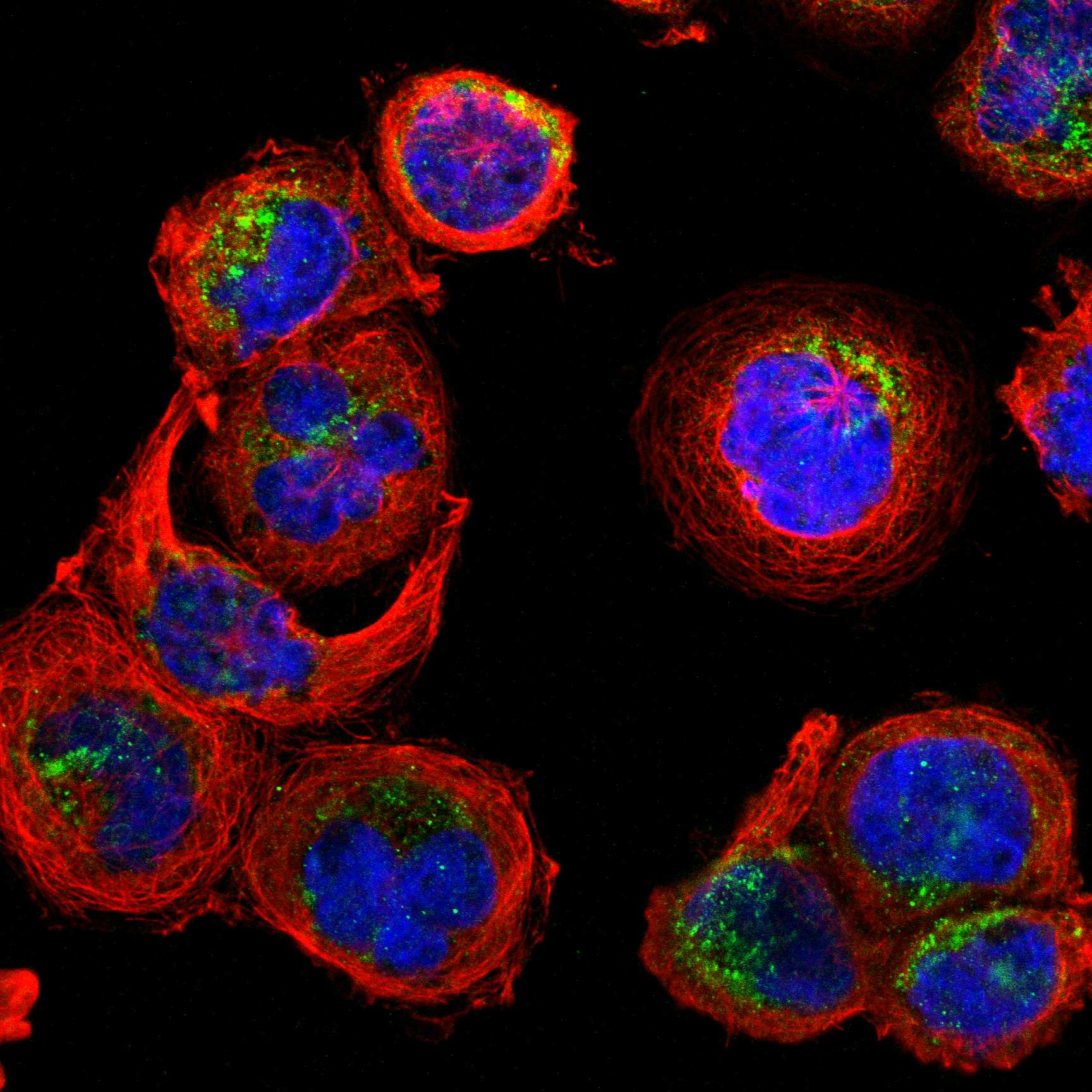
| IF/ICC expectation? (HPA: subcellular ICC-IF). | For the separate IF/ICC application, expect mitochondrial localization: HPA reports Mitochondria (supported) and lists images from A-431, U-251MG, and U2OS (HPA: subcellular ICC-IF). This localization cross-check does not supply an IHC-P protocol or predict fluorescence intensity in paraffin tissue. |
| Situation | Likely cause | Next action |
|---|---|---|
| Hepatocyte staining is absent or very weak. | Hepatocytes should be High (HPA: liver IHC); the cause of a failed run is not specified by that rating. | Verify control-tissue identity and morphology, then review the catalog antibody's IHC-P instructions, retrieval, dilution, and detection records (standard IHC practice). Avoid attributing failure to OTC-specific fixation effects without evidence. |
| Staining appears mainly nuclear or along cell borders. | That location conflicts with mitochondrial matrix OTC and its lack of a transmembrane segment (UniProt P00480 topology). | Recheck compartment against stained hepatocytes, inspect counterstain and section morphology, and repeat with appropriate detection controls if the pattern persists (HPA: High in hepatocytes; standard IHC practice). |
| An HPA-negative cell population stains strongly. | For example, adipose-tissue adipocytes are Not detected (HPA: adipose tissue IHC); cross-reactivity or endogenous detection activity may contribute (standard IHC practice). | Confirm the stained cell type, compare the same run's positive tissue, and use a primary-antibody omission control to assess detection-derived color (standard IHC practice). Do not score unexpected color as OTC solely from intensity. |
| The whole section has diffuse chromogenic background. | Diffuse color can hide the reported granular cytoplasmic profile (HPA: tissue IHC); nonspecific binding or detection background is possible (standard IHC practice). | Inspect the omission control and review blocking, antibody concentration, washing, and chromogen development under the established IHC-P workflow (standard IHC practice). Reassess cellular localization after background is controlled. |
| Intestinal staining seems inconsistent between sections or cell groups. | HPA reports High duodenal and small-intestinal endocrine cells, but Medium rectal enterocytes and appendix endocrine cells (HPA: tissue IHC). | Record the tissue site and identified cell population before comparing intensity; include hepatocytes as a High reference when available (HPA: tissue IHC). Avoid treating all intestinal cells as an equivalent positive control. |
| A granular signal appears, but its specificity remains uncertain. | Granularity agrees with the HPA profile, yet morphology alone cannot establish antibody specificity (HPA: tissue IHC profile; standard IHC practice). | Compare positive and HPA Not detected reference populations, inspect the omission control, and document whether staining follows the expected cells and compartment (HPA: tissue IHC; UniProt P00480 topology; standard IHC practice). |


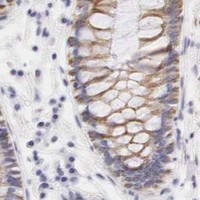


Comprehensive Human Protein Atlas IHC scoring per tissue (reliability: Enhanced — High consistency between antibody staining and RNA expression data.). Rows are taken directly from the HPA tissue chart — click any row's HPA link to view the source.
| Tissue | Cell type | Level | Evidence | Source |
|---|---|---|---|---|
| Duodenum | Endocrine cells | High | Protein (IHC) | HPA → |
| Liver | Hepatocytes | High | Protein (IHC) | HPA → |
| Small intestine | Endocrine cells | High | Protein (IHC) | HPA → |
| Appendix | Endocrine cells | Medium | Protein (IHC) | HPA → |
| Rectum | Enterocytes | Medium | Protein (IHC) | HPA → |
| Tissue | Cell type | Level | Evidence | Source |
|---|---|---|---|---|
| Adipose tissue | Adipocytes | Not detected | Protein (IHC) | HPA → |
| Adrenal gland | Glandular cells | Not detected | Protein (IHC) | HPA → |
| Bone marrow | Hematopoietic cells | Not detected | Protein (IHC) | HPA → |
| Breast | Adipocytes | Not detected | Protein (IHC) | HPA → |
| Bronchus | Respiratory epithelial cells | Not detected | Protein (IHC) | HPA → |
Troubleshoot chromogenic OTC staining in paraffin sections by checking retrieval, cell type, mitochondrial pattern, and matched controls.
A00721-2 has real IHC data from formalin-fixed, paraffin-embedded human hepatoarcinoma (IHC image caption); the catalog lists Human, Mouse, and Rat reactivity (catalog: reactivity).
A00721-2 will render with an IHC-P image of formalin-fixed, paraffin-embedded human hepatoarcinoma stained with a peroxidase-conjugated secondary antibody and DAB (IHC image caption). The catalog lists IHC-P and Human, Mouse, and Rat reactivity for A00721-2; it provides no IF image or IF/ICC application for this SKU (catalog: applications, reactivity, IF images).
Which to pick: Choose A00721-2 for tissue IHC-P: its own image documents staining in formalin-fixed, paraffin-embedded human hepatoarcinoma, and the antibody is rabbit polyclonal (IHC image caption; catalog: host, dilution_raw). For cross-species work, the catalog lists Human, Mouse, and Rat reactivity, while the supplied IHC image documents only the human sample (catalog: reactivity; IHC image caption). No IF/ICC choice is supported by this payload because A00721-2 has no listed IF/ICC application or IF image (catalog: applications, IF images).