This website uses cookies to ensure you get the best experience on our website.


- Table of Contents
Plan chromogenic OTUB2 IHC in paraffin sections with the catalog antibody at 1:100–1:300 (datasheet A10542-1). Use testis as a positive reference and score cytoplasmic staining in elongated or late spermatids by cell type (HPA tissue IHC).

Expected localisation, validated protocols, controls and antibodies — the at-a-glance facts below, then the full design guide.
| Expected localisation | Cytoplasmic in elongated or late spermatids (HPA tissue IHC) | |
| Staining pattern | Distinct cytoplasmic staining in elongated or late spermatids (HPA tissue IHC) | |
| Antigen retrieval | Tris-EDTA pH 9.0 HIER, heat-mediated (datasheet A10542-1) | |
| Positive control | Testis+1 more · see all | |
| Negative control | Adipose tissue+4 more · see all |
| Fixation | Keep fixation consistent across sections. (standard IHC practice; not target-specific) | |
| Caveat | Staining depends on the presence of late spermatids (HPA tissue IHC) | |
| Regulation | Staining-linked regulation is unreported (UniProt) | |
| Isoform / epitope | 2 isoforms; epitope differences are unspecified (UniProt) |
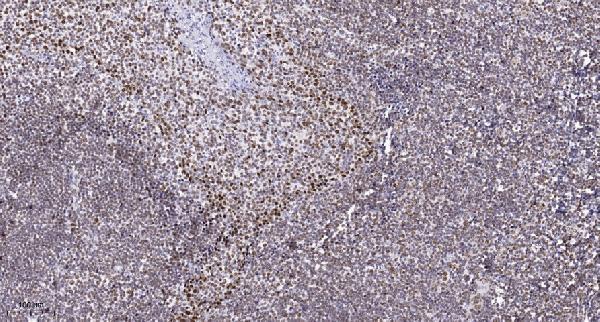
Start with the catalog antibody’s IHC-P protocol, then compare the 4 published OTUB2 IHC protocols below (datasheet A10542-1; PMC9290284; PMC7563931; PMC12034018; PMC11446981).
| Sample | Paraffin-embedded human tonsil tissue; fixative not specified (datasheet A10542-1) |
| Fixation | Image fixative and duration unreported (datasheet A10542-1); verify before use. |
| Sectioning | 4–5 µm sections on charged slides (standard) |
| Deparaffinisation | Xylene, graded ethanol series to water (standard) |
| Antigen retrieval | Heat retrieval: Tris-EDTA pH 9.0 (datasheet A10542-1); 20 min, 95–100 °C (standard) |
| Peroxidase block | 3% H2O2, 10 min, room temperature (standard) |
| Blocking | 10% normal serum of the secondary host, 30 min, room temperature (standard) |
| Primary antibody | Rabbit anti-OTUB2, 1:100-1:300 (datasheet A10542-1) |
| Primary incubation | Overnight at 4 °C (standard) |
| Detection | HRP-polymer secondary, DAB chromogen 5–10 min (standard) |
| Counterstain | Hematoxylin, blue, dehydrate and mount (standard) |
| Expected result | OTUB2-positive staining in elongated or late spermatids of testis (HPA tissue IHC: High). HPA tissue profile: Distinct cytoplasmic expression in spermatids in testis. No signal in the no-primary control. |
OTUB2 should show predominantly cytoplasmic staining in elongated or late spermatids in testis, with weaker staining in placental trophoblastic cells (HPA tissue IHC). This compartment assignment comes from the observed testis pattern; UniProt does not annotate a subcellular location, although OTUB2 has no transmembrane segment (UniProt Q96DC9). HPA rates the tissue IHC evidence Enhanced for consistency with RNA data, pending external verification (HPA tissue IHC).
| Strong cytoplasmic staining in elongated or late spermatids. | This matches the most distinctive reported positive pattern: High staining in those testicular cells (HPA tissue IHC). Judge the result by both cell type and compartment; widespread staining elsewhere in the section does not strengthen this match. HPA's Enhanced rating reflects consistency with RNA data and remains pending external verification (HPA tissue IHC). |
| Predominantly nuclear staining in spermatids, with little cytoplasmic signal. | This conflicts with the distinct cytoplasmic testis pattern (HPA tissue IHC). Treat it as a possible staining artefact and check the controls and detection conditions (standard IHC practice). UniProt gives no subcellular location, so these data do not prove that every nuclear signal is impossible (UniProt Q96DC9). |
| Strong staining in an unexpected cell type, such as testicular cells outside the reported spermatid population. | The reported High testis signal is assigned to elongated or late spermatids (HPA tissue IHC). An unexpected distribution may reflect cross-reactivity or endogenous detection activity; examine a negative control before assigning it to OTUB2 (standard IHC practice). The HPA summary alone cannot establish the identity of every additional stained cell. |
| Diffuse colour across cells and tissue spaces without a clear spermatid pattern. | A uniform haze does not reproduce the distinct cytoplasmic spermatid pattern (HPA tissue IHC). It may arise from background in antibody incubation, blocking or chromogenic detection (standard IHC practice). Assess signal against a matched negative control and inspect whether cellular boundaries and the expected positive population remain discernible. |
| No visible staining in elongated or late spermatids in testis. | Those cells are a reported High positive population, so a blank result warrants a technical check before concluding that OTUB2 is absent (HPA tissue IHC; standard IHC practice). Review section quality, antigen retrieval, antibody incubation and detection with appropriate run controls. HPA's tissue profile is a reference pattern, not a guarantee for every specimen. |
| Tissue and cell selection | Testis provides a reported High positive population in elongated or late spermatids; placental trophoblastic cells are Medium (HPA tissue IHC). Adipocytes in adipose tissue are Not detected in the supplied profile (HPA tissue IHC). Compare the specified cell populations rather than treating an entire tissue as uniformly positive or negative. |
| Antibody validation | Both listed rabbit polyclonal antibodies, HPA002329 and HPA075090, have Enhanced IHC status (HPA antibodies). Enhanced denotes agreement with independent antibodies or orthogonal data in the supplied validation definition; the tissue reliability summary specifically notes consistency with RNA and pending external verification (HPA tissue IHC). |
| Topology and processing | OTUB2 has no annotated transmembrane segment, signal peptide or propeptide; its annotated chain spans residues 1–234 (UniProt Q96DC9). These facts do not specify an epitope or prove a subcellular compartment. Use the observed cytoplasmic spermatid pattern for slide interpretation (HPA tissue IHC). |
| Isoforms and epitope scope | UniProt lists 2 isoforms, but the supplied record does not locate either antibody's epitope or distinguish isoform staining (UniProt Q96DC9; HPA antibodies). Do not infer isoform-specific tissue patterns from these IHC observations; check documented antibody information if an isoform-specific conclusion is required. |
| Can IF/ICC confirm the compartment? | HPA provides no main IF/ICC location, no cell lines with ICC-IF images and no ICC validation status for the listed antibodies (HPA subcellular; HPA antibodies). The cytoplasmic expectation here rests on tissue IHC (HPA tissue IHC). Interpret any separate IF/ICC experiment with its own controls (standard IF practice). |
| Situation | Likely cause | Next action |
|---|---|---|
| Testis positive control is blank. | The observed High spermatid signal has not been reproduced (HPA tissue IHC); the failed step is unknown. | Check tissue preservation and the run control, then review retrieval, primary incubation and chromogenic detection one step at a time (standard IHC practice). Do not attribute the failure to OTUB2-specific fixation sensitivity: none is reported in the supplied sources. |
| Signal is mainly nuclear in the expected spermatids. | The compartment differs from the reported cytoplasmic testis pattern (HPA tissue IHC); a specific cause is unproven. | Compare with a negative control and reassess localisation at high magnification (standard IHC practice). Record the discrepancy rather than assigning nuclear OTUB2 from this result alone; UniProt supplies no location annotation (UniProt Q96DC9). |
| Unexpected cells stain as strongly as spermatids. | Cross-reactivity or endogenous detection activity is possible (standard IHC practice); the reported High population is elongated or late spermatids (HPA tissue IHC). | Inspect a matched negative control and review blocking and detection controls (standard IHC practice). Identify the stained cell type before scoring it as OTUB2 positive; HPA's summary does not validate that new cell-specific pattern. |
| The section shows diffuse brown background. | Nonspecific antibody binding or chromogenic detection background is possible (standard IHC practice). | Compare background with a negative control, then review blocking, primary antibody concentration, washing and detection conditions (standard IHC practice). Score only interpretable cellular staining; the reference pattern is distinct cytoplasmic spermatid signal (HPA tissue IHC). |
| Placenta stains less strongly than testis. | That difference can fit the supplied profile: trophoblastic cells are Medium and elongated or late spermatids are High (HPA tissue IHC). | Compare the named cell populations and assess each against its own background (standard IHC practice). Avoid requiring equal colour intensity across tissues to accept a run; the HPA categories describe different observed levels (HPA tissue IHC). |
| A designated HPA negative cell population shows colour. | Adipocytes in adipose tissue are reported Not detected, so convincing staining there needs investigation (HPA tissue IHC). | Check morphology and the matched negative control, then reassess nonspecific binding or endogenous detection activity (standard IHC practice). Treat a discordant result as unresolved until controls support it; Not detected is the reported observation, not proof of universal absence. |


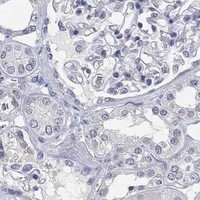
Comprehensive Human Protein Atlas IHC scoring per tissue (reliability: Enhanced — High consistency between antibody staining and RNA expression data. Pending external verification.). Rows are taken directly from the HPA tissue chart — click any row's HPA link to view the source.
| Tissue | Cell type | Level | Evidence | Source |
|---|---|---|---|---|
| Testis | Elongated or late spermatids | High | Protein (IHC) | HPA → |
| Placenta | Trophoblastic cells | Medium | Protein (IHC) | HPA → |
| Tissue | Cell type | Level | Evidence | Source |
|---|---|---|---|---|
| Adipose tissue | Adipocytes | Not detected | Protein (IHC) | HPA → |
| Adrenal gland | Glandular cells | Not detected | Protein (IHC) | HPA → |
| Bone marrow | Hematopoietic cells | Not detected | Protein (IHC) | HPA → |
| Breast | Adipocytes | Not detected | Protein (IHC) | HPA → |
| Caudate | Glial cells | Not detected | Protein (IHC) | HPA → |
Troubleshoot OTUB2 staining in paraffin sections using the catalog antibody’s tissue image and cell-specific expression references; assess IF as a separate application.
A10542-1 lists IHC and IF applications and human, mouse, and rat reactivity (catalog). Its IHC figure shows paraffin-embedded human tonsil (image caption).
A10542-1 will render with an IHC figure of paraffin-embedded human tonsil (image caption). IF is listed as an application, but no IF figure is supplied (catalog).
Which to pick: For tissue IHC, choose A10542-1: its human tonsil caption reports Tris-EDTA retrieval at pH 9.0 and primary antibody at 1:200 overnight at 4°C; the fixative is unreported (image caption). For IF, A10542-1 lists a 1:50 dilution, while ICC validation and an IF figure are unreported (catalog). For cross-species work, A10542-1 lists mouse and rat reactivity; its clone type is unreported (catalog).