This website uses cookies to ensure you get the best experience on our website.


- Table of Contents
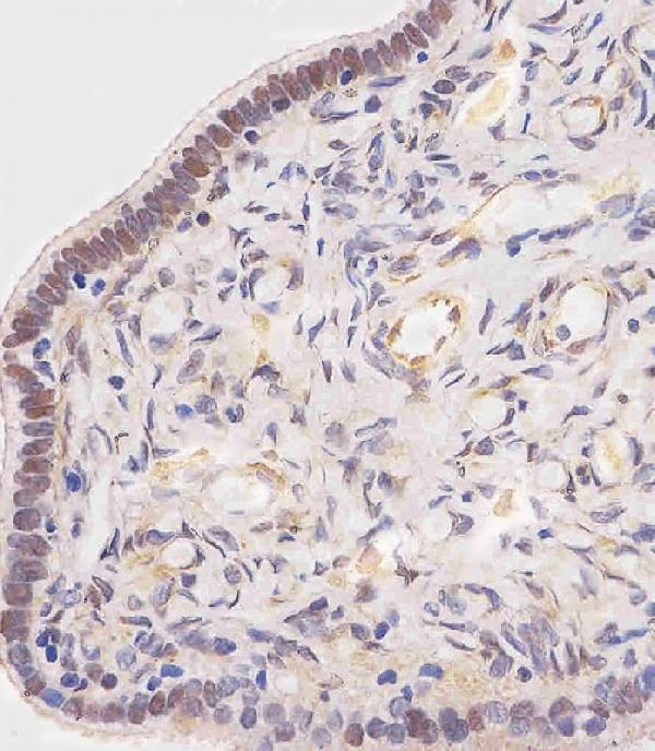
Plan OVGP1 IHC-P using fallopian tube non-ciliated cells as the positive reference (HPA tissue IHC). The catalog antibody has a documented formaldehyde-fixed paraffin workflow at 1:25 primary dilution (datasheet M07911); account for secretion when interpreting tissue staining (UniProt).

Expected localisation, validated protocols, controls and antibodies — the at-a-glance facts below, then the full design guide.
| Expected localisation | Secretory vesicles expected (UniProt); tissue compartment unassigned (HPA tissue IHC) | |
| Staining pattern | High in fallopian tube non-ciliated cells; compartment unspecified (HPA tissue IHC) | |
| Antigen retrieval | Citrate pH 6 HIER, heat-mediated (datasheet M07911) | |
| Positive control | Fallopian tube | |
| Negative control | Adipose tissue+4 more · see all |
| Fixation | Formaldehyde-fixed paraffin sections (selected-SKU IHC image M07911) | |
| Caveat | Secreted protein may stain away from its RNA site (HPA tissue IHC) | |
| Regulation | Estrogen-dependent (UniProt) | |
| Isoform / epitope | No isoforms; signal peptide 1–21 cleaved, no cytoplasmic tail (UniProt) |
The catalog antibody’s IHC-P protocol is accompanied by four published OVGP1 IHC protocols (PMC7457448; PMC7661263; PMC11954873; PMC11447938).
| Sample | Formaldehyde-fixed, paraffin-embedded human fallopian tube tissue (datasheet M07911) |
| Fixation | Image formalin-fixed; duration unreported (datasheet M07911); verify before use. |
| Sectioning | 4–5 µm sections on charged slides (standard) |
| Deparaffinisation | Xylene, graded ethanol series to water (standard) |
| Antigen retrieval | Heat retrieval: Citrate pH 6 (datasheet M07911); 20 min, 95–100 °C (standard) |
| Peroxidase block | 3% H2O2, 10 min, room temperature (standard) |
| Blocking | 10% normal serum of the secondary host, 30 min, room temperature (standard) |
| Primary antibody | Rabbit anti-OVGP1, 1:25 (datasheet M07911) |
| Primary incubation | 1 hours at 37°C (datasheet M07911) |
| Detection | HRP-polymer secondary, DAB chromogen 5–10 min (standard) |
| Counterstain | Hematoxylin, blue, dehydrate and mount (standard) |
| Expected result | OVGP1-positive staining in non-ciliated cells of fallopian tube (HPA tissue IHC: High). HPA tissue profile: Selective expression in fallopian tube. No signal in the no-primary control. |
OVGP1 is a secretory-vesicle protein with no annotated transmembrane segment (UniProt Q12889 subcellular location; topology). In paraffin-section IHC, expect strong staining in fallopian-tube non-ciliated cells (HPA: High in non-ciliated cells; tissue IHC reliability: Enhanced). As a secreted protein, its staining location need not exactly match the cells with OVGP1 RNA (HPA tissue IHC reliability description).
| Strong staining in fallopian-tube non-ciliated cells, with a cytoplasmic, vesicle-associated appearance (HPA: High in non-ciliated cells; UniProt Q12889 subcellular location). | This fits the reported cell-type pattern and secretory-vesicle annotation (HPA tissue IHC; UniProt Q12889). Judge the cell type and compartment together; staining intensity alone does not establish specificity (general IHC practice). |
| Predominantly nuclear or sharp plasma-membrane staining in non-ciliated cells (UniProt Q12889 subcellular location; topology). | Those compartments do not match the annotated secretory vesicles or the absence of a transmembrane segment (UniProt Q12889). Review controls and antibody specificity before calling this OVGP1 (general IHC practice). |
| Strong staining in adipocytes or adrenal glandular cells (HPA: Not detected in these sampled cell types). | This conflicts with the reported tissue IHC pattern (HPA tissue IHC). Consider cross-reactivity or endogenous detection activity; an HPA ‘Not detected’ result is a reference observation, not proof that every specimen must be negative (HPA tissue IHC; general IHC practice). |
| Diffuse staining across tissue compartments, without a discernible non-ciliated-cell pattern (HPA: High in fallopian-tube non-ciliated cells). | A broadly distributed signal is difficult to assign to the reported positive cells (HPA tissue IHC). Check background in a control lacking primary antibody and reassess blocking, detection and washes (general IHC practice). |
| No detectable signal in a fallopian-tube section expected to contain non-ciliated cells (HPA: High in non-ciliated cells). | First confirm that the expected cells are present and the tissue and detection controls worked (general IHC practice). Then review the antibody’s IHC validation and the chosen retrieval and detection conditions; this result alone does not establish absent OVGP1 (general IHC practice). |
| Tissue and cell selection (HPA tissue IHC). | Fallopian tube supplies the reported positive pattern, specifically high staining in non-ciliated cells (HPA tissue IHC). HPA reports no detection in several sampled cell types, including adipocytes and adrenal glandular cells (HPA tissue IHC). |
| Secretory location and topology (UniProt Q12889). | OVGP1 is annotated in secretory vesicles and lacks a transmembrane segment (UniProt Q12889). Interpret intracellular staining in that context; HPA cautions that RNA and protein tissue locations can differ for secreted proteins (HPA tissue IHC). |
| Protein processing and glycosylation (UniProt Q12889). | The annotated signal peptide spans residues 1–21 and the mature chain spans 22–678; five glycosylation sites are listed (UniProt Q12889). These annotations do not identify an antibody epitope or establish a target-specific retrieval requirement (UniProt Q12889; evidence scope). |
| IHC antibody evidence (HPA antibody validation). | HPA062205 has Enhanced IHC status, while HPA031666 has no listed IHC status in the supplied record (HPA antibody validation). Apply that distinction only to those antibodies; it does not validate an unspecified catalog antibody (HPA antibody validation). |
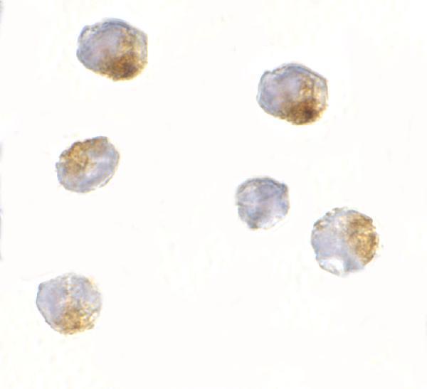
| IF/ICC Q: What location is supported? (HPA subcellular ICC-IF). | A: Vesicles are the supported main location; the additional microtubule location is uncertain (HPA subcellular ICC-IF). HPA031666 has Supported ICC status (HPA antibody validation). Use the separate IF/ICC guide for that application. |
| Situation | Likely cause | Next action |
|---|---|---|
| Fallopian-tube non-ciliated cells are unstained (HPA: High in non-ciliated cells). | The expected cells may be absent from the section, or the IHC workflow may have failed (general IHC practice). | Confirm cell identity and section quality, then check positive and detection controls and the antibody’s IHC instructions (general IHC practice). |
| Nuclei dominate the signal (UniProt Q12889: secretory vesicles). | A dominant nuclear pattern is discordant with the annotated location; its source is unresolved from this slide alone (UniProt Q12889; general IHC practice). | Compare with a control lacking primary antibody and reassess specificity before scoring nuclear signal as OVGP1 (general IHC practice). |
| Adipocytes or adrenal glandular cells stain strongly (HPA: Not detected). | The signal conflicts with HPA’s sampled-cell observations; cross-reactivity or endogenous detection activity are possibilities (HPA tissue IHC; general IHC practice). | Inspect controls lacking primary antibody and review blocking and detection reagents; compare the fallopian-tube pattern (general IHC practice; HPA tissue IHC). |
| Diffuse colour obscures cell boundaries (general IHC practice). | Background from detection or insufficient washing can obscure cell-specific interpretation (general IHC practice). | Check the control lacking primary antibody, blocking and washes; score only interpretable staining in the expected cells (general IHC practice; HPA tissue IHC). |
| Only the luminal or extracellular space appears positive (UniProt Q12889: secretory protein). | Secreted protein may be separated from the cells that express its RNA (HPA tissue IHC reliability description). Extracellular signal alone cannot identify the producing cell (general IHC practice). | Examine whether non-ciliated cells also show the reported high staining, and interpret extracellular signal with tissue morphology and controls (HPA tissue IHC; general IHC practice). |
| An IF/ICC image shows apparent microtubule staining (HPA subcellular ICC-IF). | HPA labels microtubules an uncertain additional location; vesicles are the supported main location (HPA subcellular ICC-IF). | Treat microtubule signal as provisional and consult the separate IF/ICC guide; do not use it to redefine the paraffin-section IHC pattern (HPA subcellular ICC-IF; HPA tissue IHC). |


Comprehensive Human Protein Atlas IHC scoring per tissue (reliability: Enhanced — High consistency between antibody staining and RNA expression data. Secreted protein, tissue location of RNA and protein is expected to differ.). Rows are taken directly from the HPA tissue chart — click any row's HPA link to view the source.
| Tissue | Cell type | Level | Evidence | Source |
|---|---|---|---|---|
| Fallopian tube | Non-ciliated cells | High | Protein (IHC) | HPA → |
| Tissue | Cell type | Level | Evidence | Source |
|---|---|---|---|---|
| Adipose tissue | Adipocytes | Not detected | Protein (IHC) | HPA → |
| Adrenal gland | Glandular cells | Not detected | Protein (IHC) | HPA → |
| Appendix | Glandular cells | Not detected | Protein (IHC) | HPA → |
| Bone marrow | Hematopoietic cells | Not detected | Protein (IHC) | HPA → |
| Breast | Adipocytes | Not detected | Protein (IHC) | HPA → |
Use fallopian tube non-ciliated cells and a matched negative tissue to assess OVGP1 staining (HPA tissue IHC).
The catalog pairs human fallopian tube paraffin-section IHC data with 293-cell IF/ICC data (M07911 and A07911 figure captions). Listed reactivity covers human, mouse, and rat (catalog reactivity).
M07911 is listed for human IHC-P and has an OVGP1 staining image from human fallopian tube paraffin sections at 1:25 (catalog applications and reactivity; M07911 figure caption). A07911 is listed for IF/ICC and has 293-cell images at 2.5 μg/mL; its listed reactivity is human, mouse, and rat (catalog applications and reactivity; A07911 figure captions).
Which to pick: Choose M07911 for human paraffin-section IHC: it is listed as polyclonal and IHC-P tested, with its own fallopian tube section image (M07911 dilution notes; catalog applications; M07911 figure caption). Choose A07911 for IF/ICC because those applications and 293-cell images are listed for it; the captions do not report cell fixation (catalog applications; A07911 figure captions). For mouse or rat work, A07911 is the listed option by reactivity, but its supplied IF/ICC images show 293 cells only (catalog reactivity; A07911 figure captions).