This website uses cookies to ensure you get the best experience on our website.


- Table of Contents
Use hypothalamic magnocellular neurons as a reference for OXT staining in paraffin sections (HPA tissue IHC). This guide helps plan chromogenic IHC and interpret cytoplasmic staining in light of OXT secretion (HPA tissue IHC).

Expected localisation, validated protocols, controls and antibodies — the at-a-glance facts below, then the full design guide.
| Expected localisation | Cytoplasm in hypothalamic neurosecretory neurons (HPA tissue IHC) | |
| Staining pattern | Cytoplasmic staining in magnocellular hypothalamic neurons (HPA tissue IHC) | |
| Antigen retrieval | Citrate pH 6.0 HIER, 95–98 °C, 20 min (rule: cytoplasmic / membrane antigen) | |
| Positive control | Hypothalamus+1 more · see all | |
| Negative control | Adipose tissue+4 more · see all |
| Fixation | Keep fixation consistent across paraffin sections. (standard IHC practice; not target-specific) | |
| Caveat | Secretion can separate protein staining from RNA location (HPA tissue IHC) | |
| Regulation | Regulation not annotated (UniProt) | |
| Isoform / epitope | No isoforms; cleavage separates oxytocin and neurophysin 1 (UniProt) |
The catalog antibody’s IHC-P protocol is accompanied by 2 published chromogenic OXT IHC protocols using filter-mounted tissue and mouse brain sections (PMC2637930; PMC7023993).
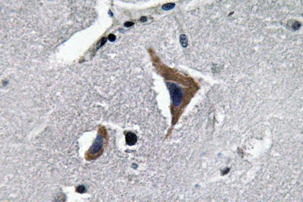
| Sample | Paraffin-embedded human brain tissue; fixative not specified (datasheet A01545) |
| Fixation | Image fixative and duration unreported (datasheet A01545); verify before use. |
| Sectioning | 4–5 µm sections on charged slides (standard) |
| Deparaffinisation | Xylene, graded ethanol series to water (standard) |
| Antigen retrieval | Heat-induced epitope retrieval in citrate buffer, pH 6.0, 20 min at 95–98 °C (standard rule: cytoplasmic / membrane antigen) |
| Peroxidase block | 3% H2O2, 10 min, room temperature (standard) |
| Blocking | 10% normal serum of the secondary host, 30 min, room temperature (standard) |
| Primary antibody | Rabbit anti-OXT, 1:50-1:200 (datasheet A01545) |
| Primary incubation | Overnight at 4 °C (standard) |
| Detection | HRP-polymer secondary, DAB chromogen 5–10 min (standard) |
| Counterstain | Hematoxylin, blue, dehydrate and mount (standard) |
| Expected result | OXT-positive staining in neuronal cells of hypothalamus (HPA tissue IHC: High). HPA tissue profile: Cytoplasmic expression mainly in magnocellular neurosecretory neurons in hypothalamus and in oocytes. No signal in the no-primary control. |
OXT precursor is packaged in neurosecretory granules and has no transmembrane segment, so cellular staining is expected in the cytoplasm rather than at the plasma membrane (UniProt P01178). In tissue IHC, HPA reports cytoplasmic staining mainly in magnocellular neurosecretory neurons of the hypothalamus; its ovarian profile also mentions oocytes (HPA: tissue IHC). The tissue pattern has Supported reliability, with medium agreement between antibody staining and RNA data (HPA: reliability Supported).
| Distinct cytoplasmic chromogen in hypothalamic neuronal cells, with little staining in adjacent cells. | This fits the principal tissue pattern: HPA rates hypothalamic neuronal staining High and describes expression mainly in magnocellular neurosecretory neurons (HPA: tissue IHC). Granule-associated cytoplasmic staining is biologically plausible because pro-OXT is packaged into neurosecretory granules (UniProt P01178). Score the stained cells and compartment together. |
| Ovarian staining appears in follicle cells, with possible staining in oocytes. | HPA's positive-cell table rates follicle-cell staining High, while its summary also mentions oocytes (HPA: tissue IHC). Record which cell type stains rather than treating the two descriptions as identical. Interpret ovarian staining alongside morphology and a negative control; this tissue pattern alone does not establish antibody specificity. |
| Signal is predominantly nuclear or sharply outlines cell membranes. | That compartment differs from HPA's cytoplasmic tissue pattern and the vesicular or secreted localization in UniProt (HPA: tissue IHC; UniProt P01178). Review morphology and controls before calling it OXT. A membrane rim should not be justified solely by UniProt's reported interactions with vasopressin receptors (UniProt P01178). |
| Strong staining occurs in an HPA negative cell population, or chromogen is spread diffusely across the section. | For example, HPA reports OXT not detected in adipose-tissue adipocytes and caudate glial cells (HPA: tissue IHC). Such staining warrants a specificity check; cross-reactivity or endogenous detection activity are possibilities, not proven causes. Diffuse deposit without cell boundaries is also difficult to score as cellular OXT (standard IHC practice). |
| The hypothalamic neuronal positive control has no detectable signal. | This conflicts with HPA's High staining report for hypothalamic neuronal cells (HPA: tissue IHC). First check that the section contains the expected neuronal population, then assess the staining run and antibody performance (standard IHC practice). Do not infer that every hypothalamic cell must stain: HPA identifies a specific neuronal pattern. |
| Precursor processing and epitope identity | OXT is a processed precursor: UniProt annotates a signal peptide at residues 1–19 and neurophysin 1 at 32–125 (UniProt P01178). The payload does not identify the catalog antibody's epitope. Consequently, the observed stain cannot be assigned specifically to mature oxytocin, precursor, or neurophysin without epitope information. |
| Secretion and tissue location | Pro-OXT enters neurosecretory granules, and OXT products are secreted (UniProt P01178). HPA cautions that protein and RNA tissue locations can differ for a secreted protein (HPA: reliability description). Use the observed IHC cell pattern for slide interpretation; tissue RNA enrichment in brain is supporting context, not a map of every stained cell (HPA: RNA specificity). |
| Strength of IHC evidence | HPA assigns tissue staining Supported reliability and lists antibody HPA071892 as IHC Supported (HPA: tissue IHC; HPA: antibodies). HPA also notes medium staining–RNA consistency and cautions that the antibody targets protein from more than one gene (HPA: reliability description). These limits make controls and cell-level interpretation important. |
| Translation to IF/ICC | HPA's subcellular entry says secreted, but gives no main ICC/IF location or cell-line images; HPA071892 has no listed ICC status (HPA: subcellular; HPA: antibodies). The IHC tissue pattern therefore does not establish an IF/ICC appearance or validation. Evaluate IF/ICC on its own guide page using that application's evidence. |
| Situation | Likely cause | Next action |
|---|---|---|
| The expected hypothalamic neurons are negative. | The relevant neuronal population may be absent from the section, or the staining run may have failed; HPA reports High neuronal staining in hypothalamus (HPA: tissue IHC). | Confirm tissue anatomy and inspect a known-positive section processed in the same run. Then review the antibody and detection controls and the validated IHC-P workflow (standard IHC practice). |
| Many unrelated cell types show uniform brown signal. | Broad signal is inconsistent with HPA's mainly hypothalamic neuronal cytoplasmic pattern; nonspecific binding or endogenous detection activity may contribute (HPA: tissue IHC; standard IHC practice). | Compare a negative detection control and inspect whether signal follows cells or tissue deposits. Review blocking and detection conditions within the established chromogenic IHC workflow (standard IHC practice). |
| Nuclei or membrane rims dominate the stain. | The compartment conflicts with HPA's cytoplasmic tissue pattern and UniProt's secretory-vesicle localization; OXT has no annotated transmembrane segment (HPA: tissue IHC; UniProt P01178). | Check the counterstain and cell boundaries, then compare positive and negative controls. Withhold an OXT-positive call until a plausible cytoplasmic cellular pattern is demonstrated. |
| Adipose-tissue adipocytes or caudate glia stain strongly. | HPA reports OXT not detected in those cell populations; the signal may reflect cross-reactivity or a detection artifact, although the image alone cannot determine which (HPA: tissue IHC). | Verify cell identity, compare the same run's hypothalamic positive control, and inspect a negative detection control before assigning the unexpected signal to OXT (standard IHC practice). |
| Ovarian results seem inconsistent between cell annotations. | HPA's positive table names follicle cells, while its profile also mentions oocytes (HPA: tissue IHC). These are distinct cell descriptions and should not be merged when scoring. | Annotate follicle cells and oocytes separately on the slide. Report the compartment and intensity for each, and qualify any conclusion that depends on which population is stained. |
| Can the IHC pattern be used as an IF/ICC positive-control specification? | HPA provides no ICC/IF images or main subcellular location for OXT, and lists no ICC status for HPA071892 (HPA: subcellular; HPA: antibodies). | Use the separate IF/ICC guide to define that application's controls and interpretation. Treat the IHC cytoplasmic pattern as biological context, not as IF/ICC validation. |


Comprehensive Human Protein Atlas IHC scoring per tissue (reliability: Supported — Medium consistency between antibody staining and RNA expression data. Caution, targets protein from more than one gene. Secreted protein, tissue location of RNA and protein is expected to differ. External characterization data supports antibody staining.). Rows are taken directly from the HPA tissue chart — click any row's HPA link to view the source.
| Tissue | Cell type | Level | Evidence | Source |
|---|---|---|---|---|
| Hypothalamus | Neuronal cells | High | Protein (IHC) | HPA → |
| Ovary | Follicle cells | High | Protein (IHC) | HPA → |
| Tissue | Cell type | Level | Evidence | Source |
|---|---|---|---|---|
| Adipose tissue | Adipocytes | Not detected | Protein (IHC) | HPA → |
| Adrenal gland | Glandular cells | Not detected | Protein (IHC) | HPA → |
| Appendix | Lymphoid tissue | Not detected | Protein (IHC) | HPA → |
| Bone marrow | Hematopoietic cells | Not detected | Protein (IHC) | HPA → |
| Breast | Adipocytes | Not detected | Protein (IHC) | HPA → |
Troubleshoot paraffin-section chromogenic IHC for OXT by checking retrieval, cellular distribution, processing and controls before interpreting staining intensity.
A01545 has IHC data from paraffin-embedded human brain tissue (A01545 IHC image caption). Listed reactivity covers Human, Mouse, and Rat (catalog: reactivity); no IF image is supplied (catalog: IF image alts).
A01545 is listed for IHC in Human, Mouse, and Rat (catalog: applications, reactivity). Its IHC image shows paraffin-embedded human brain tissue (A01545 IHC image caption).
Which to pick: Choose A01545 for paraffin-section tissue IHC: its own image documents human brain tissue, but does not report the fixative (A01545 IHC image caption). The rabbit pAb is listed as reactive with Mouse and Rat, although its supplied IHC image shows only human tissue (catalog: host, reactivity; A01545 IHC image caption). No SKU has listed IF/ICC validation or an IF image, so the payload does not support an IF/ICC pick (catalog: applications, IF image alts).