This website uses cookies to ensure you get the best experience on our website.


- Table of Contents
Plan chromogenic IHC-P with the catalog antibody at 1:50 (datasheet M10087-1). Compare high staining in tonsil squamous epithelial cells with adipocytes where staining was not detected (HPA tissue IHC).

Expected localisation, validated protocols, controls and antibodies — the at-a-glance facts below, then the full design guide.
| Expected localisation | Cell membrane expected (UniProt); tissue compartment unassigned (HPA tissue IHC) | |
| Staining pattern | Immune cell subsets in spleen, intestine and placental stroma (HPA tissue IHC) | |
| Antigen retrieval | EDTA pH 8.0 HIER, heat-mediated (datasheet M10087-1) | |
| Positive control | Tonsil+4 more · see all | |
| Negative control | Adipose tissue+4 more · see all |
| Fixation | Keep fixation consistent across paraffin sections. (standard IHC practice; not target-specific) | |
| Caveat | Staining in unannotated cells complicates assignment (HPA tissue IHC) | |
| Regulation | Lymphoid tissue–enhanced RNA (HPA tissue RNA) | |
| Isoform / epitope | No isoforms or processing annotated; epitope side matters (UniProt) |
Compare the catalog antibody’s IHC-P protocol with three published P2RY6 chromogenic IHC workflows (datasheet; PMC10601148; PMC6844642; PMC13473056).
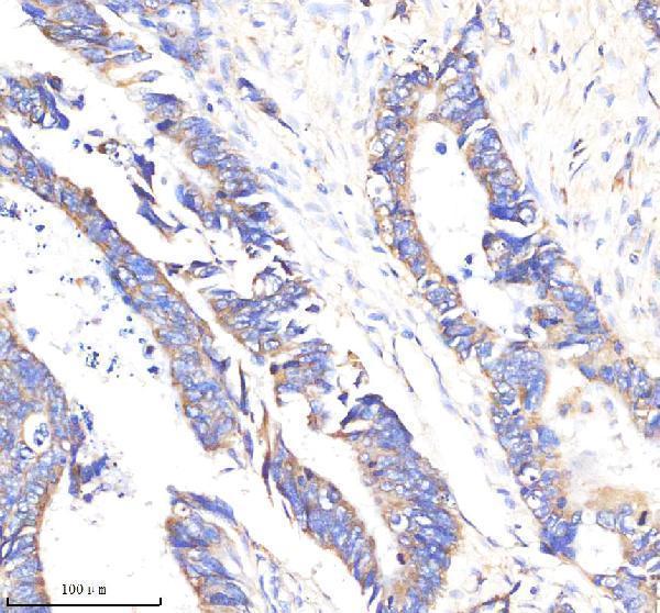
| Sample | Paraffin-embedded human colon cancer tissue; fixative not specified (datasheet M10087-1) |
| Fixation | Image fixative and duration unreported (datasheet M10087-1); verify before use. |
| Sectioning | 4–5 µm sections on charged slides (standard) |
| Deparaffinisation | Xylene, graded ethanol series to water (standard) |
| Antigen retrieval | Heat retrieval: EDTA pH 8.0 (datasheet M10087-1); 20 min, 95–100 °C (standard) |
| Peroxidase block | 3% H2O2, 10 min, room temperature (standard) |
| Blocking | 10% goat serum (datasheet M10087-1) |
| Primary antibody | Rabbit monoclonal (clone 19P56) anti-P2RY6, 1:50 (datasheet M10087-1) |
| Primary incubation | Overnight at 4 °C (datasheet M10087-1) |
| Detection | HRP-conjugated secondary, DAB chromogen (datasheet M10087-1) |
| Counterstain | Hematoxylin, blue, dehydrate and mount (standard) |
| Expected result | P2RY6-positive staining in squamous epithelial cells of tonsil (HPA tissue IHC: High). HPA tissue profile: Expressed in a subset of immune cells in spleen, intestine and placental stroma. Also expressed in immune celler in other tissues. No signal in the no-primary control. |
P2RY6 is a seven-transmembrane receptor annotated at the cell membrane (UniProt Q15077 topology; UniProt Q15077 subcellular location). In IHC, expect staining in selected cells rather than every cell in a tissue: HPA reports positive immune-cell populations, along with several named epithelial and other cell populations (HPA tissue IHC). Treat these as reference patterns with limited certainty: HPA rates the tissue IHC evidence Enhanced but reports medium consistency with RNA expression (HPA tissue IHC).
| Selected cells show membrane-associated chromogenic staining; splenic red-pulp cells or tonsillar squamous epithelial cells are conspicuous. | This fits the predicted compartment (UniProt Q15077 cell membrane) and reported staining: medium in splenic red-pulp cells and high in tonsillar squamous epithelial cells (HPA tissue IHC). Evaluate the named cell population, since staining elsewhere in the same section does not establish the expected pattern (HPA tissue IHC). |
| The dominant signal is nuclear or uniformly fills cytoplasm, with no convincing membrane-associated component. | That distribution conflicts with the cell-membrane annotation (UniProt Q15077 subcellular location). Treat it as suspect staining and compare with a positive tissue and detection controls (general IHC practice); the supplied HPA tissue data do not establish a nuclear or diffuse cytoplasmic P2RY6 pattern (HPA tissue IHC). |
| Strong staining appears in cells HPA lists as not detected, such as adipocytes in adipose tissue or respiratory epithelial cells in bronchus. | This conflicts with those cell-specific reference observations (HPA tissue IHC). Check cell identification and controls for nonspecific primary binding or endogenous chromogen-generating activity (general IHC practice). A negative HPA observation is a comparator, not proof that every specimen of that tissue must be negative (HPA tissue IHC). |
| Chromogen spreads across tissue structures, including expected negative cell populations, without a cell-associated pattern. | Diffuse deposition is hard to reconcile with the selected-cell tissue profile (HPA tissue IHC) or predicted membrane location (UniProt Q15077 subcellular location). Assess background with an appropriate no-primary control, then review blocking, washing and detection chemistry (general IHC practice). |
| A known-positive cell population has no convincing signal, for example tonsillar squamous epithelial cells or splenic red-pulp cells. | That result disagrees with the reported high or medium HPA observations, respectively (HPA tissue IHC). First verify that the relevant cells are present and identifiable; then inspect the IHC run and its controls (general IHC practice). One negative section does not overturn an Enhanced but medium-consistency reference pattern (HPA tissue IHC). |
| Predicted compartment and epitope side | P2RY6 has seven transmembrane segments with extracellular and cytoplasmic regions (UniProt Q15077 topology). Its annotation supports a membrane expectation, but the supplied record gives no antibody epitope; do not infer which face the IHC-validated antibody recognizes or prescribe permeabilisation from topology alone (UniProt Q15077 topology; HPA antibodies). |
| Cell population within the section | HPA reports medium staining in appendix germinal-center cells, bone-marrow hematopoietic cells, lymph-node non-germinal-center cells and splenic red-pulp cells (HPA tissue IHC). Score the reported cell population, since a whole-section impression can miss a localized positive population (general IHC practice). |
| Reference strength | The listed antibody HPA074751 has Enhanced IHC validation, while HPA describes medium consistency between antibody staining and RNA expression (HPA antibodies; HPA tissue IHC). This supports using the reported pattern as a guide while investigating discordant cells rather than treating every listed intensity as a guaranteed outcome (HPA tissue IHC). |
| Lower-signal and negative comparators | HPA reports low staining in small-intestine goblet cells and no detection in several specified cell populations, including adrenal glandular cells and cerebellar granular-layer cells (HPA tissue IHC). These comparisons concern named cells, not blanket negativity throughout each tissue (HPA tissue IHC). |
| Sequence annotation limits | UniProt annotates one glycosylation site at residue 5 and no isoforms, signal peptide or propeptide in the supplied record (UniProt Q15077 processing and glycosylation). Those annotations do not identify the antibody epitope or establish a P2RY6-specific retrieval, fixation-sensitivity or shedding effect (UniProt Q15077 annotations). |
| Situation | Likely cause | Next action |
|---|---|---|
| The expected positive cells are absent or cannot be identified in the section. | The sampled area may lack the cell population needed to compare with the HPA reference (HPA tissue IHC). | Choose a section containing a reported positive population, such as tonsillar squamous epithelial cells or splenic red-pulp cells, and score those cells specifically (HPA tissue IHC; general IHC practice). |
| Reported positive cells are present, but the chromogenic IHC run shows no signal. | A failed or weak staining run is possible; the HPA reference alone cannot identify a target-specific fixation or retrieval cause (HPA tissue IHC). | Check section integrity, primary-antibody application, detection reagents and a run-level positive control; adjust general IHC conditions only against appropriate controls (general IHC practice). |
| Brown signal appears in unexpected negative-reference cells. | Nonspecific binding or endogenous detection activity can mimic a positive chromogenic result (general IHC practice); HPA lists some of these cell types as not detected (HPA tissue IHC). | Confirm cell identity, compare a no-primary control, and review blocking and detection steps before assigning P2RY6 positivity (general IHC practice). |
| Signal is widespread and obscures cell boundaries. | High background or deposited chromogen can make the membrane pattern unreadable (general IHC practice; UniProt Q15077 cell membrane). | Compare no-primary and positive controls; review primary-antibody concentration, washing and detection time as general IHC variables (general IHC practice). No target-specific dilution is supplied here. |
| A cell stains, but the signal is predominantly nuclear or diffuse cytoplasmic. | The compartment differs from the annotated cell-membrane location (UniProt Q15077 subcellular location). | Score it as inconclusive for the expected pattern; inspect morphology and controls, then seek membrane-associated signal in a reported positive population (UniProt Q15077 subcellular location; HPA tissue IHC; general IHC practice). |
| Q: Can the HPA ICC-IF entry confirm an IF pattern or supply an IF/ICC protocol? | HPA summarizes the location as Membrane but provides no main location or cell-line ICC-IF images in the supplied entry (HPA subcellular ICC-IF). | A: Use that entry only as limited localisation context alongside UniProt's cell-membrane annotation; assess IF/ICC on its separate guide page, since these payloads provide no IF/ICC protocol (HPA subcellular ICC-IF; UniProt Q15077 subcellular location). |

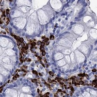


Comprehensive Human Protein Atlas IHC scoring per tissue (reliability: Enhanced — Medium consistency between antibody staining and RNA expression data. Antibody staining in cells/structures not annotated, view images.). Rows are taken directly from the HPA tissue chart — click any row's HPA link to view the source.
| Tissue | Cell type | Level | Evidence | Source |
|---|---|---|---|---|
| Tonsil | Squamous epithelial cells | High | Protein (IHC) | HPA → |
| Appendix | Germinal center cells | Medium | Protein (IHC) | HPA → |
| Bone marrow | Hematopoietic cells | Medium | Protein (IHC) | HPA → |
| Duodenum | Goblet cells | Medium | Protein (IHC) | HPA → |
| Epididymis | Glandular cells | Medium | Protein (IHC) | HPA → |
| Tissue | Cell type | Level | Evidence | Source |
|---|---|---|---|---|
| Adipose tissue | Adipocytes | Not detected | Protein (IHC) | HPA → |
| Adrenal gland | Glandular cells | Not detected | Protein (IHC) | HPA → |
| Breast | Adipocytes | Not detected | Protein (IHC) | HPA → |
| Bronchus | Respiratory epithelial cells | Not detected | Protein (IHC) | HPA → |
| Caudate | Glial cells | Not detected | Protein (IHC) | HPA → |
Troubleshoot P2RY6 staining in paraffin sections by checking retrieval, cellular context and membrane localisation before scoring chromogenic signal.
The catalog antibody has a human paraffin-section IHC image and lists IF/ICC among its applications; no IF image is supplied (M10087-1 catalog and IHC caption).
M10087-1 is listed for human IHC and IF/ICC (M10087-1 catalog). Its IHC image shows staining in a paraffin-embedded human colon cancer section; no IF image is supplied (M10087-1 IHC caption and catalog).
Which to pick: Choose M10087-1 for paraffin-section IHC because its own caption documents that preparation; the fixative is unreported (M10087-1 IHC caption). For IF/ICC, M10087-1 is listed as applicable, but the payload supplies no IF image (M10087-1 catalog). Cross-species use is unsupported here because human is the only listed reactivity (M10087-1 catalog).