P4HB / Protein disulfide-isomerase · IHC design guide

Design Immunohistochemistry for P4HB

Plan paraffin-section P4HB IHC around the cytoplasmic staining seen in most tissues (HPA tissue IHC). Use pancreatic exocrine cells as a high-staining reference and start the catalog antibody at 2–5 μg/mL (HPA tissue IHC; datasheet A02335-1).

Evidence assembled Oct 2026 · For research use; verify linked source records and product datasheet before use
Immunohistochemistry protocol sheet for P4HB (IHC for P4HB): expected localisation Cytoplasmic staining in most tissues (HPA tissue IHC), antibody A02335-1, validated IHC image, and IHC protocol steps
Printable P4HB IHC protocol sheet — expected localisation Cytoplasmic staining in most tissues (HPA tissue IHC), antibody A02335-1, controls and protocol steps. Open the full P4HB IHC guide →

P4HB Immunohistochemistry Experimental Design Guide

Expected localisation, validated protocols, controls and antibodies — the at-a-glance facts below, then the full design guide.

Must know before staining
Expected localisation Cytoplasmic staining in most tissues (HPA tissue IHC)
Staining pattern Cytoplasmic in most tissues; high in pancreatic exocrine cells (HPA tissue IHC)
Antigen retrieval EDTA pH 8.0 HIER, heat-mediated (datasheet A02335-1)
Positive control ⓘ Appendix+4 more · see all
Negative control ⓘ Cerebellum+3 more · see all
Important caveats
Reasons your staining may differ from the expected pattern.
Fixation Keep fixation conditions consistent across sections. (standard IHC practice; not target-specific)
Caveat Surface-associated P4HB may be shed in some cell types (UniProt)
Regulation No abundance regulator specified (UniProt)
Isoform / epitope No isoforms listed; signal peptide 1–17 is removed (UniProt)
Section 1

Recommended P4HB IHC & IF Protocols

Start with the catalog antibody’s IHC-P protocol, then compare the published P4HB staining conditions for kidney, prostate, gastric and bladder tissue (PMC9548632; PMC11304455; PMC6000295; PMC7751343).

Recommended immunohistochemistry (IHC-P) protocol parameters
SampleParaffin-embedded mouse spleen tissue; fixative not specified (datasheet A02335-1)
FixationImage fixative and duration unreported (datasheet A02335-1); verify before use.
Sectioning4–5 µm sections on charged slides (standard)
DeparaffinisationXylene, graded ethanol series to water (standard)
Antigen retrievalHeat retrieval: EDTA pH 8.0 (datasheet A02335-1); 20 min, 95–100 °C (standard)
Peroxidase block3% H2O2, 10 min, room temperature (standard)
Blocking10% goat serum (datasheet A02335-1)
Primary antibodyRabbit anti-P4HB, 2-5μg/ml (datasheet A02335-1)
Primary incubationOvernight at 4 °C (datasheet A02335-1)
DetectionStreptavidin-biotin complex (SABC), DAB chromogen (datasheet A02335-1)
CounterstainHematoxylin, blue, dehydrate and mount (standard)
Expected resultP4HB-positive staining in endocrine cells of appendix (HPA tissue IHC: High). HPA tissue profile: Cytoplasmic expression in most tissues. No signal in the no-primary control.
💡Decision noteTry heat-mediated EDTA pH 8.0 retrieval first (datasheet A02335-1); the prostate and bladder protocols used citrate retrieval (PMC11304455; PMC7751343).
Section 2

What Is the Expected P4HB Staining Pattern?

P4HB is primarily an endoplasmic reticulum lumen protein with no transmembrane segment (UniProt P07237: location and topology). In paraffin section IHC, expect cytoplasmic staining across many tissues (HPA: cytoplasmic expression in most tissues). Strong reference cells include pancreatic exocrine glandular cells, kidney proximal tubule cell bodies and lung type II alveolar cells (HPA: High). HPA rates the tissue IHC profile Enhanced, reflecting consistency between antibody staining and RNA expression (HPA: Enhanced).

What am I looking at on my slide?
Strong cytoplasmic staining in pancreatic exocrine glandular cells or kidney proximal tubule cell bodies.This fits two documented High reference patterns (HPA: pancreas and kidney). Compare staining among the named cells on the same section; the tissue name alone does not establish that every cell should be equally positive. The expected compartment is cytoplasmic in tissue IHC (HPA: tissue profile).
Signal appears exclusively nuclear, with little or no cytoplasmic staining in otherwise positive cells.An exclusively nuclear pattern conflicts with the reported cytoplasmic tissue pattern and ER localization (HPA: tissue profile and subcellular location; UniProt P07237: ER lumen). Treat it as suspect staining and review counterstain, detection background and antibody specificity before scoring it as P4HB.
Strong staining predominates in skeletal muscle myocytes or parathyroid glandular cells.Those specific cell populations are reported Not detected by HPA tissue IHC (HPA: skeletal muscle and parathyroid gland). Unexpected signal may reflect cross-reactivity or endogenous detection activity (general IHC practice). Check a detection-only control; do not assume that every cell in either tissue is a negative reference.
Diffuse colour covers cells and spaces without a clear cellular pattern.This is difficult to reconcile with HPA's cell-associated cytoplasmic profile (HPA: tissue profile). In chromogenic IHC, nonspecific binding, excessive detection signal or residual endogenous activity can obscure localization (general IHC practice). Assess background on a matched control before assigning intensity scores.
No convincing signal appears in pancreatic exocrine glandular cells.HPA reports High staining in that cell population (HPA: pancreas). First verify that those cells are present and recognizable, then review the staining run and controls (general IHC practice). One blank section does not establish biological absence or a P4HB-specific sensitivity to fixation.
💡Expected P4HB appearanceCall positive a clear cytoplasmic signal in the documented High cell populations, especially pancreatic exocrine glandular cells (HPA: tissue IHC); regard exclusively nuclear staining or uniform diffuse colour as suspect against the reported ER and cytoplasmic pattern (HPA: subcellular and tissue profiles).
How each factor affects the staining
Compartment and topologyP4HB has an ER lumen assignment and no transmembrane segment (UniProt P07237: location and topology). For chromogenic tissue IHC, interpret a cellular cytoplasmic pattern in context (HPA: tissue profile); topology alone cannot identify the epitope recognized by a particular antibody.
Processing and possible surface signalThe precursor has a signal peptide at residues 1–17, with the annotated chain spanning 18–508 (UniProt P07237: processing). Surface association or secretion is described as possible in some cell types (UniProt P07237: location). It does not make widespread membrane-only tissue staining the expected IHC result (HPA: tissue profile).
Tissue reference selectionHPA reports High staining in pancreatic exocrine glandular cells and Not detected in skeletal muscle myocytes (HPA: tissue IHC). Use the named cell populations when judging a run; these observations are not a universal positive or negative designation for all cells in those organs.
Antibody evidenceTwo listed antibodies, HPA018884 and CAB012463, have Enhanced IHC status (HPA: antibody validation). This supports the reported tissue pattern but does not provide an epitope, a working dilution or evidence that a particular antigen retrieval condition is required.
IF/ICC Q&A: where is P4HB expected?The supported main location is the endoplasmic reticulum in the HPA ICC-IF record (HPA: subcellular profile). This localization answers the IF/ICC interpretation question; it does not specify an IF/ICC staining protocol or predict identical image contrast across assays.
Why is my staining missing, weak or wrong?
SituationLikely causeNext action
Known-positive pancreatic cells are blank.The result conflicts with the reported High pancreatic exocrine cell staining (HPA: pancreas); a failed or weak staining run is possible (general IHC practice).Confirm the correct cells are in the section, inspect a run control, and review antibody dilution, antigen retrieval and detection steps (general IHC practice). No P4HB-specific retrieval condition is established here.
Only nuclei appear positive.That distribution conflicts with cytoplasmic tissue staining and ER localization (HPA: tissue and subcellular profiles). Counterstain or detection artefact is possible (general IHC practice).Review the chromogen and counterstain separately, then compare with a control stained using the same detection system (general IHC practice). Score P4HB only where the cellular pattern is credible.
Unexpected staining appears in skeletal muscle myocytes.HPA reports these myocytes as Not detected (HPA: skeletal muscle). Cross-reactivity or endogenous detection activity is possible (general IHC practice).Check a detection-only control and compare the suspect cells with a documented positive population (general IHC practice; HPA: pancreas). Investigate the unexpected signal before treating it as biological expression.
Brown colour is widespread, including between cells.Diffuse background can hide the cell-associated cytoplasmic pattern reported by HPA (HPA: tissue profile); nonspecific binding or excessive detection signal are possible (general IHC practice).Compare a matched control, then review blocking, antibody dilution, washing and chromogen development (general IHC practice). Reassess localization after background is controlled.
Staining is present but weak in a reference-positive cell population.A weak result differs from HPA's High category for the named cells (HPA: positive tissue entries). Section and assay performance can also affect apparent intensity (general IHC practice).Verify cell identity and compare a control from the same run; review dilution, retrieval and detection settings (general IHC practice). Do not infer a P4HB-specific fixation effect from intensity alone.
A tissue shows a mixture of strongly stained and unstained cells.HPA assigns levels to particular cell populations, including High pancreatic exocrine cells and Not detected skeletal muscle myocytes (HPA: tissue IHC). A whole-tissue average can conceal this distinction.Identify and score the relevant cell types and their cytoplasmic staining separately (HPA: tissue profile; general IHC practice). Compare results with the matching HPA cell-level entry.

Sample controls for P4HB IHC & IF

🧪Run colon first: endocrine cells should stain strongly (HPA: High in colon endocrine cells). Use skeletal muscle myocytes as the biological comparison (HPA: Not detected in skeletal muscle myocytes); within the colon slide, nuclei should lack specific signal, while neighboring cells should be judged against reagent-control background rather than assumed negative (HPA: endoplasmic reticulum localization).
Positive control tissue: Appendix (Endocrine cells, HPA High)
Negative control tissue: Cerebellum (HPA Not detected)
ICC-IF cell lines (HPA subcellular resource): HPA ICC-IF images show P4HB in A-431, U-251MG, U2OS, NIH 3T3, with annotated localisation: Endoplasmic reticulum (supported) (HPA subcellular).
Technical controls: Include no-primary (secondary-only), a concentration-matched rabbit IgG isotype control appropriate to the primary antibody, and P4HB knockout tissue if available (selected-SKU caption: rabbit primary; standard IHC practice). For chromogenic colon sections, quench endogenous peroxidase and check for endogenous biotin if using the caption’s biotin–streptavidin detection system (selected-SKU caption: biotinylated secondary and SABC–DAB; standard IHC practice).
⚠️Feasibility: A target-specific fixation window or fixation effect is unreported, and the selected-SKU paraffin-section caption does not state a fixative (selected-SKU caption: fixative unreported). Heat retrieval with EDTA at pH 8.0 is a documented starting point for paraffin IHC in mouse spleen; its performance in colon needs verification (selected-SKU caption: EDTA pH 8.0, mouse spleen). HPA ICC-IF supports an endoplasmic reticulum localization check, but the supplied evidence does not establish whether frozen sections or IF are easier than paraffin IHC; colon DAB interpretation should account for endogenous peroxidase and, with SABC detection, biotin background (HPA: supported endoplasmic reticulum localization; selected-SKU caption: SABC–DAB; standard IHC practice).

HPA tissue IHC evidence for P4HB

Comprehensive Human Protein Atlas IHC scoring per tissue (reliability: Enhanced — High consistency between antibody staining and RNA expression data.). Rows are taken directly from the HPA tissue chart — click any row's HPA link to view the source.

Positive expression · recommended positive controls

TissueCell typeLevelEvidenceSource
Appendix Endocrine cells High Protein (IHC) HPA →
Colon Endocrine cells High Protein (IHC) HPA →
Duodenum Endocrine cells High Protein (IHC) HPA →
Epididymis Glandular cells High Protein (IHC) HPA →
Gallbladder Glandular cells High Protein (IHC) HPA →

Undetected expression · recommended negative controls

TissueCell typeLevelEvidenceSource
Cerebellum Cells in granular layer Not detected Protein (IHC) HPA →
Parathyroid gland Glandular cells Not detected Protein (IHC) HPA →
Skeletal muscle Myocytes Not detected Protein (IHC) HPA →
Smooth muscle Smooth muscle cells Not detected Protein (IHC) HPA →
Section 3

Advanced P4HB IHC Tips

Troubleshoot P4HB staining in paraffin sections by checking retrieval, compartment, cell identity and assay controls before comparing signal intensity (datasheet A02335-1; HPA tissue IHC; UniProt P07237).

Which retrieval condition should I start with for P4HB IHC?
Use heat-mediated retrieval in EDTA at pH 8.0 for paraffin sections (datasheet A02335-1). Keep heating and cooling conditions consistent across the comparison set, then assess staining in the positive-control section before changing retrieval intensity (standard IHC practice; HPA tissue IHC: high in pancreatic exocrine glandular cells). Excessive retrieval can damage morphology or increase diffuse signal, so evaluate staining alongside intact cell boundaries and counterstain (standard IHC practice). If signal remains weak, vary retrieval duration in a small controlled series while holding antibody concentration and chromogenic development constant (standard IHC practice; datasheet A02335-1: 2 µg/mL primary antibody).
How should I troubleshoot fixation-related loss of P4HB staining?
The selected tissue caption identifies a paraffin section but does not report its fixative, so target-specific fixation sensitivity is unknown (datasheet A02335-1 caption). Record the actual fixative and fixation time for each specimen, and compare sections processed together when investigating weak staining (standard IHC practice). Maintain the documented EDTA pH 8.0 retrieval condition while testing fixation or processing differences, so several variables do not change at once (datasheet A02335-1; standard IHC practice). Judge recovery by cellular staining and preserved morphology in a control section; cytoplasmic expression is expected in most tissues, but that pattern does not establish a fixation requirement (HPA tissue IHC; standard IHC practice).
Where should convincing P4HB staining appear in a paraffin section?
Expect predominantly cytoplasmic staining consistent with an abundant endoplasmic reticulum luminal protein (HPA tissue IHC: cytoplasmic expression in most tissues; UniProt P07237: ER lumen). A reticular cytoplasmic pattern can support this assignment when section quality permits, but chromogenic resolution alone may not establish organelle identity (UniProt P07237: ER lumen; standard IHC practice). P4HB has no transmembrane segment, while surface association or secretion is described as probable in some cell types; isolated membrane staining therefore needs independent confirmation (UniProt P07237 topology and subcellular annotation). Compare the suspected compartment with adjacent cellular morphology, a positive-control section and a negative reagent control before assigning unusual signal to P4HB (standard IHC practice).
Could epitope location or P4HB variants explain inconsistent staining?
The supplied record lists 0 isoforms, and the selected tissue caption does not map the antibody epitope (UniProt P07237 isoform record; datasheet A02335-1 caption). P4HB has a signal peptide at residues 1–17 and a mature chain spanning 18–508, so an unmapped epitope cannot be assigned confidently to either region (UniProt P07237 processing). Its thioredoxin domains span 18–134 and 349–475, and the record includes modified residues; these features alone do not demonstrate an effect on this antibody’s staining (UniProt P07237 domains and modified residues). If discrepant samples share processing conditions, seek epitope documentation or compare an independently validated antibody while retaining the same tissue controls (standard IHC practice).
How can IF help assess an ambiguous P4HB IHC pattern?
Use IF as a separate localisation check: an endoplasmic reticulum pattern is expected for P4HB, while the IHC tissue profile is mainly cytoplasmic (HPA subcellular; HPA tissue IHC). Multiplex with a marker identifying the cell population under study, and check whether P4HB signal belongs to those cells rather than adjacent cells (standard IF practice). Choose fluorophores after examining unstained tissue autofluorescence, and include single-label controls to assess spectral overlap (standard IF practice). Because P4HB is principally luminal and has no transmembrane segment, permeabilise fixed cells for an intracellular epitope; establish any surface-accessible epitope experimentally before omitting permeabilisation (UniProt P07237 topology and subcellular annotation; standard IF practice).
What should I check when P4HB DAB staining is diffuse?
First compare signal with a no-primary control and inspect section edges, folds and damaged areas, where chromogenic background can accumulate (standard IHC practice). The selected caption used 10% goat serum blocking, 2 µg/mL primary antibody overnight at 4°C, a biotinylated secondary and DAB development (datasheet A02335-1 caption). Check peroxidase blocking, secondary-only signal and development time as general chromogenic controls; these checks do not establish a P4HB-specific background mechanism (standard IHC practice). Because P4HB is highly abundant and principally associated with the endoplasmic reticulum, widespread cytoplasmic signal can be genuine, but it should retain cellular boundaries and differ from control background (UniProt P07237 subcellular annotation; HPA tissue IHC).
How should I score P4HB IHC across tissue samples? ⚠ ANSWER MARKED FOR VERIFICATION
Define the cell population and compartment before scoring, because P4HB is predominantly cytoplasmic and expression varies by cell type (HPA tissue IHC). For each annotated population, report the percentage of positive cells and an H-score using intensity categories 0–3; use positive-cell density per mm² when cell counts per tissue area answer the study question (standard IHC quantification practice). Normalise counts to viable tissue area or the number of eligible cells, and apply the same threshold and DAB exposure criteria across slides (standard IHC quantification practice). Keep endocrine and glandular populations distinct where relevant, since the tissue profile reports different cell-specific staining levels (HPA tissue IHC).
How do I distinguish true P4HB signal from staining artefact?
Prioritise cellular cytoplasmic staining that fits P4HB’s endoplasmic reticulum localisation, and assess the expected cell population before interpreting intensity (HPA tissue IHC; UniProt P07237 subcellular annotation). Pancreatic exocrine glandular cells and kidney proximal tubule cell bodies are reported high, whereas skeletal-muscle myocytes are reported undetected; use these contrasts as contextual checks, not universal thresholds (HPA tissue IHC). Treat isolated nuclear or sharp membrane staining cautiously, especially near section edges or necrotic tissue, and compare it with no-primary and peroxidase-block controls (UniProt P07237 subcellular annotation; standard IHC practice). Confirm unusual patterns across intact regions and replicate sections before attributing them to surface-associated P4HB, which is described as probable in some cell types (UniProt P07237 subcellular annotation; standard IHC practice).
Boster reagents

Best P4HB / Protein disulfide-isomerase IHC Antibodies

Catalog anti-P4HB antibodies cover paraffin-section IHC in human, mouse and rat tissues (A02335-1 and M02335-3 image captions), plus cell-based IF in T-47D cells (A02335-1 IF caption).

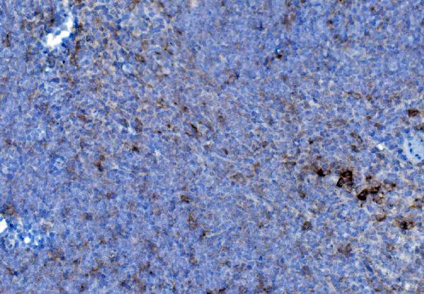
Real IHC data IHC analysis of P4HB using anti-P4HB antibody (A02335-1). P4HB was detected in paraffin-embedded section of mouse spleen tissue. Heat mediated antigen retrieval was performed in EDTA buffer (pH8.0, epitope retrieval solution). The tissue section was blocked with 10% goat serum. The tissue section was then incubated with 2μg/ml rabbit anti-P4HB Antibody (A02335-1) overnight at 4°C. Biotinylated goat anti-rabbit IgG was used as secondary antibody and incubated for 30 minutes at 37°C. The tissue section was developed using Strepavidin-Biotin-Complex (SABC) (Catalog # SA1022) with DAB as the chromogen.
Anti-P4HB Antibody ®
Cat # A02335-1
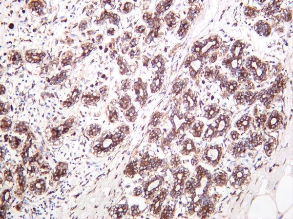
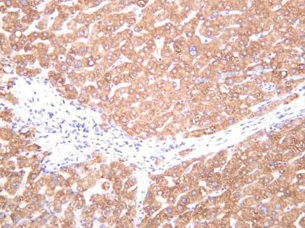
Real IHC data Human breast carcinoma was stained with anti-PDI rabbit antibody
Anti-PDI Rabbit Monoclonal Antibody
Cat # M02335-3
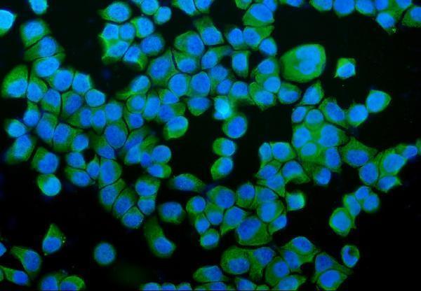
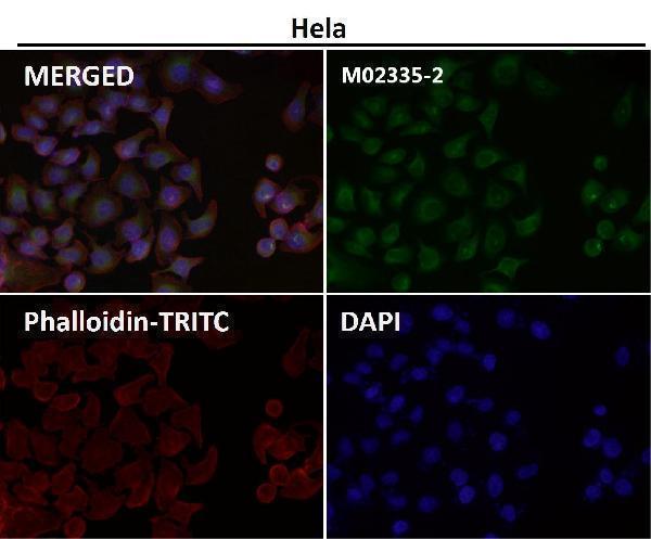
Real IF data Immunofluorescent analysis using the Antibody at 1:50 dilution.
Anti-P4HB Rabbit Monoclonal Antibody
Cat # M02335-2

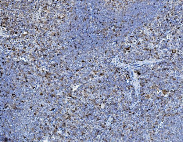
A02335-1 has paraffin-section IHC images from mouse and rat spleen, human lymphoma and human renal clear cell carcinoma, plus IF in T-47D cells (A02335-1 image captions); M02335-3 has IHC images from human breast carcinoma, human liver and rat liver (M02335-3 image captions). M02335-2 lists IHC and ICC/IF applications, but its supplied figures show IF without an identified sample type (M02335-2 applications and IF captions).

Which to pick: For tissue IHC, choose A02335-1 when the documented paraffin-section workflow fits: its mouse spleen image used EDTA retrieval at pH 8.0 and 2 μg/mL primary antibody; the fixative is unreported (A02335-1 IHC caption). For IF/ICC, A02335-1 has a T-47D IF image at 5 μg/mL, while rabbit monoclonal M02335-2 lists ICC/IF and shows IF at 1:50 without identifying the sample (A02335-1 IF caption; M02335-2 catalog and IF caption). For broader listed species reactivity, choose A02335-1, which lists human, monkey, mouse and rat; its IHC images document human, mouse and rat only (A02335-1 catalog and IHC captions).

Each figure is that product's own IHC / IF validation image from its datasheet.

References

  1. UniProt Consortium. UniProt entry P07237 (PDIA1_HUMAN, Protein disulfide-isomerase).
  2. Human Protein Atlas. P4HB tissue IHC expression (reliability: Enhanced).
  3. Human Protein Atlas. P4HB subcellular location (ICC-IF): Localized to the endoplasmic reticulum..
  4. Human Protein Atlas. P4HB antibody validation summary (2 antibodies).
  5. Identification and validation of P4HB as a novel autophagy-related biomarker in diabetic nephropathy. Frontiers in genetics 2022 — PMC9548632.
  6. The Role of P4HB and SOX4 in Prostatic Carcinoma and Their Clinical Significance. Iranian journal of pathology 2024 — PMC11304455.
  7. Prognostic value of hypoxia-inducible factor-1 alpha and prolyl 4-hydroxylase beta polypeptide overexpression in gastric cancer. World journal of gastroenterology 2018 — PMC6000295.
  8. P4HB: A novel diagnostic and prognostic biomarker for bladder carcinoma. Oncology letters 2021 — PMC7751343.
  9. PubMed PMID:3034602 — UniProt-cited evidence.
  10. PubMed PMID:3611107 — UniProt-cited evidence.
  11. PubMed PMID:2846539 — UniProt-cited evidence.