PA2G4 / Proliferation-associated protein 2G4 · IHC design guide

Design Immunohistochemistry for PA2G4

This guide uses catalog antibody A02791 at 0.5–1 μg/ml for chromogenic IHC on paraffin sections (datasheet: A02791). Use glandular cells as a high-staining comparator and expect broad cytoplasmic tissue staining (HPA tissue IHC).

Evidence assembled Oct 2026 · For research use; verify linked source records and product datasheet before use
Immunohistochemistry protocol sheet for PA2G4 (IHC for PA2G4): expected localisation Tissue: cytoplasmic (HPA tissue IHC); molecular: nuclear/nucleolar (UniProt), antibody A02791, validated IHC image, and IHC protocol steps
Printable PA2G4 IHC protocol sheet — expected localisation Tissue: cytoplasmic (HPA tissue IHC); molecular: nuclear/nucleolar (UniProt), antibody A02791, controls and protocol steps. Open the full PA2G4 IHC guide →

PA2G4 Immunohistochemistry Experimental Design Guide

Expected localisation, validated protocols, controls and antibodies — the at-a-glance facts below, then the full design guide.

Must know before staining
Expected localisation Tissue: cytoplasmic (HPA tissue IHC); molecular: nuclear/nucleolar (UniProt)
Staining pattern Broad cytoplasmic staining across tissue cell types (HPA tissue IHC)
Antigen retrieval Citrate pH 6 HIER, heat-mediated (datasheet A02791)
Positive control ⓘ Appendix+4 more · see all
Negative control ⓘ None in HPA (detected in all 45 tissues); use no-primary + isotype controls
Important caveats
Reasons your staining may differ from the expected pattern.
Fixation Matched tissue-IHC evidence does not establish the fixation claim. Validate the specimen-specific method before use. (selected-SKU IHC image A02791)
Caveat Muscle cells can stain weakly despite broad expression (HPA tissue IHC)
Regulation Isoform 1 is strong in cancer cells (UniProt)
Isoform / epitope 2 isoforms; antibody epitope coverage is unspecified (UniProt; datasheet A02791)
Section 1

Recommended PA2G4 IHC & IF Protocols

The catalog antibody protocol (datasheet: A02791) is followed by a published PA2G4 chromogenic IHC protocol for nasopharyngeal paraffin sections (PMC8354561).

Recommended immunohistochemistry (IHC-P) protocol parameters
SampleParaffin-embedded rat intestine tissues; fixative not specified (datasheet A02791)
FixationImage fixative and duration unreported (datasheet A02791); verify before use.
Sectioning4–5 µm sections on charged slides (standard)
DeparaffinisationXylene, graded ethanol series to water (standard)
Antigen retrievalHeat retrieval: Citrate pH 6, 20 min (datasheet A02791)
Peroxidase block3% H2O2, 10 min, room temperature (standard)
Blocking10% goat serum (datasheet A02791)
Primary antibodyRabbit anti-PA2G4, 0.5-1μg/ml (datasheet A02791)
Primary incubationOvernight at 4 °C (datasheet A02791)
DetectionStreptavidin-biotin complex (SABC), DAB chromogen (datasheet A02791)
CounterstainHematoxylin, blue, dehydrate and mount (standard)
Expected resultPA2G4-positive staining in glandular cells of appendix (HPA tissue IHC: High). HPA tissue profile: Ubiquitous cytoplasmic expression. No signal in the no-primary control.
💡Decision noteStart with heat-mediated citrate pH 6 retrieval (datasheet: A02791); the published protocol also uses heated citrate, but does not state its pH (PMC8354561).
Section 2

What Is the Expected PA2G4 Staining Pattern?

PA2G4 should show predominantly cytoplasmic staining in many tissues, with strong staining reported in colon and duodenum glandular cells, bronchial respiratory epithelial cells, and cerebral cortex neurons (HPA tissue IHC: ubiquitous cytoplasmic expression; High in those cells). Interpret the pattern with moderate confidence because staining and RNA data have medium consistency (HPA tissue IHC: Supported). Nuclear or nucleolar localization is also possible (UniProt Q9UQ80: subcellular location). PA2G4 has no transmembrane segment (UniProt Q9UQ80: topology).

What am I looking at on my slide?
Clear cytoplasmic staining in colon or duodenum glandular cells, with recognizable tissue structure.This fits the reported strong glandular signal (HPA tissue IHC: High in colon and duodenum glandular cells). Assess the proportion of stained cells and intensity within the named compartment; a dark section alone does not establish a specific result (general IHC practice).
A membrane-only rim dominates, with little cytoplasmic staining in an expected positive cell population.That distribution conflicts with the reported cytoplasmic pattern and the absence of a transmembrane segment (HPA tissue IHC: ubiquitous cytoplasmic expression; UniProt Q9UQ80: topology). Check morphology and controls before calling it PA2G4. Nuclear or nucleolar staining requires separate assessment because those locations are annotated (UniProt Q9UQ80: subcellular location).
Strong staining appears chiefly in an unexpected cell population, while the expected cells are weak.Treat this as a specificity question, not proof of cross-reactivity: PA2G4 is broadly distributed (HPA tissue IHC: ubiquitous cytoplasmic expression). Compare a second validated antibody where available and inspect detection-only controls for endogenous activity (HPA antibodies: IHC Supported; general IHC practice).
Color spreads across stroma, blank areas, or many cell compartments without clear boundaries.Diffuse deposition is difficult to score as cellular PA2G4. Recheck blocking, washing, detection, and counterstain using matched control sections (general IHC practice). HPA describes a cytoplasmic tissue pattern, not diffuse extracellular staining (HPA tissue IHC: profile).
The expected glandular or epithelial cells show no signal in an otherwise interpretable section.A negative call needs a working positive control: colon or duodenum glandular cells and bronchial respiratory epithelial cells are reported High (HPA tissue IHC). If a parallel positive section also fails, review retrieval, antibody dilution, and detection as general IHC variables; the sources do not establish PA2G4-specific fixation sensitivity.
💡Expected PA2G4 appearanceCall the IHC result positive when the expected cells show distinct, often strong cytoplasmic color—such as glandular cells in colon or duodenum (HPA tissue IHC: High); isolated membrane rims or diffuse extracellular color warrant investigation rather than a PA2G4 call (HPA tissue IHC: cytoplasmic profile; UniProt Q9UQ80: topology; general IHC practice).
How each factor affects the staining
Tissue and cell selectionHPA reports High staining in appendix, colon, duodenum, endometrium, and fallopian tube glandular cells; bronchial respiratory epithelium and cortical neurons are also High (HPA tissue IHC). Cholangiocytes and smooth or skeletal muscle cells are Low, so weak staining there is an unsuitable sole failure criterion (HPA tissue IHC).
Compartment and biological stateCytoplasmic staining is the tissue-wide reference pattern (HPA tissue IHC). UniProt also annotates nucleus and nucleolus, nuclear translocation after HRG treatment, and regulation of nucleolar localization by Ser-361 phosphorylation (UniProt Q9UQ80). These annotations support considering a real nuclear component, but do not predict its intensity in a given paraffin section.
Isoforms and antibody recognitionTwo isoforms are listed, and both are described as widely expressed; isoform 1 is reported strongly expressed at protein level in cancer cells, while isoform 2 is undetectable there (UniProt Q9UQ80: tissue specificity). The supplied antibody summary does not specify recognized epitopes or isoform selectivity, so do not assign a staining pattern to either isoform.
Strength of reference evidenceThe tissue profile is Supported with medium consistency between antibody staining and RNA data (HPA tissue IHC). Three listed antibodies have IHC Supported status, while two also have ICC Supported status (HPA antibodies). These ratings guide comparison; they do not validate every compartment or exclude nonspecific color in an individual section.
Paraffin-section workflowAntigen retrieval, blocking, dilution, and detection can be checked as general IHC variables when controls fail (general IHC practice). No target-specific fixation or retrieval effect, epitope position, or recommended dilution is supplied here; avoid attributing a weak result to a particular PA2G4 epitope or fixation response.
Why is my staining missing, weak or wrong?
SituationLikely causeNext action
Colon or duodenum glandular positive control is blank (HPA tissue IHC: High).The run may have failed at retrieval, antibody incubation, or chromogenic detection; the record does not identify a PA2G4-specific cause.Verify the control section and run records, then check retrieval conditions, primary dilution, reagent integrity, and detection on a repeat run (general IHC practice).
Tissue and empty spaces both show diffuse color.Background from incomplete blocking, washing, or detection chemistry is possible (general IHC practice).Inspect a section without primary antibody, adjust general blocking or washing steps, and score only color tied to intact cells (general IHC practice).
A sharp membrane rim is the dominant pattern.This conflicts with cytoplasmic tissue staining and PA2G4's lack of a transmembrane segment (HPA tissue IHC; UniProt Q9UQ80: topology).Compare with a validated positive section and detection-only control; review cell boundaries before interpreting the rim as specific (general IHC practice).
Unexpected cells stain much more strongly than the expected cell population.Cross-reactivity or endogenous detection activity is possible, but broad PA2G4 expression means cell identity alone cannot settle specificity (HPA tissue IHC: ubiquitous expression; general IHC practice).Confirm cell identity and compartment; compare a second IHC Supported antibody if available and use a detection-only control (HPA antibodies; general IHC practice).
Cholangiocytes or myocytes show faint staining.Low staining is reported in liver cholangiocytes, smooth muscle cells, and skeletal myocytes (HPA tissue IHC: Low).Treat faint signal as compatible with the reference profile. Judge assay performance against a High cell population in a control section (HPA tissue IHC; general IHC practice).
Q: Does cytosolic IF/ICC signal support the IHC interpretation?A: HPA lists cytosol as the supported main ICC-IF location, consistent with its cytoplasmic tissue IHC profile (HPA subcellular; HPA tissue IHC).Use that agreement as a localization check, while interpreting the paraffin-section result and its controls on their own terms; consult the separate IF/ICC guide for that application.

Sample controls for PA2G4 IHC & IF

🧪Run colon first: glandular cells should stain (HPA: High in colon glandular cells); cells without specific staining should show only background chromogen, but no colon cell type is established here as an internal PA2G4-negative control. HPA detects PA2G4 in all 45 scored tissues (HPA: no negative tissues), so use no-primary and isotype controls for the negative comparison.
Positive control tissue: Appendix (Glandular cells, HPA High)
Negative control tissue: None in HPA: PA2G4 is detected in all 45 scored tissues. Use a no-primary (secondary-only) and an isotype control instead.
ICC-IF cell lines (HPA subcellular resource): HPA ICC-IF images show PA2G4 in A-431, U2OS, U-251MG, with annotated localisation: Cytosol (supported) (HPA subcellular).
Technical controls: Include a secondary-only slide, a concentration-matched rabbit IgG isotype control for the catalog rabbit antibody (A02791 caption: rabbit primary), and, if available, matched PA2G4-knockout material as a biological specificity control. In colon sections, quench endogenous peroxidase and check for endogenous biotin signal when using the caption’s biotin-based DAB detection (A02791 caption: biotinylated secondary, streptavidin–biotin complex and DAB).
⚠️Feasibility: No target-specific fixation window or fixation effect is reported, and the selected A02791 paraffin-section caption does not state a fixative (A02791 caption: fixative unreported). Heat retrieval in citrate buffer at pH 6 for 20 minutes was used for rat intestine, but retrieval dependence in colon is unreported (A02791 caption: rat intestine protocol). Frozen sections are not shown to be easier; HPA ICC-IF images in A-431, U2OS and U-251MG offer an IF localisation reference (HPA: cytosol supported), while colon IF requires its own autofluorescence check.

HPA tissue IHC evidence for PA2G4

Comprehensive Human Protein Atlas IHC scoring per tissue (reliability: Supported — Medium consistency between antibody staining and RNA expression data.). Rows are taken directly from the HPA tissue chart — click any row's HPA link to view the source.

Positive expression · recommended positive controls

TissueCell typeLevelEvidenceSource
Appendix Glandular cells High Protein (IHC) HPA →
Bronchus Respiratory epithelial cells High Protein (IHC) HPA →
Cerebellum Cells in molecular layer High Protein (IHC) HPA →
Cerebral cortex Neuronal cells High Protein (IHC) HPA →
Colon Glandular cells High Protein (IHC) HPA →

Undetected expression · recommended negative controls

TissueCell typeLevelEvidenceSource
None in HPA: PA2G4 is detected in all 45 scored tissues. Use a no-primary (secondary-only) and an isotype control instead.
Section 3

Advanced PA2G4 IHC Tips

Use the validated paraffin-section workflow as a starting point, then assess PA2G4 staining by compartment, cell type and controls.

What retrieval conditions should I try when PA2G4 staining is weak?
Start with heat-mediated antigen retrieval in citrate buffer at pH 6 (datasheet A02791). The selected paraffin-section image used 20 minutes of retrieval, so match that duration before changing the primary antibody concentration (caption A02791). If signal remains weak, compare a longer or shorter heating interval on adjacent sections while holding detection and exposure constant; excessive retrieval can damage morphology or increase background (standard IHC practice). Check that positive-control cells stain before judging an experimental section negative: PA2G4 shows high staining in colon glandular cells (HPA: High in colon glandular cells). Record nuclear and cytoplasmic signal separately because both are biologically plausible locations (UniProt Q9UQ80 subcellular location).
How should I troubleshoot fixation-related loss of PA2G4 signal?
The selected image identifies a paraffin-embedded section but gives no fixative, so target-specific fixation sensitivity is unknown (caption A02791: fixative not stated). Compare sections with documented fixation histories using the same pH 6 citrate retrieval and staining run, then judge signal alongside preserved morphology (datasheet A02791; standard IHC practice). Excessive fixation can mask epitopes, while inadequate fixation can impair tissue preservation; these are general IHC considerations, not demonstrated PA2G4 effects (standard IHC practice). Include a known-staining control section in each comparison, and avoid attributing a negative result to fixation alone when processing history or antibody performance is uncertain (standard IHC practice).
Should PA2G4 appear in cytoplasm, nucleus, or nucleoli?
Expect substantial cytoplasmic staining in routine tissue IHC, consistent with the reported ubiquitous cytoplasmic pattern (HPA: tissue IHC profile). Nuclear and nucleolar signal can also be plausible: PA2G4 is annotated in the cytoplasm, nucleus and nucleolus, with nuclear translocation after HRG treatment (UniProt Q9UQ80 subcellular location). Phosphorylation at Ser-361 regulates nucleolar localisation, but a chromogenic section cannot establish that modification by appearance alone (UniProt Q9UQ80 modified residues). Score cytoplasmic and nuclear compartments separately against the same counterstained cell population, and use matched controls before interpreting a shift in distribution (standard IHC practice).
Can this IHC stain distinguish PA2G4 isoforms or phosphorylation states?
PA2G4 has 2 annotated isoforms, but the supplied IHC caption does not map the A02791 epitope or establish isoform selectivity (UniProt Q9UQ80 isoforms; caption A02791). Treat chromogenic staining as total antibody-reactive PA2G4 unless isoform specificity is independently established for this reagent (standard IHC interpretation). Isoform 1 is reported strongly expressed in cancer cells while isoform 2 is undetectable there at protein level; that context does not prove which isoform produces an individual stain (UniProt Q9UQ80 tissue specificity). Likewise, PA2G4 has annotated phosphosites, including Ser-361, but this antibody's staining cannot identify a phosphorylation state without epitope evidence and appropriate controls (UniProt Q9UQ80 modified residues; standard IHC practice).
How can I assess PA2G4 by multiplex IF after seeing an IHC pattern?
For IF, pair PA2G4 with a marker that identifies the expected cell population, such as glandular cells in colon, where tissue IHC reports high PA2G4 staining (HPA: High in colon glandular cells). Choose a fluorophore channel after checking that tissue's autofluorescence, and include single-stain and no-primary controls to assess bleed-through and background (standard IF practice). PA2G4 has no annotated transmembrane segment and is found in the cytoplasm and nucleus, so use controlled permeabilisation to access intracellular epitopes while preserving compartment boundaries (UniProt Q9UQ80 topology and subcellular location; standard IF practice). Optimise IF fixation and permeabilisation on matched samples; the paraffin-section caption does not establish IF conditions (caption A02791).
What should I check when PA2G4 DAB staining is diffuse?
First compare a no-primary section with the stained section to separate antibody-associated signal from endogenous enzyme activity or nonspecific detection (standard IHC practice). The selected image used 10% goat serum blocking, 1 μg/mL primary antibody overnight at 4°C, and biotin-based detection with DAB (caption A02791). Check the peroxidase block, blocking, wash steps and antibody concentration systematically; endogenous biotin can also matter in a biotin-based workflow (standard IHC practice). Because PA2G4 has a reported ubiquitous cytoplasmic tissue profile, broad staining alone is not evidence of background; assess cell boundaries, morphology and control-section contrast (HPA: tissue IHC profile; standard IHC practice).
How should I score PA2G4 staining across paraffin sections? ⚠ ANSWER MARKED FOR VERIFICATION
Choose a predefined readout, such as cytoplasmic H-score, percentage of positive cells or positive-cell density per mm², and apply identical thresholds across slides (standard IHC practice). Score nuclear staining separately because PA2G4 can occupy the nucleus as well as the cytoplasm (UniProt Q9UQ80 subcellular location). Normalise cell-based scores to the number of eligible, viable cells in the defined compartment, or density to the measured viable tissue area; exclude folds and necrosis consistently (standard IHC practice). Stratify by cell type when composition varies, since HPA reports high staining in colon glandular cells but low staining in skeletal-muscle myocytes (HPA: colon and skeletal-muscle tissue IHC).
How do I distinguish genuine PA2G4 staining from artefact?
A credible pattern has interpretable cell boundaries and matches expected compartments: PA2G4 is reported predominantly cytoplasmic in tissue IHC, with nuclear and nucleolar localisation also annotated (HPA: tissue IHC profile; UniProt Q9UQ80 subcellular location). Check the cell population before calling a section negative or positive; colon glandular cells show high staining, whereas skeletal-muscle myocytes show low staining in the supplied tissue survey (HPA: colon and skeletal-muscle tissue IHC). Discount staining restricted to section edges, folds or necrotic areas, and compare no-primary and peroxidase-block controls for nonspecific DAB deposition (standard IHC practice). Treat an unexpected compartment or cell pattern as a finding requiring replication and control review, not proof of altered PA2G4 biology (standard IHC interpretation).
Boster reagents

Best PA2G4 / Proliferation-associated protein 2G4 IHC Antibodies

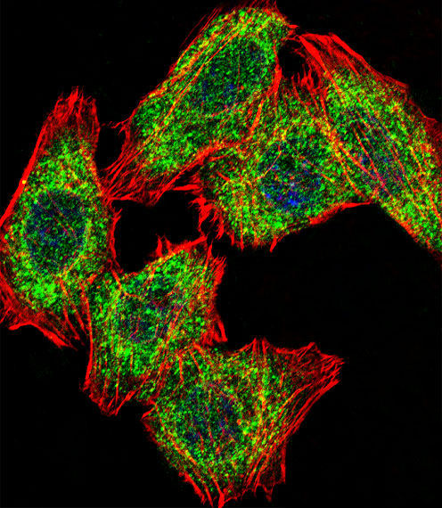
The catalog includes PA2G4 antibodies with tissue IHC images from human, mouse, and rat samples (catalog IHC captions) and a cellular IF image from U251 cells (A02791-1 IF caption).

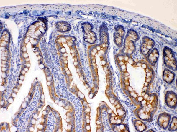
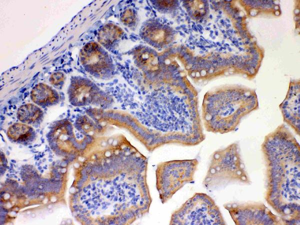
Real IHC data IHC analysis of EBP1 using anti-EBP1 antibody (A02791). EBP1 was detected in paraffin-embedded section of rat intestine tissues. Heat mediated antigen retrieval was performed in citrate buffer (pH6, epitope retrieval solution) for 20 mins. The tissue section was blocked with 10% goat serum. The tissue section was then incubated with 1μg/ml rabbit anti-EBP1 Antibody (A02791) overnight at 4°C. Biotinylated goat anti-rabbit IgG was used as secondary antibody and incubated for 30 minutes at 37°C. The tissue section was developed using Strepavidin-Biotin-Complex (SABC)(Catalog # SA1022) with DAB as the chromogen.
Anti-EBP1/PA2G4 Antibody ®
Cat # A02791
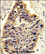
Real IHC data Formalin-fixed and paraffin-embedded human lung carcinoma reacted with PA2G4 Antibody (Center R243), which was peroxidase-conjugated to the secondary antibody, followed by DAB staining. This data demonstrates the use of this antibody for immunohistochemistry; clinical relevance has not been evaluated.
Anti-PA2G4 Antibody (Center R243)
Cat # A02791-1
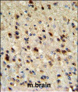
Real IHC data EBP1 Antibody (C-term) IHC analysis in formalin fixed and paraffin embedded mouse brain tissue followed by peroxidase conjugation of the secondary antibody and DAB staining. This data demonstrates the use of the EBP1 Antibody (C-term) for immunohistochemistry. Clinical relevance has not been evaluated.
Anti-EBP1 Antibody (C-term)
Cat # A02791-2

A02791 has paraffin-section IHC images from rat intestine, mouse intestine and brain, and human stomach cancer; A02791-1 has an IHC image from formalin-fixed, paraffin-embedded human lung carcinoma (catalog IHC captions). A02791-2 has an IHC image from formalin-fixed, paraffin-embedded mouse brain (A02791-2 IHC caption).

Which to pick: For tissue IHC, choose A02791 when its paraffin-section examples fit the study; its captions do not report a fixative (A02791 IHC captions). For cellular IF, choose A02791-1, which lists IF and shows staining in U251 cells (A02791-1 applications; A02791-1 IF caption). For work spanning human, mouse, and rat, A02791 lists IHC reactivity in all three species, while A02791-2 lists human and mouse IHC-P reactivity (catalog applications and reactivity).

Each figure is that product's own IHC / IF validation image from its datasheet.

References

  1. UniProt Consortium. UniProt entry Q9UQ80 (PA2G4_HUMAN, Proliferation-associated protein 2G4).
  2. Human Protein Atlas. PA2G4 tissue IHC expression (reliability: Supported).
  3. Human Protein Atlas. PA2G4 subcellular location (ICC-IF): Localized to the cytosol..
  4. Human Protein Atlas. PA2G4 antibody validation summary (3 antibodies).
  5. Increased PA2G4 Expression Is an Unfavorable Factor in Nasopharyngeal Carcinoma. Applied immunohistochemistry & molecular morphology : AIMM 2021 — PMC8354561.
  6. PA2G4 promotes the metastasis of hepatocellular carcinoma by stabilizing FYN mRNA in a YTHDF2-dependent manner. Cell & bioscience 2022 — PMC9080163.
  7. Evidence for a fragile X messenger ribonucleoprotein 1 (FMR1) mRNA gain-of-function toxicity mechanism contributing to the pathogenesis of fragile X-associated premature ovarian insufficiency. FASEB journal : official publication of the Federation of American Societies for Experimental Biology 2022 — PMC9828574.
  8. PA2G4 Functions as a Cofactor for MYC Family Oncoproteins in MYC-Driven Malignancies. Cells 2025 — PMC12468391.
  9. PubMed PMID:9345902 — UniProt-cited evidence.
  10. PubMed PMID:10682683 — UniProt-cited evidence.
  11. PubMed PMID:15489334 — UniProt-cited evidence.