This website uses cookies to ensure you get the best experience on our website.


- Table of Contents
Plan PABPC1 staining in paraffin sections around its widespread cytoplasmic tissue pattern (HPA tissue IHC). Human esophageal squamous cells and testicular pachytene spermatocytes show high staining and can guide positive-control selection (HPA tissue IHC).

Expected localisation, validated protocols, controls and antibodies — the at-a-glance facts below, then the full design guide.
| Expected localisation | Cytoplasmic staining across tissues (HPA tissue IHC) | |
| Staining pattern | Cytoplasmic signal in many tissue cell types (HPA tissue IHC) | |
| Antigen retrieval | EDTA pH 8.0 HIER, heat-mediated (datasheet A01877-1) | |
| Positive control | Bone marrow+4 more · see all | |
| Negative control | Adipose tissue+4 more · see all |
| Fixation | Keep fixation conditions consistent across sections. (standard IHC practice; not target-specific) | |
| Caveat | Staining may reflect proteins from more than one gene (HPA tissue IHC) | |
| Regulation | Ubiquitous expression (UniProt) | |
| Isoform / epitope | 2 isoforms; antibody epitope coverage is unknown (UniProt) |
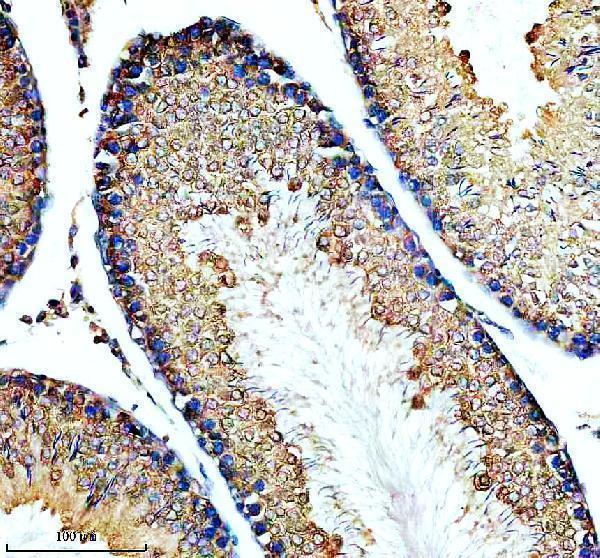
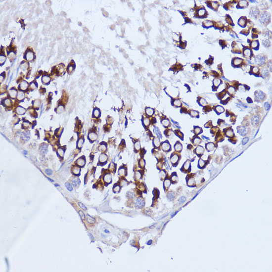
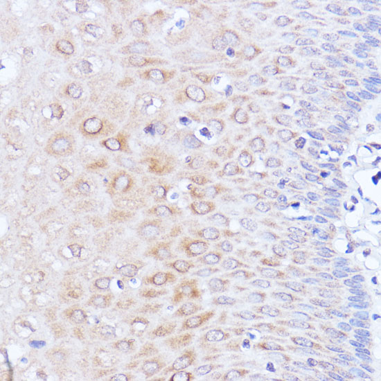
The catalog antibody protocol uses EDTA retrieval (datasheet: A01877-1). The published IHC options below report PABPC1 staining in esophageal carcinoma, mouse spermatogenesis, and nasopharyngeal carcinoma (PMC8962095; PMC12040762; PMC8275044; PMC9940331).
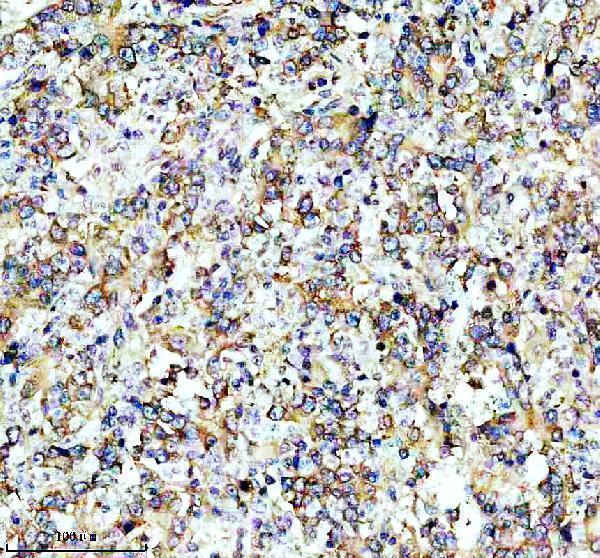
| Sample | Paraffin-embedded human stomach cancer tissue; fixative not specified (datasheet A01877-1) |
| Fixation | Image fixative and duration unreported (datasheet A01877-1); verify before use. |
| Sectioning | 4–5 µm sections on charged slides (standard) |
| Deparaffinisation | Xylene, graded ethanol series to water (standard) |
| Antigen retrieval | Heat retrieval: EDTA pH 8.0 (datasheet A01877-1); 20 min, 95–100 °C (standard) |
| Peroxidase block | 3% H2O2, 10 min, room temperature (standard) |
| Blocking | 10% goat serum (datasheet A01877-1) |
| Primary antibody | Rabbit anti-PABPC1, 1:50 recommended; image 1:200 (datasheet A01877-1) |
| Primary incubation | Overnight at 4 °C (datasheet A01877-1) |
| Detection | HRP-conjugated secondary, DAB chromogen (datasheet A01877-1) |
| Counterstain | Hematoxylin, blue, dehydrate and mount (standard) |
| Expected result | PABPC1-positive staining in hematopoietic cells of bone marrow (HPA tissue IHC: High). HPA tissue profile: Ubiquitous cytoplasmic expression. No signal in the no-primary control. |
PABPC1 should appear predominantly in the cytoplasm of many cell types: HPA describes ubiquitous cytoplasmic tissue expression, with high staining in selected cell populations and a Supported IHC reliability rating (HPA tissue IHC). UniProt also lists nuclear and stress granule localisation; PABPC1 has no transmembrane segment, so membrane-restricted staining is unexpected (UniProt P11940 localisation and topology). HPA cautions that its tissue antibodies may target protein from more than one gene (HPA tissue IHC).
| Cytoplasmic staining in esophageal squamous epithelial cells, tonsillar germinal center cells or bone marrow hematopoietic cells. | This matches HPA's High staining in those cell populations (HPA tissue IHC). Compare signal within the identified cells rather than treating the whole section as uniformly positive; HPA describes broad cytoplasmic expression but reports different levels by cell type (HPA tissue IHC). |
| Signal confined to cell borders or nuclei, with little cytoplasmic staining. | A membrane-only pattern conflicts with the reported cytoplasmic pattern and lack of a transmembrane segment (HPA tissue IHC; UniProt P11940 topology). Nuclear PABPC1 is possible, including during stress, so nuclear signal alone is not proof of an artefact; assess its context and controls (UniProt P11940 localisation; general IHC practice). |
| Strong staining in adipocytes, cholangiocytes or skeletal myocytes while expected positive cells are unstained. | HPA reports these cell populations as Not detected (HPA tissue IHC). Check cell identification and controls; staining there could reflect cross-reactivity or endogenous detection activity, especially given HPA's caution that the tissue antibodies may target protein from more than one gene (HPA tissue IHC; general IHC practice). |
| Even chromogenic haze across cells and spaces, obscuring cytoplasmic boundaries. | This cannot establish the cell-associated cytoplasmic pattern reported by HPA (HPA tissue IHC). Evaluate reagent-only controls, blocking, washes and detection background before assigning positive cells; these checks address general staining artefacts and do not establish a PABPC1-specific cause (general IHC practice). |
| No staining in an otherwise interpretable section containing HPA High cells. | Absent signal in an expected positive population warrants a run-level check before interpreting other cells as negative (HPA tissue IHC; general IHC practice). Confirm tissue identity, antibody and detection controls, then review the validated IHC-P workflow; the supplied sources do not identify a PABPC1-specific fixation sensitivity (general IHC practice). |
| Cell population and staining level (HPA tissue IHC) | HPA reports High staining in testis pachytene spermatocytes and urinary bladder urothelial cells, Medium in stomach glandular cells, and Not detected in adipocytes (HPA tissue IHC). Use those observations to choose comparison areas; ubiquitous expression does not imply equal intensity in every cell (HPA tissue IHC; UniProt P11940 tissue specificity). |
| Antibody interpretation (HPA tissue IHC; HPA antibodies) | HPA rates tissue IHC Supported, describes medium agreement between staining and RNA data, and cautions that the antibodies may target protein from more than one gene (HPA tissue IHC). HPA045423 and CAB011536 have Supported IHC status; HPA067156 has no listed IHC status in the supplied record (HPA antibodies). |
| Isoforms and epitope coverage (UniProt P11940) | UniProt lists 2 isoforms and four RRM domains plus a PABC domain (UniProt P11940). The supplied record gives no antibody epitope or isoform-specific IHC evidence, so a staining difference cannot be assigned to an isoform or domain without further validation (UniProt P11940; HPA antibodies). |
| IF/ICC Q: What localisation should an IF image show? | A: Predominantly cytosolic signal is supported by HPA ICC-IF images; UniProt also reports nuclear shuttling and stress-associated cytoplasmic granules (HPA subcellular; UniProt P11940 localisation). Interpret those conditional locations in context; this IHC section does not establish an IF staining workflow (HPA subcellular; UniProt P11940). |
| Situation | Likely cause | Next action |
|---|---|---|
| Expected positive cells show no chromogenic signal. | Tissue or cell identification, reagent performance, or an IHC workflow step may be at issue (general IHC practice). | Check an identifiable HPA High population, the run controls and detection reagents; review the antibody's validated IHC-P instructions before changing conditions (HPA tissue IHC; general IHC practice). No target-specific fixation effect is established by the supplied sources. |
| The entire section has diffuse color. | Background from detection chemistry, incomplete blocking or insufficient washing can obscure cellular staining (general IHC practice). | Inspect reagent-only and detection controls, then review blocking and washes (general IHC practice). Score PABPC1 only where cell-associated cytoplasmic signal is distinguishable from background (HPA tissue IHC; general IHC practice). |
| Signal is strongest along membranes. | This distribution is inconsistent with the reported cytoplasmic pattern and no-transmembrane topology (HPA tissue IHC; UniProt P11940 topology). | Recheck focus, morphology and control sections; withhold a PABPC1-positive call if the membrane pattern persists without convincing cytoplasmic staining (general IHC practice; HPA tissue IHC). |
| A nucleus-only pattern appears. | UniProt reports nuclear shuttling and nuclear localisation under some stress conditions, while HPA describes the usual tissue pattern as cytoplasmic (UniProt P11940 localisation; HPA tissue IHC). | Check whether cytoplasmic staining is present in expected positive cells and compare controls before calling artefact or a condition-related change; the section alone cannot establish the cause (HPA tissue IHC; general IHC practice). |
| Cells HPA calls Not detected stain strongly. | Cell misidentification, nonspecific binding or endogenous detection activity are possibilities; HPA also flags potential cross-gene targeting (HPA tissue IHC; general IHC practice). | Verify the cell type against morphology, compare expected positive cells in the same run, and examine reagent controls. Avoid assigning unexpected staining specifically to PABPC1 from this result alone (HPA tissue IHC; general IHC practice). |
| Only weak staining appears in an HPA Low population. | HPA reports Low staining in thyroid glandular cells, pancreatic exocrine glandular cells and kidney tubular cells (HPA tissue IHC). | Compare with an HPA High population and the run controls before treating weak signal as assay failure; record the cell type and background separately when scoring (HPA tissue IHC; general IHC practice). |


Comprehensive Human Protein Atlas IHC scoring per tissue (reliability: Supported — Medium consistency between antibody staining and RNA expression data. Caution, targets protein from more than one gene.). Rows are taken directly from the HPA tissue chart — click any row's HPA link to view the source.
| Tissue | Cell type | Level | Evidence | Source |
|---|---|---|---|---|
| Bone marrow | Hematopoietic cells | High | Protein (IHC) | HPA → |
| Esophagus | Squamous epithelial cells | High | Protein (IHC) | HPA → |
| Nasopharynx | Ciliated cells (cell body) | High | Protein (IHC) | HPA → |
| Stomach | Glandular cells | Medium | Protein (IHC) | HPA → |
| Testis | Pachytene spermatocytes | High | Protein (IHC) | HPA → |
| Tissue | Cell type | Level | Evidence | Source |
|---|---|---|---|---|
| Adipose tissue | Adipocytes | Not detected | Protein (IHC) | HPA → |
| Liver | Cholangiocytes | Not detected | Protein (IHC) | HPA → |
| Ovary | Ovarian stroma cells | Not detected | Protein (IHC) | HPA → |
| Skeletal muscle | Myocytes | Not detected | Protein (IHC) | HPA → |
| Smooth muscle | Smooth muscle cells | Not detected | Protein (IHC) | HPA → |
Troubleshoot chromogenic PABPC1 staining in paraffin sections using the selected antibody’s tissue image, protein localization, and tissue expression evidence (A01877-1 caption; UniProt P11940; HPA).
Both antibodies have real IHC images from paraffin sections of human and rat tissues (catalog IHC captions); both list IF/ICC, but no IF images are supplied (catalog applications and IF image fields).
A01877-1 will render with a human stomach cancer IHC image; its captions also describe rat testis staining (A01877-1 IHC captions). A01877 will render with a rat testis IHC image; its captions also describe human esophageal staining (A01877 IHC captions).
Which to pick: For tissue IHC, choose A01877-1 if its detailed paraffin-section procedure is useful: the caption specifies EDTA retrieval at pH 8.0 and a 1:200 primary incubation (A01877-1 IHC caption); A01877 also has paraffin-section images (A01877 IHC captions). For IF/ICC, either SKU lists those applications, but neither supplies an IF image (catalog applications and IF image fields). Both list human, mouse and rat reactivity (catalog reactivity); the IHC captions report paraffin sections without identifying the fixative (catalog IHC captions).