This website uses cookies to ensure you get the best experience on our website.


- Table of Contents
Plan paraffin-section PABPC4 staining around cytoplasmic signal in most tissues, including immune-cell subsets (HPA tissue IHC). The catalog antibody has a documented chromogenic IHC-P application; compare staining by cell type with the HPA tissue profile (datasheet A07960-2; HPA tissue IHC).

Expected localisation, validated protocols, controls and antibodies — the at-a-glance facts below, then the full design guide.
| Expected localisation | Cytoplasmic in most tissues (HPA tissue IHC) | |
| Staining pattern | Cytoplasmic staining in most tissues, including immune-cell subsets (HPA tissue IHC) | |
| Antigen retrieval | EDTA pH 8.0 HIER, heat-mediated (datasheet A07960-2) | |
| Positive control | Cerebellum+4 more · see all | |
| Negative control | No source-confirmed negative tissue/cell row; use no-primary + isotype controls |
| Fixation | Keep fixation consistent across sections (standard IHC practice; not target-specific) | |
| Caveat | Staining and RNA show medium consistency (HPA tissue IHC) | |
| Regulation | T-cell activation rapidly raises mRNA (UniProt) | |
| Isoform / epitope | 3 isoforms; epitope coverage is unknown (UniProt) |
The catalog antibody’s IHC-P protocol (datasheet: A07960-2) is accompanied by published PABPC4 staining methods for prostate cancer TMAs (PMC12336731) and colorectal tissues (PMC13496150).
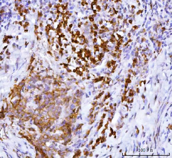
| Sample | Paraffin-embedded human cervical cancer tissue; fixative not specified (datasheet A07960-2) |
| Fixation | Image fixative and duration unreported (datasheet A07960-2); verify before use. |
| Sectioning | 4–5 µm sections on charged slides (standard) |
| Deparaffinisation | Xylene, graded ethanol series to water (standard) |
| Antigen retrieval | Heat retrieval: EDTA pH 8.0 (datasheet A07960-2); 20 min, 95–100 °C (standard) |
| Peroxidase block | 3% H2O2, 10 min, room temperature (standard) |
| Blocking | 10% goat serum (datasheet A07960-2) |
| Primary antibody | Rabbit anti-PABPC4, 2-5 μg/ml (datasheet A07960-2) |
| Primary incubation | Overnight at 4 °C (datasheet A07960-2) |
| Detection | HRP-conjugated secondary, DAB chromogen (datasheet A07960-2) |
| Counterstain | Hematoxylin, blue, dehydrate and mount (standard) |
| Expected result | PABPC4-positive staining in purkinje cells of cerebellum (HPA tissue IHC: High). HPA tissue profile: Cytoplasmic expression in most tissues, including subsets of immune cells. No signal in the no-primary control. |
PABPC4 is a cytoplasmic protein with no transmembrane segment (UniProt Q13310 topology). In paraffin sections, expect cytoplasmic staining in many tissues, including subsets of immune cells (HPA: tissue IHC profile). Strong examples include cerebellar Purkinje cells, pancreatic exocrine glandular cells, and placental cytotrophoblasts (HPA: High). HPA rates the tissue IHC pattern Supported, with medium consistency between antibody staining and RNA expression (HPA: tissue IHC reliability).
| Cytoplasmic stain in Purkinje cells, pancreatic exocrine cells, or placental cytotrophoblasts. | This matches reported High staining in those cell types (HPA: tissue IHC). Compare each cell population with nearby tissue before scoring: PABPC4 has a broad cytoplasmic profile, so staining outside these examples is not automatically unexpected (HPA: tissue IHC profile). |
| A strong nuclear-only, membrane-rim, or extracellular pattern dominates the section. | That compartment conflicts with the expected cytoplasmic localization (UniProt Q13310 subcellular location; HPA: tissue IHC profile). Treat it as suspect and review morphology, detection controls, and staining conditions (general IHC practice). UniProt reports cytoplasmic mRNP granules, but that does not establish a required punctate IHC pattern (UniProt Q13310 subcellular location). |
| The dominant stain is in cells other than the expected population for the chosen tissue. | Compare the stained cells with the tissue-specific HPA entry before calling the result positive (HPA: tissue IHC). An unexpected distribution can reflect antibody cross-reactivity or endogenous detection activity (general IHC practice); it cannot be diagnosed from location alone, especially given the broad reported expression (HPA: tissue IHC profile). |
| Diffuse color covers tissue and blank areas, obscuring cell boundaries. | A field-wide haze prevents a reliable cytoplasmic call (general IHC practice). Check background on a no-primary control, then review blocking, washes, and chromogen development (general IHC practice). HPA reports staining by cell population; it does not identify a PABPC4-specific cause of diffuse background (HPA: tissue IHC). |
| No discernible stain appears in a section selected for reported High expression. | Confirm that the section contains the relevant cells: HPA reports High staining in pancreatic exocrine glandular cells and cerebellar Purkinje cells (HPA: tissue IHC). Then check the staining run and tissue integrity (general IHC practice). A blank result alone does not establish PABPC4 absence (HPA: Supported, medium RNA–protein consistency). |
| Tissue and cell population | HPA reports High staining in Purkinje cells, ovarian follicle cells, pancreatic exocrine glandular cells, placental cytotrophoblasts, and spermatogonia (HPA: tissue IHC). It lists Low staining in hepatocytes and several epithelial or glandular populations (HPA: tissue IHC); compare like cells when judging intensity. |
| Expected compartment and topology | Cytoplasm is the primary location, with cytoplasmic mRNP granules also described (UniProt Q13310 subcellular location). The protein has no transmembrane segment or signal peptide (UniProt Q13310 topology and processing), so a dominant membrane-rim or secreted-looking pattern needs scrutiny. |
| Evidence strength | The tissue profile is Supported, with medium consistency between antibody staining and RNA expression (HPA: tissue IHC reliability). HPA lists IHC as Supported for HPA027301 and HPA056496; their Enhanced status applies to ICC, not IHC (HPA: antibody validation). |
| Isoforms and antibody recognition | UniProt lists three PABPC4 isoforms (UniProt Q13310 isoforms). The supplied record gives no epitope location or isoform-specific IHC result, so it cannot establish which isoforms an antibody detects in sections (UniProt Q13310 isoforms; HPA: antibody validation). |
| IF/ICC cross-check: what location is expected? | HPA reports enhanced cytosol localization in ICC-IF and Enhanced ICC validation for HPA027301 and HPA056496 (HPA: subcellular; HPA: antibody validation). Use that as compartment context; it does not supply an IHC-P retrieval condition or establish equal performance between applications. |
| Situation | Likely cause | Next action |
|---|---|---|
| Reported High tissue is blank. | The relevant cells may be absent from the section, or the staining run may have failed (HPA: tissue IHC; general IHC practice). | Check for the specified cells, then review tissue integrity, the run control, detection reagents, and retrieval against the IHC-validated antibody's instructions (general IHC practice). No PABPC4-specific retrieval setting is supplied. |
| Signal is weak in a reported Low population. | HPA lists Low staining in hepatocytes, lung type II alveolar cells, and several glandular or epithelial populations (HPA: tissue IHC). | Compare the same cell type across sections and include a reported High population when available (HPA: tissue IHC; general IHC practice). Do not turn a weak Low-population result into an assay failure without a run control. |
| Nuclear-only or membrane-rim stain dominates. | The distribution conflicts with cytoplasmic localization and lack of a transmembrane segment (UniProt Q13310 subcellular location and topology). | Review morphology and a no-primary control; check antibody dilution and detection conditions under the applicable IHC protocol (general IHC practice). Do not score the discordant compartment as the expected PABPC4 pattern. |
| Many unrelated structures carry similar diffuse chromogen. | Nonspecific binding, endogenous detection activity, or excess chromogen development are possible general IHC causes; this appearance is not a PABPC4-specific finding (general IHC practice). | Compare a no-primary control, verify appropriate blocking, and review wash and development steps (general IHC practice). Interpret cell-specific cytoplasmic staining only after background is controlled. |
| An unexpected cell type stains more strongly than the reported High population. | Cross-reactivity or endogenous detection activity is possible, but HPA also reports cytoplasmic expression in most tissues (general IHC practice; HPA: tissue IHC profile). | Identify the stained cells histologically, compare their HPA tissue entry, and inspect detection controls (HPA: tissue IHC; general IHC practice). Seek independent antibody support before assigning an unexpected pattern to PABPC4 (general IHC practice). |
| Results vary between sections after retrieval changes. | Retrieval is a general IHC variable; the supplied sources do not report PABPC4-specific fixation sensitivity or an optimal retrieval condition (general IHC practice; HPA: tissue IHC; UniProt Q13310). | Hold tissue and detection conditions consistent while following the IHC-validated antibody's protocol, and compare run controls (general IHC practice). Report the observed conditions without attributing the difference to a proven PABPC4 fixation effect. |


Comprehensive Human Protein Atlas IHC scoring per tissue (reliability: Supported — Medium consistency between antibody staining and RNA expression data.). Rows are taken directly from the HPA tissue chart — click any row's HPA link to view the source.
| Tissue | Cell type | Level | Evidence | Source |
|---|---|---|---|---|
| Cerebellum | Purkinje cells | High | Protein (IHC) | HPA → |
| Ovary | Follicle cells | High | Protein (IHC) | HPA → |
| Pancreas | Exocrine glandular cells | High | Protein (IHC) | HPA → |
| Placenta | Cytotrophoblasts | High | Protein (IHC) | HPA → |
| Testis | Spermatogonia cells | High | Protein (IHC) | HPA → |
| Tissue | Cell type | Level | Evidence | Source |
|---|---|---|---|---|
| No source-confirmed negative tissue/cell row among selected HPA candidates. Use a no-primary (secondary-only) and an isotype control instead. | ||||
Use cytoplasmic staining, tissue morphology and matched controls to evaluate PABPC4 in paraffin section IHC (UniProt Q13310 localisation; standard IHC practice).
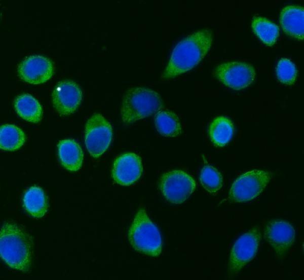
A07960-2 has IHC images from human paraffin sections and IF/ICC images from human tissue and SiHa cells (catalog image captions). Listed reactivity includes human, mouse, and rat (catalog).
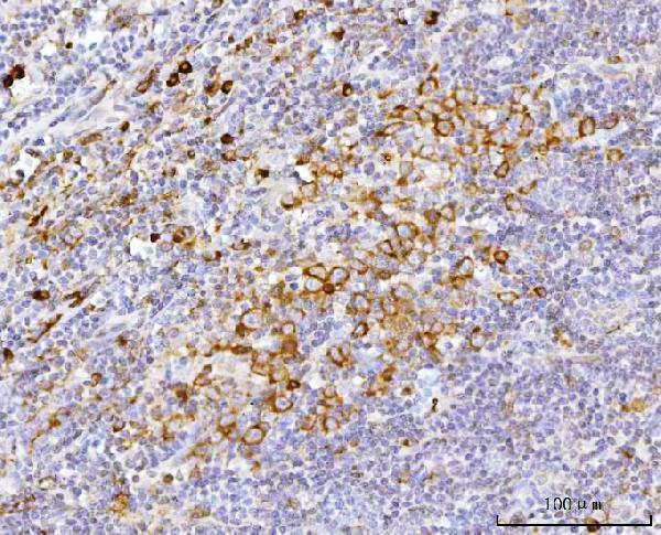
A07960-2 was demonstrated by IHC on paraffin sections of human cervical cancer and tonsil tissue (catalog IHC image captions). Its IF images show SiHa cells and paraffin sections of human chronic tonsillitis and ovary cancer tissue (catalog IF image captions).
Which to pick: For tissue IHC, choose A07960-2: its paraffin-section captions document EDTA retrieval at pH 8.0 and primary antibody at 2 μg/ml; the fixative is unreported (catalog IHC image captions). For IF/ICC, the same SKU has human cell and tissue images at 5 μg/ml (catalog IF image captions). For mouse or rat samples, A07960-2 lists reactivity with both species, but the supplied IHC and IF images document human samples (catalog reactivity; catalog image captions).