This website uses cookies to ensure you get the best experience on our website.


- Table of Contents
Plan PABPN1 paraffin IHC around widespread nuclear staining (HPA tissue IHC) and a catalog antibody range of 2–5 μg/ml (datasheet A02445-2). Compare high staining appendix glandular cells with low staining skeletal myocytes (HPA tissue IHC), and account for HPA’s multi-gene targeting caution (HPA tissue IHC).

Expected localisation, validated protocols, controls and antibodies — the at-a-glance facts below, then the full design guide.
| Expected localisation | Predominantly nuclear; tissue IHC shows nuclear staining (UniProt; HPA tissue IHC) | |
| Staining pattern | Widespread nuclear staining across cell types (HPA tissue IHC) | |
| Antigen retrieval | EDTA pH 8.0 HIER, heat-mediated (datasheet A02445-2) | |
| Positive control | Appendix+4 more · see all | |
| Negative control | None in HPA (detected in all 45 tissues); use no-primary + isotype controls |
| Fixation | Keep fixation consistent across paraffin sections. (standard IHC practice; not target-specific) | |
| Caveat | Staining may reflect proteins from more than one gene (HPA tissue IHC) | |
| Regulation | Ubiquitously expressed (UniProt) | |
| Isoform / epitope | 3 isoforms; epitope differences unspecified (UniProt) |
The catalog antibody’s IHC-P protocol is supplemented by published PABPN1 staining methods for muscle biopsies (PMC9727945) and paraffin-embedded hepatoblastoma tissues (PMC10520360).
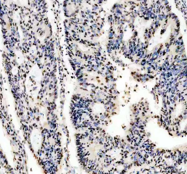
| Sample | Paraffin-embedded human colon cancer tissue; fixative not specified (datasheet A02445-2) |
| Fixation | Image fixative and duration unreported (datasheet A02445-2); verify before use. |
| Sectioning | 4–5 µm sections on charged slides (standard) |
| Deparaffinisation | Xylene, graded ethanol series to water (standard) |
| Antigen retrieval | Heat retrieval: EDTA pH 8.0 (datasheet A02445-2); 20 min, 95–100 °C (standard) |
| Peroxidase block | 3% H2O2, 10 min, room temperature (standard) |
| Blocking | 10% goat serum (datasheet A02445-2) |
| Primary antibody | Rabbit anti-PABPN1, 2-5 μg/ml (datasheet A02445-2) |
| Primary incubation | Overnight at 4 °C (datasheet A02445-2) |
| Detection | HRP-conjugated secondary, DAB chromogen (datasheet A02445-2) |
| Counterstain | Hematoxylin, blue, dehydrate and mount (standard) |
| Expected result | PABPN1-positive staining in glandular cells of appendix (HPA tissue IHC: High). HPA tissue profile: Ubiquitous nuclear expression. No signal in the no-primary control. |
PABPN1 is predominantly nuclear, with nucleoplasmic and nuclear-speckle localisation; UniProt also reports cytoplasmic shuttling (UniProt Q86U42; HPA: supported subcellular locations). Expect staining across many cell types, with strong staining in several glandular and epithelial populations (UniProt: ubiquitous; HPA: ubiquitous nuclear expression; HPA: High in appendix glandular and bronchial respiratory epithelial cells). HPA rates tissue IHC reliability Supported, with medium agreement between staining and RNA data and a warning that the antibodies may recognise proteins from more than one gene (HPA: tissue IHC reliability).
| Nuclear staining in glandular or respiratory epithelial cells, with visible unstained tissue structures. | This fits the reported distribution: HPA records High staining in appendix glandular cells and bronchial respiratory epithelial cells and describes ubiquitous nuclear expression (HPA: tissue IHC). Judge the signal within identified cells and their nuclei; a uniformly coloured section gives less confidence than a pattern with discernible cellular boundaries (standard IHC practice). |
| Predominantly cytoplasmic or extracellular staining, with little nuclear staining. | This is discordant with the main expected compartment (UniProt Q86U42: predominantly nuclear; HPA: nucleoplasm and nuclear speckles). Cytoplasmic signal alone cannot prove artefact because UniProt also reports cytoplasmic shuttling and mRNP granules (UniProt Q86U42). Check morphology, detection controls and an independently validated antibody before assigning the signal to PABPN1 (standard IHC practice; HPA: cross-gene recognition warning). |
| Strong staining in an unexpected cell population, especially when expected nuclei are faint. | Treat an isolated cell-type claim cautiously: UniProt calls expression ubiquitous, and HPA lists no negative tissue in this payload (UniProt Q86U42: tissue specificity; HPA: tissue IHC). Antibody cross-reactivity or endogenous detection activity can mimic a positive population (HPA: cross-gene recognition warning; standard IHC practice). Confirm cell identity by morphology and inspect appropriate reagent controls (standard IHC practice). |
| Diffuse colour over nuclei, cytoplasm and tissue spaces, without clear cellular boundaries. | This cannot be scored confidently as the expected nuclear pattern (HPA: ubiquitous nuclear expression). General IHC causes include excess primary antibody, insufficient washing, nonspecific detection or residual endogenous activity (standard IHC practice). Compare a no-primary control and review reagent concentrations, washing and blocking before interpreting intensity (standard IHC practice). |
| No nuclear signal in a known-positive appendix or bronchus section. | Absence conflicts with the reported High staining in the specified cell populations (HPA: High in appendix glandular and bronchial respiratory epithelial cells). First verify that those cells are present and that a positive control processed in the same run works (standard IHC practice). A failed control points to assay performance; a working control calls for closer review of the test section and antibody specificity (standard IHC practice; HPA: cross-gene recognition warning). |
| Compartment and topology | PABPN1 has no transmembrane segment and is predominantly nuclear (UniProt Q86U42: topology and localisation). Expect a cellular nuclear readout rather than membrane outlining (UniProt Q86U42; HPA: ubiquitous nuclear expression). UniProt-reported cytoplasmic shuttling means minor cytoplasmic signal needs context, not automatic rejection (UniProt Q86U42). |
| Tissue and cell selection | Appendix glandular cells and bronchial respiratory epithelial cells are reported High; heart cardiomyocytes and skeletal myocytes are reported Low (HPA: tissue IHC). Low is not a negative control, and no negative tissue is listed in this payload (HPA: tissue IHC). Use a reported High population as the positive reference when assessing a weak section (standard IHC practice). |
| Antibody evidence and specificity | HPA000637 has Supported IHC status; HPA079093 has no IHC status in the supplied list (HPA: antibody validation). HPA also warns that tissue staining may detect proteins from more than one gene and reports only medium consistency with RNA expression (HPA: tissue IHC reliability). An apparently correct location alone therefore does not establish target-specific staining (HPA: tissue IHC reliability). |
| Isoforms and epitope knowledge | UniProt lists three isoforms and an RRM at residues 172–249 (UniProt Q86U42: isoforms and domains). The payload gives no antibody epitope, so it cannot establish which isoforms either antibody detects or predict a retrieval response (UniProt Q86U42; HPA: antibody validation). Record the actual reagent and its IHC validation when comparing sections (standard IHC practice). |
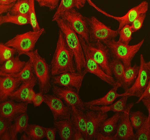
| IF/ICC Q&A: what pattern should I expect? | HPA supports nucleoplasmic and nuclear-speckle localisation in ICC-IF (HPA: subcellular localisation). That agrees with predominant nuclear localisation in UniProt (UniProt Q86U42). This answers the localisation question only; the separate IF/ICC guide covers its assay workflow. |
| Situation | Likely cause | Next action |
|---|---|---|
| High control tissue is blank. | The expected cells may be absent from the section, or the staining run may have failed (HPA: High in appendix glandular and bronchial respiratory epithelial cells; standard IHC practice). | Find the relevant cells on the counterstain, then check a same-run positive control and the primary-antibody and detection steps (standard IHC practice). |
| Signal is mostly cytoplasmic. | PABPN1 can shuttle, but a chiefly cytoplasmic pattern conflicts with its predominant nuclear localisation (UniProt Q86U42; HPA: ubiquitous nuclear expression). Nonspecific staining is another possibility (standard IHC practice). | Check nuclear morphology and no-primary controls; confirm any claimed cytoplasmic pattern with independent target evidence (standard IHC practice; HPA: cross-gene recognition warning). |
| The whole section looks brown or hazy. | Nonspecific detection, excess reagent or incomplete washing can obscure nuclear detail (standard IHC practice). | Inspect a no-primary control and review blocking, antibody concentration, washes and detection timing using the reagent's IHC instructions (standard IHC practice). |
| A surprising cell population stains strongly. | Expression is widespread, while HPA flags possible recognition of proteins from more than one gene; endogenous detection activity can also produce apparent positives (UniProt Q86U42: ubiquitous; HPA: tissue IHC reliability; standard IHC practice). | Confirm cell identity, inspect reagent controls and compare localisation with a separately validated antibody where available (standard IHC practice). |
| Heart or skeletal muscle has weak staining. | HPA reports Low staining in cardiomyocytes and skeletal myocytes (HPA: tissue IHC). Weak staining there alone does not show that the assay failed. | Compare a reported High tissue population processed in the same run before changing assay conditions (HPA: tissue IHC; standard IHC practice). |
| A retrieval change alters staining. | The supplied UniProt and HPA records do not establish a PABPN1-specific fixation or retrieval effect (UniProt Q86U42; HPA: tissue IHC and antibody validation). | Compare the conditions using the same tissue and controls; assess whether nuclear definition and background improve, without attributing the change to a proven PABPN1-specific mechanism (standard IHC practice). |
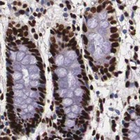


Comprehensive Human Protein Atlas IHC scoring per tissue (reliability: Supported — Medium consistency between antibody staining and RNA expression data. Caution, targets protein from more than one gene.). Rows are taken directly from the HPA tissue chart — click any row's HPA link to view the source.
| Tissue | Cell type | Level | Evidence | Source |
|---|---|---|---|---|
| Appendix | Glandular cells | High | Protein (IHC) | HPA → |
| Bronchus | Respiratory epithelial cells | High | Protein (IHC) | HPA → |
| Cerebral cortex | Neuropil | High | Protein (IHC) | HPA → |
| Cervix | Glandular cells | High | Protein (IHC) | HPA → |
| Duodenum | Glandular cells | High | Protein (IHC) | HPA → |
| Tissue | Cell type | Level | Evidence | Source |
|---|---|---|---|---|
| None in HPA: PABPN1 is detected in all 45 scored tissues. Use a no-primary (secondary-only) and an isotype control instead. | ||||
Use nuclear staining as the primary readout for PABPN1 in chromogenic paraffin-section IHC, with attention to retrieval, tissue controls and compartment-specific scoring (UniProt Q86U42; HPA tissue IHC).
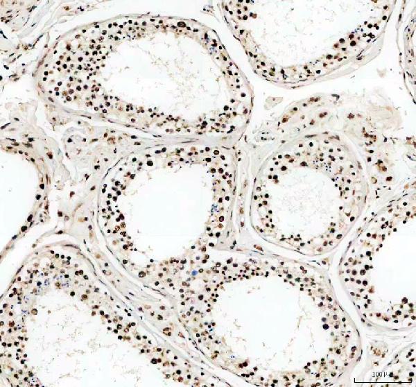
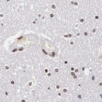
A02445-2 has IHC images from human colon cancer and testis paraffin sections and mouse and rat brain paraffin sections, plus IF images from human cells and tissue and mouse brain (catalog image captions).
A02445-2 will render with a human colon cancer paraffin-section IHC figure; its other captions show IHC in human testis and mouse and rat brain, and IF in human cells and tissue and mouse brain (A02445-2 image captions). M02445 will render with IHC, IF and ICC listed for human, mouse and rat, but no IHC or IF image captions are supplied (M02445 catalog applications and reactivity; image captions absent).
Which to pick: Choose A02445-2 for paraffin-section IHC because its own captions document staining in human, mouse and rat tissue; the fixative is unreported (A02445-2 IHC image captions). For IF/ICC, A02445-2 has images from human cells and paraffin sections, while M02445 is a rabbit monoclonal with IF/ICC listed but no image captions supplied (A02445-2 IF image captions; M02445 catalog clone, applications and image captions). Both list human, mouse and rat reactivity, but A02445-2 has IHC images across all three species (catalog reactivity; A02445-2 IHC image captions).