PAK3 / Serine/threonine-protein kinase PAK 3 · IHC design guide

Design Immunohistochemistry for PAK3

Plan PAK3 paraffin IHC around cerebellar Purkinje-cell cytoplasmic staining (HPA tissue IHC). Use the catalog antibody at 2–5 μg/ml (datasheet A03124-1), with consistent fixation and chromogenic controls.

Evidence assembled Oct 2026 · For research use; verify linked source records and product datasheet before use
Immunohistochemistry protocol sheet for PAK3 (IHC for PAK3): expected localisation Cytoplasmic in Purkinje cells (HPA tissue IHC), antibody A03124-1, validated IHC image, and IHC protocol steps
Printable PAK3 IHC protocol sheet — expected localisation Cytoplasmic in Purkinje cells (HPA tissue IHC), antibody A03124-1, controls and protocol steps. Open the full PAK3 IHC guide →

PAK3 Immunohistochemistry Experimental Design Guide

Expected localisation, validated protocols, controls and antibodies — the at-a-glance facts below, then the full design guide.

Must know before staining
Expected localisation Cytoplasmic in Purkinje cells (HPA tissue IHC)
Staining pattern Cerebellar Purkinje-cell cytoplasmic staining (HPA tissue IHC)
Antigen retrieval EDTA pH 8.0 HIER, heat-mediated (datasheet A03124-1)
Positive control ⓘ Cerebellum
Negative control ⓘ Adipose tissue+4 more · see all
Important caveats
Reasons your staining may differ from the expected pattern.
Fixation Matched tissue-IHC evidence does not establish the fixation claim. Validate the specimen-specific method before use. (selected-SKU IHC image A03124-1)
Caveat Antibody staining has low consistency with RNA data (HPA tissue IHC)
Regulation High in developing and postnatal neurons (UniProt)
Isoform / epitope 4 isoforms; epitope coverage needs checking (UniProt)
Section 1

Recommended PAK3 IHC & IF Protocols

The catalog antibody uses EDTA pH 8.0 retrieval (datasheet: A03124-1). Two published PAK3 IHC protocols cover hepatocellular carcinoma and neuroendocrine tumor tissue arrays (PMC8692680; PMC13615580).

Recommended immunohistochemistry (IHC-P) protocol parameters
SampleParaffin-embedded human thyroid papillary carcinom tissue; fixative not specified (datasheet A03124-1)
FixationImage fixative and duration unreported (datasheet A03124-1); verify before use.
Sectioning4–5 µm sections on charged slides (standard)
DeparaffinisationXylene, graded ethanol series to water (standard)
Antigen retrievalHeat retrieval: EDTA pH 8.0 (datasheet A03124-1); 20 min, 95–100 °C (standard)
Peroxidase block3% H2O2, 10 min, room temperature (standard)
Blocking10% goat serum (datasheet A03124-1)
Primary antibodyRabbit anti-PAK3, 2-5 μg/ml (datasheet A03124-1)
Primary incubationOvernight at 4 °C (datasheet A03124-1)
DetectionStreptavidin-biotin complex (SABC), DAB chromogen (datasheet A03124-1)
CounterstainHematoxylin, blue, dehydrate and mount (standard)
Expected resultPAK3-positive staining in purkinje cells of cerebellum (HPA tissue IHC: Medium). HPA tissue profile: Cytoplasmic expression restricted to Purkinje cells. No signal in the no-primary control.
💡Decision noteStart with heat-mediated EDTA pH 8.0 for the catalog antibody (datasheet: A03124-1); citrate pH 6.0 is a published alternative with a different PAK3 antibody (PMC13615580).
Section 2

What Is the Expected PAK3 Staining Pattern?

For paraffin-section IHC, expect cytoplasmic PAK3 staining in cerebellar Purkinje cells (HPA tissue IHC; UniProt O75914 subcellular location). HPA reports medium staining there and describes the observed tissue pattern as restricted to Purkinje cells (HPA tissue IHC). Its IHC assessment is Approved, but antibody staining has low consistency with RNA expression; external characterization supports the staining (HPA tissue IHC). PAK3 has no transmembrane segment (UniProt O75914 topology).

What am I looking at on my slide?
Medium cytoplasmic staining outlines Purkinje cell bodies in cerebellum, with limited staining elsewhere on the section.This matches the observed positive tissue and cell compartment (HPA tissue IHC: cerebellar Purkinje cells, Medium; UniProt O75914: cytoplasm). Judge the cell pattern alongside morphology and a matched negative control (standard IHC practice).
Strong nuclear or surface-rim staining dominates the Purkinje cell signal.That distribution does not match the reported cytoplasmic tissue pattern (HPA tissue IHC; UniProt O75914 subcellular location). Check whether the same pattern appears in the negative control, then review antibody specificity and detection settings (standard IHC practice).
Prominent staining appears in adipocytes or other cells recorded as negative by HPA.HPA reports adipocytes as Not detected in adipose tissue (HPA tissue IHC). Consider cross-reactivity or endogenous detection activity; inspect cell identity, the negative control and reagent controls before assigning the signal to PAK3 (standard IHC practice).
Brown signal spreads evenly across cells and surrounding tissue without a recognizable Purkinje cell pattern.Diffuse staining is difficult to interpret as the reported restricted cytoplasmic pattern (HPA tissue IHC). Review background in a control lacking primary antibody, then assess blocking, washing and detection exposure (standard IHC practice).
Purkinje cells show no detectable signal in a cerebellar section.The expected HPA reference is Medium staining in those cells (HPA tissue IHC). Confirm tissue identity and run a known-positive section in the same batch; then review retrieval, antibody conditions and detection performance (standard IHC practice).
💡Expected PAK3 appearanceA credible positive is medium cytoplasmic staining in cerebellar Purkinje cells (HPA tissue IHC); widespread nuclear staining or uniform background is discordant with that pattern (HPA tissue IHC; UniProt O75914 subcellular location).
How each factor affects the staining
Tissue pattern and evidence strengthHPA records Medium staining in cerebellar Purkinje cells and calls the tissue pattern restricted to them (HPA tissue IHC). Its Approved assessment includes low staining-to-RNA consistency, so interpret unexpected cells with controls rather than treating the tissue profile as an absolute exclusion.
Antibody validation by applicationHPA060219 is Approved for IHC, whereas HPA044791 is Approved for ICC and has no listed IHC status (HPA antibodies). An ICC result does not establish how an antibody performs in paraffin-section IHC (standard assay-specific validation practice).
Isoforms and epitope coverageUniProt lists 4 PAK3 isoforms (UniProt O75914). The supplied evidence does not map either antibody's epitope to those isoforms; avoid claiming that a staining difference identifies a particular isoform.
Retrieval and detection conditionsAntigen retrieval, blocking and chromogen development are general paraffin-section IHC variables (standard IHC practice). No supplied source establishes PAK3-specific fixation sensitivity or a required retrieval condition; optimize against positive tissue and negative controls.
IF/ICC Q&A: where is PAK3 seen?HPA reports approved vesicular localization in ICC-IF images (HPA subcellular ICC-IF). That cell-based observation should be read in its own assay context; the paraffin-section tissue reference is cytoplasmic staining in Purkinje cells (HPA tissue IHC).
Why is my staining missing, weak or wrong?
SituationLikely causeNext action
No staining in the cerebellar positive sectionA failed staining run or unsuitable assay conditions are possible; HPA reports Medium Purkinje cell staining, without a PAK3-specific fixation claim (HPA tissue IHC).Confirm Purkinje cells are present, check the same-batch positive control, and review retrieval, primary antibody and detection steps one at a time (standard IHC practice).
All compartments look uniformly brownBackground from detection reagents, inadequate blocking or excess development can obscure cell-specific staining (standard IHC practice).Compare a control lacking primary antibody; adjust blocking, washes or chromogen development before scoring the section (standard IHC practice).
Nuclei stain more strongly than cytoplasmA nuclear-dominant pattern conflicts with the cytoplasmic tissue pattern (HPA tissue IHC; UniProt O75914 subcellular location).Inspect morphology and negative controls, then reassess antibody specificity and detection settings; do not score nuclear signal alone as the expected PAK3 result (standard IHC practice).
Adipocytes stain stronglyAdipocytes are Not detected in HPA's adipose tissue IHC record (HPA tissue IHC); cross-reactivity or detection background may explain discordant staining.Check a control lacking primary antibody and verify the cell identity; compare the result with Purkinje cell staining before interpreting it as PAK3 (standard IHC practice).
Only an ICC-approved antibody is available for paraffin IHCHPA044791 has ICC Approved status but no listed IHC status; HPA060219 has IHC Approved status (HPA antibodies).Use an IHC-validated antibody when available, or establish performance in paraffin sections with positive tissue and negative controls before interpreting staining (standard assay validation practice).
Vesicular ICC-IF images appear different from tissue IHCHPA reports vesicles in ICC-IF and cytoplasmic Purkinje cell staining in tissue IHC (HPA subcellular ICC-IF; HPA tissue IHC).Interpret each observation within its application; use the tissue IHC record as the reference for paraffin-section scoring (HPA tissue IHC).

Sample controls for PAK3 IHC & IF

🧪Run cerebellum first; Purkinje cells should stain (HPA: Medium in cerebellar Purkinje cells). Use adipose tissue as the negative tissue, with adipocytes expected to lack detectable staining (HPA: Not detected in adipocytes); on the cerebellar slide, use cells showing only background signal as internal negative profiles without assuming a particular neighboring cell type is PAK3-negative.
Positive control tissue: Cerebellum (Purkinje cells, HPA Medium)
Negative control tissue: Adipose tissue (HPA Not detected)
ICC-IF cell lines (HPA subcellular resource): HPA ICC-IF images show PAK3 in EFO-21, HAP1, U2OS, with annotated localisation: Vesicles (approved) (HPA subcellular).
Technical controls: Include a no-primary, secondary-only control; a concentration-matched rabbit IgG isotype control for the rabbit primary; and, if available, a PAK3 knockout specimen or immunizing-peptide competition control (caption: rabbit anti-PAK3; standard IHC control practice). For chromogenic IHC, block endogenous peroxidase and assess endogenous biotin before using the reported streptavidin–biotin/DAB detection; for cerebellar IF, check autofluorescence with the no-primary control (caption: SABC/DAB; standard IHC/IF practice).
⚠️Feasibility: A target-specific fixation window or fixation effect is unreported, and the selected A03124-1 paraffin-section caption does not state the fixative (caption: fixative not stated). The caption uses heat retrieval in EDTA at pH 8.0, but does not establish that retrieval is required; paraffin IHC is demonstrated, while the supplied evidence does not establish whether frozen sections or IF are easier (caption: EDTA retrieval and paraffin section). In cerebellum, use morphology and a counterstain to assign signal to Purkinje cells, and assess tissue autofluorescence if using IF (HPA: Medium in cerebellar Purkinje cells; standard IHC/IF practice).

HPA tissue IHC evidence for PAK3

Comprehensive Human Protein Atlas IHC scoring per tissue (reliability: Approved — Low consistency between antibody staining and RNA expression data. External characterization data supports antibody staining.). Rows are taken directly from the HPA tissue chart — click any row's HPA link to view the source.

Positive expression · recommended positive controls

TissueCell typeLevelEvidenceSource
Cerebellum Purkinje cells Medium Protein (IHC) HPA →

Undetected expression · recommended negative controls

TissueCell typeLevelEvidenceSource
Adipose tissue Adipocytes Not detected Protein (IHC) HPA →
Adrenal gland Glandular cells Not detected Protein (IHC) HPA →
Appendix Glandular cells Not detected Protein (IHC) HPA →
Bone marrow Hematopoietic cells Not detected Protein (IHC) HPA →
Breast Adipocytes Not detected Protein (IHC) HPA →
Section 3

Advanced PAK3 IHC Tips

Troubleshoot chromogenic PAK3 staining in paraffin sections using the catalog image conditions and tissue and localisation evidence (datasheet A03124-1; HPA; UniProt O75914).

How should I adjust retrieval when PAK3 staining is weak or diffuse?
Start with heat-mediated retrieval in EDTA at pH 8.0 for paraffin-section PAK3 IHC (datasheet A03124-1). The selected image paired this retrieval with 2 μg/ml primary antibody overnight at 4°C; hold antibody conditions constant while comparing retrieval cycles (datasheet A03124-1). If staining remains weak, compare shorter and longer heating within your instrument's validated range, then cool and process sections identically (standard IHC practice). Judge improvement by cytoplasmic staining in Purkinje cells and low staining in negative tissue, while rejecting diffuse staining from damaged sections (HPA: tissue IHC; standard IHC practice).
Could fixation explain weak PAK3 staining in my paraffin sections?
The selected tissue caption does not report a fixative, so target-specific PAK3 fixation sensitivity is unknown (datasheet A03124-1). Record the fixative, concentration, fixation interval and processing history for each specimen before comparing staining between blocks (standard IHC practice). If fixation varies, test matched sections under the stated EDTA pH 8.0 retrieval and 2 μg/ml primary antibody conditions, changing one preparation variable at a time (datasheet A03124-1; standard IHC practice). Include a consistently processed positive control and assess preserved morphology alongside cytoplasmic staining; an inconsistent signal alone cannot establish fixation as the cause (HPA: tissue IHC; standard IHC practice).
Where should convincing PAK3 staining appear in a tissue section?
Prioritise cellular cytoplasmic staining: UniProt places PAK3 in the cytoplasm, and HPA reports cytoplasmic expression in cerebellar Purkinje cells (UniProt O75914; HPA: tissue IHC). HPA also reports an approved vesicular location in cell imaging, which may appear as cytoplasmic puncta under suitable resolution (HPA: subcellular). PAK3 has no transmembrane segment, so an exclusively crisp membrane outline warrants scrutiny rather than automatic acceptance as target signal (UniProt O75914 topology). Compare staining in identified Purkinje cells with adjacent cells and a negative control section under identical DAB development; assess morphology and compartment together (HPA: tissue IHC; standard IHC practice).
Could isoforms or phosphorylation change the apparent PAK3 staining pattern?
PAK3 has 4 annotated isoforms, a CRIB region at residues 70–83, and a kinase domain at 283–534 (UniProt O75914). The supplied caption gives no immunogen sequence or epitope map, so staining cannot be assigned to one isoform or phosphorylation state (datasheet A03124-1). UniProt records several phosphoserines and an autophosphorylated threonine at residue 436, but those annotations do not establish sensitivity of this antibody to phosphorylation (UniProt O75914). When discrepant sections raise an epitope concern, compare matched processing and use independently characterised reagents with known epitopes before attributing staining differences to isoforms or modification (standard IHC practice).
How can I check the chromogenic result with tissue immunofluorescence?
Treat IF as a separate validation experiment because the supplied catalog image documents chromogenic IHC in a paraffin section (datasheet A03124-1). Multiplex PAK3 with an independently validated Purkinje-cell marker, and confirm that both signals occupy the expected cells while evaluating PAK3 within their cytoplasm (HPA: tissue IHC; UniProt O75914). Choose spectrally separated fluorophores after measuring tissue autofluorescence in unstained sections, and include single-label controls to assess bleed-through (standard IF practice). Because PAK3 is cytoplasmic and lacks a transmembrane segment, use permeabilisation appropriate for an intracellular epitope, then titrate it to preserve morphology and punctate signal (UniProt O75914 topology; HPA: subcellular; standard IF practice).
What causes widespread brown signal in PAK3 IHC?
The selected image used 10% goat serum blocking, a biotinylated secondary antibody, streptavidin–biotin detection and DAB (datasheet A03124-1). With this detection scheme, compare no-primary and secondary-only controls, and assess endogenous peroxidase and biotin contributions using suitable blocking controls (standard IHC practice). Keep DAB development time identical across sections and titrate the primary around the documented 2 μg/ml condition if diffuse staining obscures cell boundaries (datasheet A03124-1; standard IHC practice). Broad staining across unrelated cells is especially suspect when the intended reference pattern is cytoplasmic staining restricted to Purkinje cells (HPA: tissue IHC).
How should I score PAK3 staining across paraffin sections? ⚠ ANSWER MARKED FOR VERIFICATION
Define the anatomical region and cell type before scoring; HPA identifies cerebellar Purkinje cells as a cytoplasmic positive reference (HPA: tissue IHC). For cell-based scoring, report the percentage of positive target cells and an H-score from 0–300, calculated from the proportions at intensity grades 0–3 (standard IHC practice). If counting stained cells per area, report density per mm² of evaluable tissue and normalise comparisons to the same cell population, section quality and DAB development conditions (standard IHC practice). Exclude folds, edges and necrotic regions consistently, and state thresholds before reviewing experimental groups (standard IHC practice).
How do I distinguish a true PAK3 positive from an artefact?
A credible positive should occur in a plausible cell and compartment: HPA reports medium cytoplasmic staining in Purkinje cells, while UniProt places PAK3 in the cytoplasm (HPA: tissue IHC; UniProt O75914). Inspect unexpected nuclear or sharply membranous staining, section-edge gradients, necrotic areas and pigment-like deposits against morphology and no-primary controls (UniProt O75914 topology; standard IHC practice). Endogenous enzyme activity can produce DAB signal, so compare peroxidase-blocked controls before assigning brown deposits to PAK3 (standard IHC practice). The catalog caption shows staining in thyroid papillary carcinoma tissue, while HPA notes low consistency between antibody staining and RNA data; interpret unexpected tissue positives cautiously (datasheet A03124-1; HPA: tissue IHC).
Boster reagents

Best PAK3 / Serine/threonine-protein kinase PAK 3 IHC Antibodies

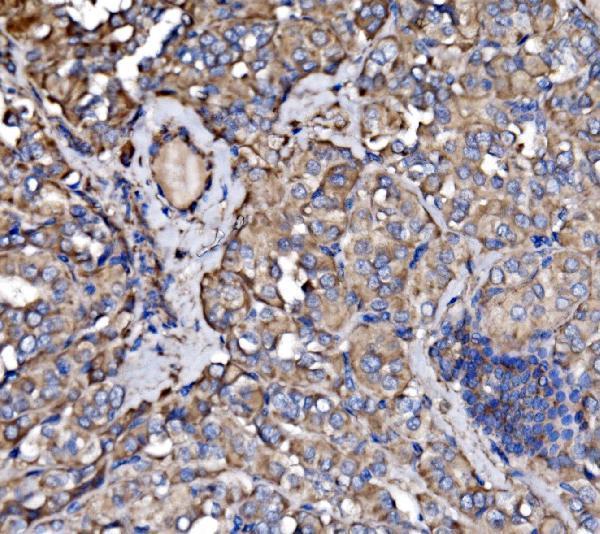
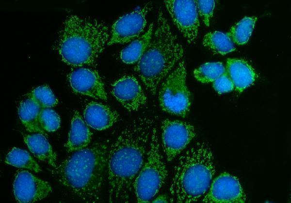
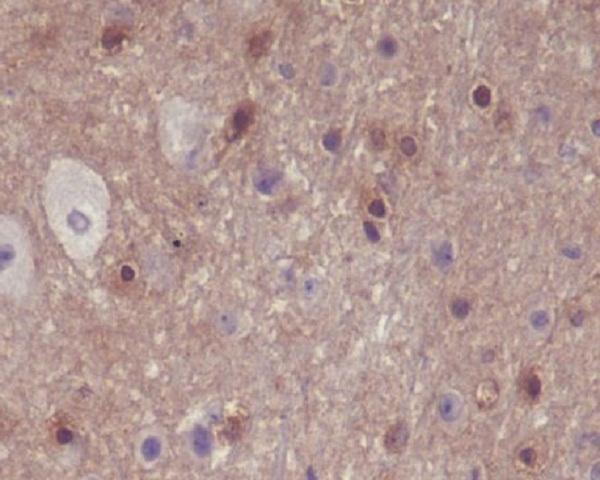
A03124-1 has paraffin-section IHC images from human, mouse and rat tissues and an IF image from MCF-7 cells; P03124 has a mouse-brain IHC image (catalog image captions).

Real IHC data IHC analysis of PAK3 using anti-PAK3 antibody (A03124-1). PAK3 was detected in a paraffin-embedded section of human thyroid papillary carcinom tissue. Heat mediated antigen retrieval was performed in EDTA buffer (pH 8.0, epitope retrieval solution). The tissue section was blocked with 10% goat serum. The tissue section was then incubated with 2 μg/ml rabbit anti-PAK3 Antibody (A03124-1) overnight at 4°C. Biotinylated goat anti-rabbit IgG was used as secondary antibody and incubated for 30 minutes at 37°C. The tissue section was developed using Strepavidin-Biotin-Complex (SABC) (Catalog # SA1022) with DAB as the chromogen.
Anti-PAK3 Antibody ®
Cat # A03124-1
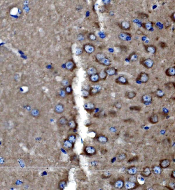
Real IHC data Immunohistochemical analysis of paraffin-embedded mouse brain, using Phospho-PAK1/2/3 (S144+S141+S139) Antibody.
Anti-Phospho-PAK1/2/3 (S144+S141+S139) PAK3 Rabbit Monoclonal Antibody
Cat # P03124

A03124-1 will render with its human thyroid papillary carcinoma IHC figure; its other captions show human cerebellum, mouse and rat brain IHC, and MCF-7 cell IF (A03124-1 image captions). P03124 will render with paraffin-embedded mouse brain IHC showing phospho-PAK1/2/3 staining (P03124 IHC caption).

Which to pick: For PAK3 tissue IHC, choose A03124-1: its own paraffin-section captions document human, mouse and rat staining with EDTA retrieval at pH 8.0 and 2 μg/ml primary antibody (A03124-1 IHC captions). For IF/ICC, A03124-1 has an MCF-7 cell IF image at 5 μg/ml (A03124-1 IF caption); P03124 lists IF/ICC but its image documents paraffin-section IHC of phospho-PAK1/2/3 in mouse brain (P03124 applications and IHC caption). Both list human, mouse and rat reactivity, while A03124-1 has IHC images for all three species (catalog reactivity; A03124-1 IHC captions); the fixative is unreported in both products’ IHC captions (catalog IHC captions).

Each figure is that product's own IHC / IF validation image from its datasheet.

References

  1. UniProt Consortium. UniProt entry O75914 (PAK3_HUMAN, Serine/threonine-protein kinase PAK 3).
  2. Human Protein Atlas. PAK3 tissue IHC expression (reliability: Approved).
  3. Human Protein Atlas. PAK3 subcellular location (ICC-IF): Localized to vesicles..
  4. Human Protein Atlas. PAK3 antibody validation summary (2 antibodies).
  5. Pak3 promotes cell cycle exit and differentiation of β-cells in the embryonic pancreas and is necessary to maintain glucose homeostasis in adult mice. Diabetes 2014 — PMC3968432.
  6. Restored autophagy is protective against PAK3-induced cardiac dysfunction. iScience 2023 — PMC10265534.
  7. PAK3 promotes the metastasis of hepatocellular carcinoma by regulating EMT process. Journal of Cancer 2022 — PMC8692680.
  8. AI-powered histopathology classifiers identify transcriptomic biomarkers associated with cell cycle regulation and increased mitotic activity in poorly differentiated neuroendocrine carcinoma. Human Genomics 2026 — PMC13615580.
  9. PubMed PMID:9731525 — UniProt-cited evidence.
  10. PubMed PMID:12777533 — UniProt-cited evidence.
  11. PubMed PMID:18507705 — UniProt-cited evidence.