This website uses cookies to ensure you get the best experience on our website.
- Table of Contents
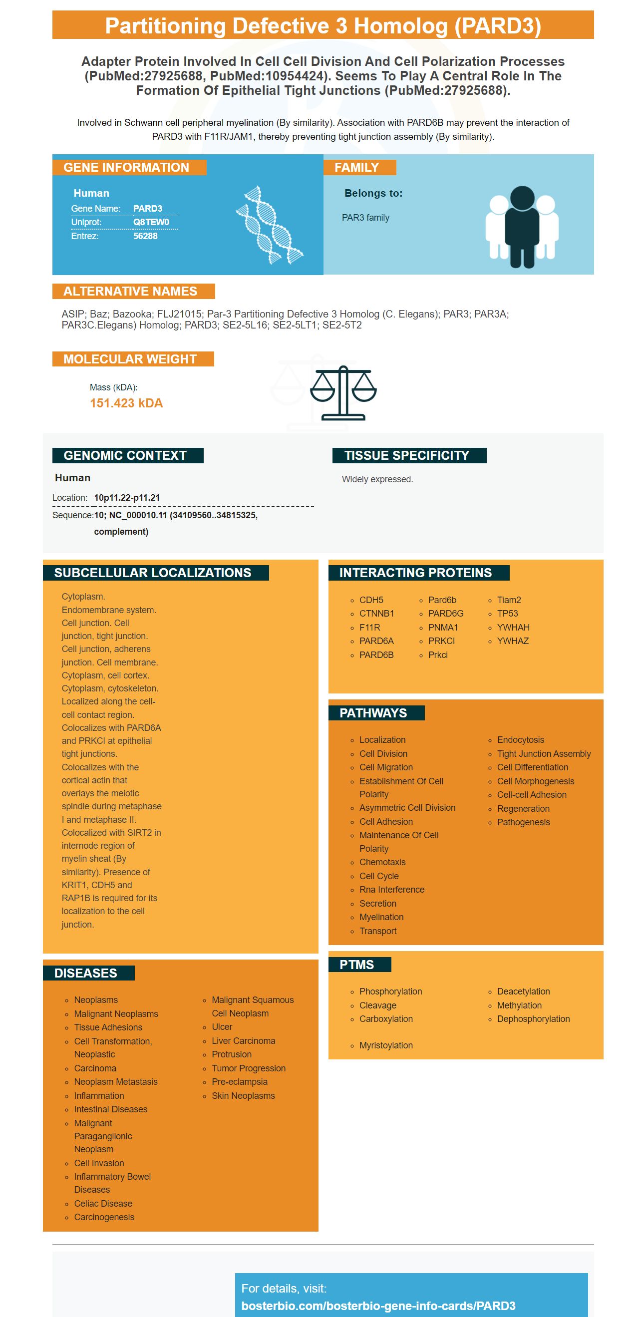
Facts about Partitioning defective 3 homolog.
Involved in Schwann cell peripheral myelination (By similarity). Association with PARD6B may prevent the interaction of PARD3 with F11R/JAM1, thereby preventing tight junction assembly (By similarity).
Human | |
---|---|
Gene Name: | PARD3 |
Uniprot: | Q8TEW0 |
Entrez: | 56288 |
Belongs to: |
---|
PAR3 family |
ASIP; Baz; Bazooka; FLJ21015; par-3 partitioning defective 3 homolog (C. elegans); PAR3; PAR3A; PAR3C.elegans) homolog; PARD3; SE2-5L16; SE2-5LT1; SE2-5T2
Mass (kDA):
151.423 kDA
Human | |
---|---|
Location: | 10p11.22-p11.21 |
Sequence: | 10; NC_000010.11 (34109560..34815325, complement) |
Widely expressed.
Cytoplasm. Endomembrane system. Cell junction. Cell junction, tight junction. Cell junction, adherens junction. Cell membrane. Cytoplasm, cell cortex. Cytoplasm, cytoskeleton. Localized along the cell-cell contact region. Colocalizes with PARD6A and PRKCI at epithelial tight junctions. Colocalizes with the cortical actin that overlays the meiotic spindle during metaphase I and metaphase II. Colocalized with SIRT2 in internode region of myelin sheat (By similarity). Presence of KRIT1, CDH5 and RAP1B is required for its localization to the cell junction.
In this article, we will be discussing the PARD3 Marker, Steven Boster, and high affinity primary antibodies. These tools will be useful for researchers who are working on special samples and species. They will also provide credit for the results of their experiments. This article is applicable to scientists around the world. It is important to understand the advantages and disadvantages of each. To find out more, continue reading!
The PARD3 Marker is a cell polarity regulator. Mutations in this gene are not likely to cause truncated proteins. PARD3 has several functional domains: N-terminal CR1, central CR2 and PDZ domains. These domains are responsible for binding and inhibition of aPKC and the coil-coil domain 10.
The PARD3 gene has several mutations. Mutations have been found in several tumor types. Western blots can detect multiple isoforms of the protein. The most prevalent transcript is NM_001184785.1. However, some cancer types harbor more than one copy of the gene. Nonetheless, these mutations do not necessarily indicate disease progression. Consequently, further studies are needed to confirm the results of these experiments.
The PARD3 protein is an adaptor protein involved in a range of cellular processes. Planar cell migration is a special example, in which orientation is triggered by cell-cell contact. The Daple protein, which forms cell junctions in epithelial cells, is a component of the polarity regulator PARD3. It interacts with the PDZ domain of PARD3 and is tyrosine phosphorylated by receptor tyrosine kinases.
Researchers have discovered that the PARD3 gene is recurrently inactivated in lung cancers that express wild-type PARD3. The presence of the protein indicates that tumors have mutated feedback mechanisms derived from the mutations. In addition to the tumors containing PARD3, they are more aggressive and likely to metastasize, which is a sign of a tumor's potential to spread.
The activity of the PARD3 gene is associated with the activation of YAP/TAZ, a transcription factor that is directly regulated by the YAP/TAZ pathway. Its knockdown reduced the phosphorylation of YAP2 at Ser127, which promotes cytoplasmic localization of YAP. By inhibiting phosphorylation, PARD3 enhances the activity of YAP/TAZ.
Don "Steve" Boster, a father of three who passed away last month in Madison, WI, is commemorated with a PARD3 marker. Born in Joliet, IL, he fought a long battle with COVID-19. His sons, Donald, Sr., and David, were named in his honor. Nina Mae Hall was his only daughter. His parents lived in Joliet, IL. He is survived by his mother, Frances, and his siblings Kimberly and Tammy in Verona, WI, and his son Jonathan and his grandson Cory in Herrin, IL.
High-affinity antibodies are produced by antibody forming cells (AFCs). During the first response, the AFCs respond to antigens and then produce a higher-affinity antibody. This process is known as affinity maturation. The antibody produced by memory B-cells has higher affinity than those produced by AFCs earlier in the primary response. Here, we will discuss the process of affinity maturation and how it affects primary antibodies.
A high-affinity antibody can bind millions of epitopes. To code for every possible antigen-binding site, an organism's genome would have to expand exponentially. To achieve this, B-cells use somatic hypermutation, in which genes coding for an antigen-binding site undergo rapid mutations. As successive generations of B cells present the antigen, only those with high affinity survive. This process is called affinity maturation.
A lower KD value means the antibody is less likely to dissociate from its target. The KD value can also be used to assess the affinity of a primary antibody to a given molecule. In this process, an antibody solution at a particular concentration is injected at time zero and then run across the peptide microarray for fifteen minutes. When the antibody dissociates from the target, it binds at a much slower rate.
During the first week of the immune response, foci of antibody-forming cells are developed. They are located along the periarteriolar lymphoid sheath. These B cells secrete IgM and then switch to downstream immunoglobulin isotypes. The production of high-affinity antibodies is promoted by the presence of non-TFH CD4+ T cells, which are responsible for antibody maturation.
As with any antibody, affinity affects the ability to detect the antigen. High-affinity antibodies bind strongly to the antigen while low-affinity ones only bind weakly to the antigen. In addition, high-affinity antibodies can maintain the bond with the antigen even under the toughest conditions. By contrast, low-affinity antibodies may not detect the antigen in vivo or in an assay.
While low-affinity antibodies are useful for in vitro studies, it may be difficult to detect proteins that are low in abundance or that are expressed at low levels. This means that capturing and detecting low-level proteins can be difficult, which increases the difficulty of the experiment. Fortunately, the development of high-affinity primary antibodies is increasingly feasible, and the possibilities for improved therapy are endless. And thanks to the latest developments in immunochemistry, high-affinity antibodies are now available for a wide range of purposes.
PMID: 10934474 by Joberty G., et al. The cell-polarity protein Par6 links Par3 and atypical protein kinase C to Cdc42.
PMID: 11642408 by Fang C.M., et al. Down-regulated expression of atypical PKC-binding domain deleted asip isoforms in human hepatocellular carcinomas.